Low-level laser therapy effects on pain perception
related to the use of orthodontic elastomeric separators
Rachel D’Aurea Furquim1, Renata Correa Pascotto2, José Rino Neto3, Jefferson Rosa Cardoso4, Adilson Luiz Ramos5
How to cite this article: Furquim RD, Pascotto RC, Rino Neto J, Cardoso JR, Ramos AL. Low-level laser therapy effects on pain perception related to the use of orthodontic elastomeric separators. Dental Press J Orthod. 2015 May-June;20(3):37-42.
DOI: http://dx.doi.org/10.1590/2176-9451.20.3.037-042.oar
Submitted: February 10, 2014
Revised and accepted: December 17, 2014
» The authors report no commercial, proprietary or financial interest in the prod-ucts or companies described in this article.
1MSc in Integrated Dentistry, Universidade Estadual de Maringá, Maringá, Paraná, Brazil.
2 Associate professor, Universidade Estadual de Maringá, Maringá, Paraná, Brazil.
3 Associate Professor, Universidade de São Paulo, São Paulo, São Paulo, Brazil. 4 Associate professor, Universidade Estadual de Londrina, Londrina, Paraná,
Brazil.
5 Associate professor, Universidade Estadual de Maringá, Maringá, Paraná, Brazil.
Contact address: Rachel D’Aurea Furquim
Av. Dr. Luiz Teixeira Mendes, 2712. CEP - 87015-180 - Maringá - PR - Brazil E-mail: [email protected]
Introduction: Some patients refer to pre-banding orthodontic separation as a painful orthodontic procedure. Low-level laser therapy (LLLT) has been reported to have local analgesic effect. Objective: The aim of this single-blind study was to investigate the perception of pain caused by orthodontic elastomeric separators with and without a single LLLT appli-cation (6J). Methods: The sample comprised 79 individuals aged between 13 and 34 years old at orthodontic treatment onset. Elastomeric separators were placed in first maxillary molars at mesial and distal surfaces and kept in place for three days. The volunteers scored pain intensity on a visual analogue scale (VAS) after 6 and 12 hours, and after the first, sec-ond and third days. One third of patients received laser applications, whereas another third received placebo applications and the remaining ones were controls. Applications were performed in a split-mouth design. Thus, three groups (laser, placebo and control) were assessed. Results: No differences were found among groups considering pain perception in all periods observed. Conclusion: The use of a single-dose of LLLT did not cause significant reduction in orthodontic pain perception. Overall pain perception due to orthodontic separator placement varied widely and was usually mild.
Keywords:Orthodontics. Laser therapy. Pain perception.
DOI: http://dx.doi.org/10.1590/2176-9451.20.3.037-042.oar
Introdução: alguns pacientes referem-se à separação ortodôntica pré-bandagem como um procedimento doloroso. Tem sido relatado que a terapia com laser de baixa intensidade (LLLT) produz um efeito analgésico local. Objetivo: o ob-jetivo deste estudo simples-cego foi investigar a percepção da dor causada por elásticos ortodônticos separadores, com ou sem uma única aplicação de LLLT (6J). Métodos: a amostra foi composta por 79 indivíduos com 13-34 anos de idade no início do tratamento ortodôntico. Elásticos separadores foram colocados nos molares superiores, nas proximais mesial e distal, e mantidos por três dias. Os voluntários marcaram a intensidade da dor em uma escala visual analógica (EVA) após 6 horas, 12 horas, 1 dia, 2 dias e 3 dias. Um terço dos dentes separados recebeu aplicações de laser; outro terço, aplicações placebo; e os demais foram usados como controle. As aplicações foram realizadas segundo um desenho metodológico de boca dividida. Portanto, foram comparados três grupos: laser, placebo e controle. Resultados: não foram encontradas di-ferenças entre os grupos, em relação à percepção de dor, em nenhum dos períodos observados. Conclusões: a utilização da LLLT em dose única não causou redução significativa na dor ortodôntica. Além disso, a percepção geral da dor devida à colocação de separadores ortodônticos variou muito e foi, geralmente, leve.
INTRODUCTION
Pain is oten associated with dental procedures. It has been reported that 28% of orthodontic patients con-sider discontinuing treatment due to fear of pain, while 39% of them claim it is the worst feature of orthodontic appliances.1 Ater placement of orthodontic accessories,
such as elastomeric separators, archwires or activation loops, the afected areas undergo a painful process trig-gered by pressure and stress.2,3 Although pain is
subjec-tive and may vary among individuals, studies show that all patients, regardless of age, have reported some degree of pain during treatment.2,3
It has been observed that, due to being mild to moderate and oten transient pain,4 medications are not
routinely prescribed in orthodontic practice, unless dis-comfort becomes intolerable.5 Moreover, medications
can produce side efects and are contraindicated for al-lergic patients.6,7 Low-level laser therapy (LLLT) has
been reported to reduce inlammation and pain by re-ducing prostaglandin and interleucine production;7 and
has, therefore, been proposed as an alternative analge-sic in Dentistry.6-14 However, few clinical LLLT trials15
have been performed with clear methods, signiicant samples, homogeneous groups and a placebo group. Furthermore, it is not clear to what extent the use of pre-banding elastomeric orthodontic separators is per-ceived by patients as painful.
In light of the above, the aim of this study was to assess pain perception associated with elastomeric separators with and without a single application of 808-nm LLLT.
MATERIAL AND METHODS
This study was approved by Universidade Es-tadual de Maringá Institutional Review Board (0315.0.093.000-09) and all volunteers and legal guard-ians signed an informed consent form.
Sample size calculation was performed with a coni-dence level of 95%, 5-mm margin of error, 8.1 mm stan-dard deviation, and an ininite population.9 Although
the results showed that each group should comprise 11 individuals, 25 subjects were initially assigned to each group, given the inclusion of the placebo group and the clinical nature of the research.
The following inclusion criteria were applied: complete permanent dentition in the maxillary arch, except for third molars, and good systemic health. Pa-tients who had undergone prior oral LLLT; those who
presented with systemic problems, such as diabetes or metabolic diseases, which may interfere in the inlam-matory process; pregnant or lactating patients; those who were using painkillers or anti-inlammatory medications and/or presented with clear signs of periodontal disease, such as bleeding or signs of inlammation (pain, heat, swelling and redness) were excluded from the study.
The initial sample comprised 100 patients and all of them had the following maxillary teeth separated with elastomeric separators (Morelli - Sorocaba, SP, Brazil): between the second premolar and irst molar (mesial of irst molar), and between the irst molar and second molar (distal of irst molar).6,12
Patients were randomly divided into four initial groups in which maxillary molars on both sides received elastomeric separators. Each group was approached dif-ferently, as follows: Group 1, LLLT applied on the let side and placebo on the right side (blind) (SOLce); Group 2, LLLT applied on the let side and control on the right side (aware) (SOLci); Group 3, control on the right side and placebo on the let side (blind) (SOce); Group 4, control on both sides (aware) (SOci). The term “blind” refers to the fact that patients were not aware of the procedure (placebo).
In the group “orthodontic separation with laser appli-cation (blind)” (SOLce), LLLT was applied immediately ater elastomeric separators placement in the maxillary let irst molars. On the right side, placebo applications were performed, with the LLLT device producing beeps without iring the laser. Since the infrared laser used is not visible and protection glasses were on, patients could not detect any diferences between the two applications.
In the group “orthodontic separation with laser application (aware)” (SOLci), laser therapy was per-formed only on the let side, as in group 1; but this time, patients were aware that the laser would be ap-plied on one side, only. On the other side, no placebo applications were performed.
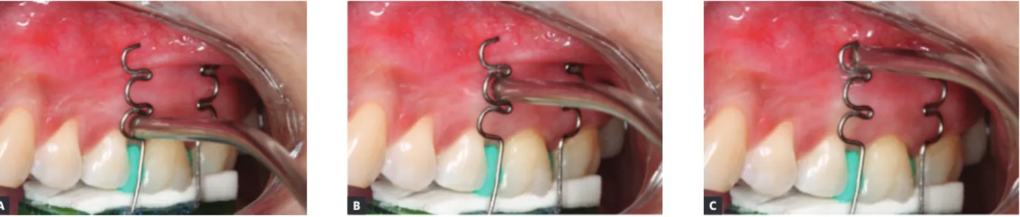
Figure 1 - Guide and scheme of laser applications used in the study. A) 10-second application in the mesio-cervical region; B) 10-second application in the mesio-medial region; C) 10-second application in the mesio-apical region. The three regions (cervical, medial and apical) also received laser applications distally, thereby totaling 60 seconds per tooth (6 J / tooth).
In the group “orthodontic separation (aware)” (SOci), recorded as group 4, the volunteers received neither placebo nor laser applications, thus fully charac-terizing it as the control group.
Twenty-one subjects dropped out of the study or provided incorrect data: ive of them reported severe pain (two from the SOLce group, one from the SOce group and two from the SOci group); and sixteen lacked complete data in one of the study periods (three from the SOce group and 13 from the SOci group). Therefore, inal data distribution (n = 79) was as fol-lows: SOLce (n = 23), SOLci (n = 25), SOce (n = 21) and SOci (n = 10).
Considering the sample in terms of the sides as-sessed (n = 158), distribution was as follows: laser = 30.37% (n = 48), placebo = 27.48% (n = 44), control = 41.77% (n = 66).
Applications were performed with a Whitening Lase II device (DMC Equipment Ltda., São Carlos, Brazil) which has two laser probes with distinct func-tions: a smaller laser probe for LLLT and a curved laser probe for teeth bleaching. The laser therapy probe in infrared mode (AsGaAl) was used.
A standard guide was used for all patients (ater disin-fection with 70% alcohol and protection with ilm paper in the foam area) based on the average size (13 mm) of the buccal roots of the maxillary irst molar.16 The device
was placed on the occlusal surface of teeth and support-ed between the marginal ridges of the teeth involvsupport-ed. The guide was fabricated so that the irst application was performed 5 mm above the gingival papilla, approach-ing patient’s bone crest region. The total length of the guide was 12 mm, allowing three applications, 4 mm apart from each other, to be performed (Fig 1).
The wavelength used was 808 nm, with a
lu-ency of 80 J/cm2, as recommended by the
manufac-turer (DMC Equipment Ltda., São Carlos, Brazil), thereby totaling approximately 6 J of energy per tooth (1 x 60 s x 100 mW).The probe of the device remained in contact with the gingival tissue during applications. Elastomeric separators were placed and laser applica-tions performed by the same previously trained and calibrated operator.
Subsequently, all patients were instructed to rate their level of spontaneous pain on a visual analogue scale (VAS). Initial scores were assigned as soon as the pa-tient arrived at the oice and before any procedure was carried out. This initial score made it possible to judge whether or not the patient already felt some pain, which was not related to the separation procedure, in the teeth involved in the study. Ater separation, patients’ pain levels were recorded 6 hours, 12 hours and 1, 2 and 3 days following separation. The scores assigned by the patient on the visual analogue scales were measured with a caliper (Mitutoyo, Japan). A zero score, located on the let side of the scale, suggested no pain; while a 100 (100 mm) score, at the right end of the scale, sug-gested maximum pain. The center of the scale corre-sponded to a score equal to 50 and suggested moderate pain. This information was provided to the subjects be-fore they started assigning scores on their dental history cards, which patients took home.
Data were tested for normality of distribution by means of the Shapiro-Wilk test. Should normal distri-bution not be found, data were presented using median and their quartiles (1st and 3rd). Pain perception was
as-sessed by analysis of variance (ANOVA) for repeated measures. Mauchly’s sphericity test was also applied
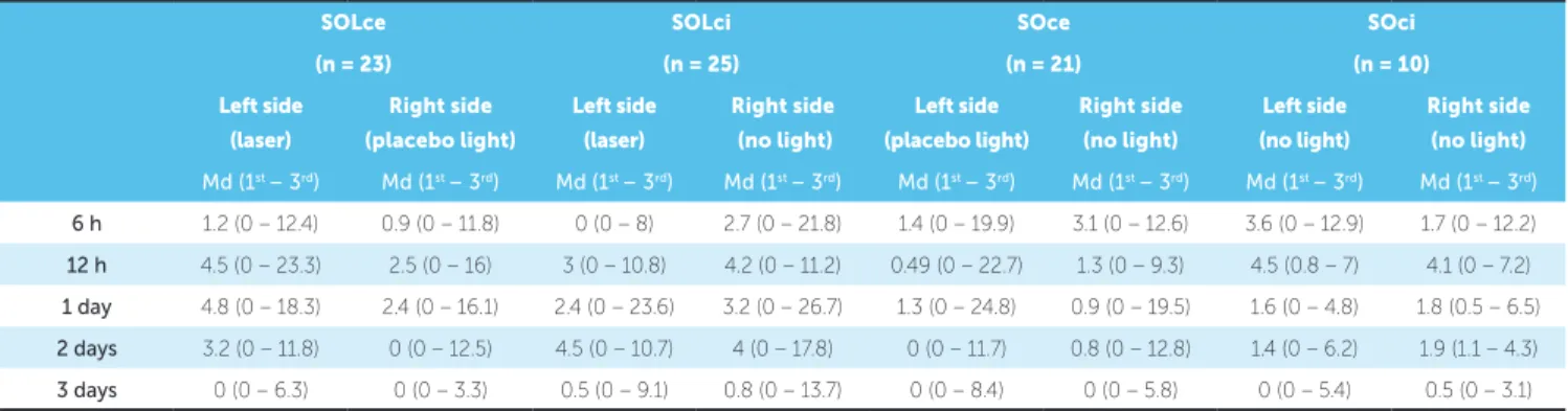
Table 2 - Median and median quartiles (1st - 3rd) of the SOLce, SOLci, SOce, SOci groups in all periods analyzed, comparing left and right sides.
Md = median; (1st - 3rd) = first and third quartiles; F Greenhouse-Geisser test= 1.78; p = 0.16.
and, whenever violated, technical corrections were performed by Greenhouse-Geisser test. Statistical sig-niicance was set at 5% and analyses were carried out by means of SPSS version 15.0.
RESULTS
Patients’ mean age was 23.4 ± 6.3 years for group SOLce (9 men and 14 women); 22.3 ± 4.1 years for group SOLci (8 men and 17 women); 23 ± 4.7 years for group SOce (6 men and 15 women) and 25.5 ± 7.8 years for group SOci (1 man and 9 women) (Table 1).
Data frequency distribution for age and sex was per-formed in a similar manner (p > 0.05), conirming the homogeneity of the sample. Female patients were pre-dominant only in the control group (Table 1). This fact did not hinder comparison among the laser, placebo and control sides (Tables 2 and 3).
All volunteers assigned zero to pain perception score at baseline. Among the 79 volunteers, 12.65% (n = 10) did not report any pain over all evaluated periods; and only 15.18% (n = 12) reported pain levels equal to or greater than 40 in at least one of the assessment periods.
No statistical diference was found (p = 0.16) between let and right sides in all periods compared across all groups (Table 2). Although the median was low, the pain peak perceived by patients occurred between 12 hours and 1 day (Tables 2 and 3).
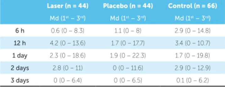
LLLT applications, placebo applications and con-trol sides were compared during the scoring periods. The three situations showed no statistical diference (p = 0.32) in terms of pain level (Table 3).
DISCUSSION
Corroborating the results of previous studies,2,3 the
pain caused by orthodontic procedures (separators or leveling archwires) reaches its peak 12 and 24 hours af-ter placement (Table 3). However, in this study, pain perception, as shown in VAS scores, was highly vari-able, with a relatively low median. It is a known fact that separators cause pain. Despite reports by some people who do not feel any pain whatsoever,6 most authors
re-port that, although pain intensity or location may vary, all patients eventually complain, which indicates that the procedures performed in orthodontic practice are
Table 1 - Demographic analysis of group data.
*P < 0.05.
SOLce SOLci SOce SOci
(n = 23) (n = 25) (n = 21) (n = 10)
Age (years) (mean ± SD) 23.4 ± 6.3 22.3 ± 4.1 23 ± 4.7 25.5 ± 7.8
Sex
Male - n (%) 9 (39.1%) 8 (32%) 6 (28.6%) 1 (10%)
Female - n (%) 14 (60.9%) 17 (68%) 15 (71.4%) 9 (90%)*
SOLce SOLci SOce SOci
(n = 23) (n = 25) (n = 21) (n = 10)
Left side
(laser)
Right side
(placebo light)
Left side
(laser)
Right side
(no light)
Left side
(placebo light)
Right side
(no light)
Left side
(no light)
Right side
(no light)
Md (1st – 3rd) Md (1st – 3rd) Md (1st – 3rd) Md (1st – 3rd) Md (1st – 3rd) Md (1st – 3rd) Md (1st – 3rd) Md (1st – 3rd)
6 h 1.2 (0 – 12.4) 0.9 (0 – 11.8) 0 (0 – 8) 2.7 (0 – 21.8) 1.4 (0 – 19.9) 3.1 (0 – 12.6) 3.6 (0 – 12.9) 1.7 (0 – 12.2)
12 h 4.5 (0 – 23.3) 2.5 (0 – 16) 3 (0 – 10.8) 4.2 (0 – 11.2) 0.49 (0 – 22.7) 1.3 (0 – 9.3) 4.5 (0.8 – 7) 4.1 (0 – 7.2)
1 day 4.8 (0 – 18.3) 2.4 (0 – 16.1) 2.4 (0 – 23.6) 3.2 (0 – 26.7) 1.3 (0 – 24.8) 0.9 (0 – 19.5) 1.6 (0 – 4.8) 1.8 (0.5 – 6.5)
2 days 3.2 (0 – 11.8) 0 (0 – 12.5) 4.5 (0 – 10.7) 4 (0 – 17.8) 0 (0 – 11.7) 0.8 (0 – 12.8) 1.4 (0 – 6.2) 1.9 (1.1 – 4.3)
always a nuisance.2,3,4,7 In the present study, 12.65%
(n = 10) did not report any pain and only 15.18% (n = 12) reported pain levels equal to or greater than 40. If the ive volunteers who dropped out of the study ater reporting too much pain were to be included, this per-centage would rise to 18% of the initial sample. Those distributions related to pain were similar among groups. Therefore, patients who claimed that the pain caused by orthodontic separation was relevant represented a mi-nority of the sample. It is worth noting that the efects of LLLT could only be noted if the majority of subjects had perceived increased pain. Nevertheless, a detailed assessment of patients reporting pain greater than or equal to 40 on VAS, in at least one of the periods, re-vealed that six of them reported feeling greater pain on the laser side, compared to placebo or control, while six of them assigned lower scores to the laser side.
Although pain is seen as a subjective and, therefore, hard-to-assess variable, the use of visual analogue scales, as it was the case in this study, has been widely reviewed and is nowadays regarded as a reliable method.6,9,17
In comparison to other investigations on orthodontic pain perception, the present study disclosed lower VAS score values. Fujiyama et al12 reported higher scores
that reached 80, 12 and 24 hours ater placing separators and when no laser was applied; and 40 when it was ap-plied; however, no placebo group was used. Our study corroborates that pain registered in VAS scores varies from mild to moderate.18-23
It is worth noting that, as performed in a variety of other studies,6,7,11,12,18 volunteers were asked to score
spon-taneous pain; however, other authors registered other situ-ations, such as biting, to which patients sometimes referred as being more painful than a spontaneous symptom.22,24
In the present study, a split-mouth, single-blind model was adopted and a placebo side was included, which al-lowed the authors to compare intrasubject pain perception with and without LLLT. Lim et al6 conducted a similar
study with separators and found no diference between the placebo and laser sides. Additionally, their scores were similar to those found in the present study, which also shows considerable variability.6 Those data also
corrobo-rate a recent study performed by Abtahi et al.18
Youssef et al,13 Tortamano et al,14 Turhani et al11 and
Harazaki et al,7 for instance, applied laser in patients
un-dergoing orthodontic treatment. The authors assessed pain during alignment and leveling or when performing canine retraction. Given that these procedures involve a higher number of teeth, they may enhance pain per-ception and underscore LLLT efects. Thus, it does not seem reasonable to compare these results with the present study which assessed pain perception in the presence of elastomeric separators.
A wide range of laser types, with different wave-lengths and energy doses, can be found in the lit-erature. AsGaAl diode laser, used in studies by Youssef et al,13 Tortamano et al14 and Lim et al,6 was
also used in the present study. Moreover, Haraza-ki et al7 used HeNe laser whereas Fujiyama et al12
used CO2 laser. At lower wavelengths, for instance, 632.8 nm7 and 670 nm11, no difference, in terms of
pain intensity, was reported between groups with or without laser applications. Nevertheless, the use of high-level laser, with wavelength of 808 nm, revealed statistically significant pain reduction in some studies.13,23 This was the wavelength used in
the present study, following the manufacturer’s rec-ommendations. However, even the use of laser with wavelength at 830 nm has yielded discrepant results,
with LLLT producing some analgesic effect,14
de-spite not being significant.6
According to the manufacturer’s instructions, we used, in this study, 6 J of energy in a single dose. Other similar studies used from 5 to 12 J of energy in single or daily applications. One single application seems more practical, as it does not rely on further ap-pointments and patient cooperation.19. Although the
amount of energy probably inluences the analgesic efect, some studies report LLLT eicacy19-22 or not6,18
with similar energy and frequency levels. Further stud-ies can clarify this point.
Table 3 - Median and median quartiles (1st - 3rd) of scores side by side with
laser, placebo and control sides applications in all periods analyzed.
Md = median; (1st - 3rd) = first and third quartiles; F Greenhouse-Geisser
test = 1.16; p = 0.32.
Laser (n = 44) Placebo (n = 44) Control (n = 66)
Md (1st – 3rd) Md (1st – 3rd) Md (1st – 3rd)
6 h 0.6 (0 – 8.3) 1.1 (0 – 8) 2.9 (0 – 14.8)
12 h 4.2 (0 – 13.6) 1.7 (0 – 17.7) 3.4 (0 – 10.7)
1 day 2.3 (0 – 18.6) 1.9 (0 – 22.3) 1.7 (0 – 19.8)
2 days 2.8 (0 – 11) 0 (0 – 11.6) 2.9 (0 – 12.9)
1. Oliver RG, Knapman YM. Attitudes to orthodontic treatment. Br J Orthod. 1985;12(4):179-88.
2. Brown DF, Moerenhout RG. The pain experience and psychological
adjustment to orthodontic treatment of preadolescents, adolescents, and adults. Am J Orthod Dentofacial Orthop. 1991;100(4):349-56.
3. Erdinç AM, Dinçer B. Perception of pain as a result of orthodontic treatment with ixed appliances. Eur J Orthod. 2004 Feb;26(1):79-85.
4. Krekmanova L, Bergius M, Robertson A, Sabel N, Hafström C, Klingberg G, et al. Everyday- and dental-pain experiences in healthy Swedish 8-19 year olds: an epidemiological study. Int J Paediatr Dent. 2009;19(6):438-47.
5. Erdinç AM, Dinçer B. Perception of pain during orthodontic treatment with ixed appliances. Eur J Orthod. 2004;26(1):79-85.
6. Lim HM, Lew KK, Tay DK. A clinical investigation of the eicacy of low level laser therapy in reducing orthodontic postadjustment pain. Am J Orthod Dentofacial Orthop. 1995;108(6):614-22.
7. Harazaki M, Isshiki Y. Soft laser irradiation efects on pain reduction in orthodontic treatment. Bull Tokyo Dent Coll. 1997;38(4):291-5
8. Shimizu N1, Yamaguchi M, Goseki T, Shibata Y, Takiguchi H, Iwasawa T, et al. Inhibition of prostaglandin E2 and interleukin 1-beta production by low-power laser irradiation in stretched human periodontal ligament cell. J Dent Res. 1995;74(7):1382-8.
9. Ngan P, Kess B, Wilson S. Perception of discomfort by patients undergoing orthodontic treatment. Am J Orthod Dentofacial Orthop. 1989;96(1):47-53. 10. Saito S, Shimizu N. Stimulatory efects of low-power laser irradiation on bone
regeneration in midpalatal suture during expansion in the rat. Am J Orthod Dentofacial Orthop. 1997;111(5):525-32.
11. Turhani D, Scheriau M, Kapral D, Benesch T, Jonke E, Bantleon HP. Pain relief by single low-level laser irradiation in orthodontic patients undergoing ixed appliance therapy. Am J Orthod Dentofacial Orthop. 2006;130(3):371-7. 12. Fujiyama K, Deguchi T, Murakami T, Fujii A, Kushima K, Takano-Yamamoto T.
Clinical efect of CO(2) laser in reducing pain in orthodontics. Angle Orthod. 2008;78(2):299-303.
REFERENCES 13. Youssef M, Ashkar S, Hamade E, Gutknecht N, Lampert F, Mir M. The efect
of low-level laser therapy during orthodontic movement: a preliminary study. Lasers Med Sci. 2008 Jan;23(1):27-33. Epub 2007 Mar 15.
14. Tortamano A, Lenzi DC, Haddad AC, Bottino MC, Dominguez GC, Vigorito JW. Low-level laser therapy for pain caused by placement of the irst orthodontic archwire: a randomized clinical trial. Am J Orthod Dentofacial Orthop. 2009;136(5):662-7.
15. Xiaoting L, Yin T, Yangxi C. Interventions for pain during ixed orthodontic appliance therapy. A systematic review. Angle Orthod. 2010;80(5):925-32. 16. Della-Serra O, Vellini FF. Anatomia dental. São Paulo: Artes Médicas; 1981. 17. Aitken R. Measurement of feelings using visual analogue scale. Proc R Soc
Med.1969;62(10):989-93.
18. Abtahi SM, Mousavi SA, Shafaee H, Tanbakuchi B. Efect of low-level laser therapy on dental pain induced by separator force in orthodontic treatment. Dent Res J (Isfahan). 2013;10(5):647-51.
19. Kim WT, Bayome M, Park JB, Park JH, Baek SH, Kook YA. Efect of frequent laser irradiation on orthodontic pain. A single-blind randomized clinical trial. Angle Orthod. 2013;83(4):611-6.
20. Artés-Ribas M, Arnabat-Dominguez J, Puigdollers A. Analgesic efect of a low-level laser therapy (830 nm) in early orthodontic treatment. Lasers Med Sci. 2013;28(1):335-41.
21. Eslamian L, Borzabadi-Farahani A, Hassanzadeh-Azhiri A, Badiee MR, Fekrazad R. The efect of 810-nm low-level laser therapy on pain caused by orthodontic elastomeric separators. Lasers Med Sci. 2013;29(2):559-64 22. Nóbrega C, Silva EM, Macedo CR. Low-level laser therapy for treatment of
pain associated with orthodontic elastomeric separator placement: a placebo-controlled randomized double-blind clinical trial. Photomed Laser Surg. 2013;31(1):10-6.
23. Doshi-Mehta G, Bhad-Patil W A. Eicacy of low-intensity laser therapy in reducing treatment time and orthodontic pain: a clinical investigation. Am J Orthod Dentofacial Orthop. 2012;141(3):289-97.
24. Shetty S, Shenoy N, Shenoy KA, Unnikrishnan B, Mogra S. Comparison of efects of preoperative piroxicam and ibuprofen on pain after separator placement: a randomized controlled trial. J Orthod Res. 2013;1(2):57-61. A systematic review has recently reported that
non-steroidal anti-inlammatory drugs (NSAIDs), such as COX-2 selective inhibitor, are still the best choice to reduce pain during orthodontic treatment, despite po-tential side efects.15 Another recent study revealed that a
single dose of Piroxicam, taken 60 minutes before sepa-rator placement, reduces pain.24
Since patients generally perceive pain as mild and transient, an analgesic regimen should only be adopted for less tolerant patients. However, should such regimen prove necessary, a single application of LLLT does not seem to provide a fully efective protocol for this purpose.
CONCLUSION
A single application (6 J) of LLLT (808 nm) did not produce signiicant efects on the perception of pain caused by orthodontic separation.