Orlando Tanaka*, Suelem Tisiane Fabianski**, Lílian Mary Karakida**, Luégya Amorin Henriques Knop***, Luciana Borges Retamoso***
Changes in pogonion and nose according to
breathing patterns
Introduction: The soft tissue profile results from complex changes in the hard and soft
tissues of the face. The pogonion and the nose are dominant facial structures that de-termine the degree of profile convexity and should, therefore, be analyzed and included in orthodontic treatment planning. Objective: To conduct a longitudinal evaluation of the anteroposterior dimensional changes of the pogonion and the nose of individu-als with Angle Class II, division 1 malocclusion at two time points during craniofacial development. Methods: Lateral cephalograms were obtained for 40 individuals — 23 nasal breathers (NB) and 17 mouth breathers (MB). Results: Linear and angular mea-sures were obtained: UL’-Pog’, UL’-B’, B’-Pog’, Pog’-PogTeg’, NB Line, Pog-NB, N’-Prn, Prn-NPog, N-Prn-Sn and Prn-Sn-UL. Two-way ANOVA was used to detect differences between mean values according to time points and/or breathing patterns. The UL’-B’, Pog’-PogTeg’, NB line and Pog-NB, N’-Prn, Prn-NPog, N-Prn-Sn and Prn-Sn-UL vari-ables had significant differences (p≤0.05) between the two time points, but there were no significant differences between breathing patterns. No interaction was found be-tween breathing patterns and time points for any variable. Conclusion: The pogonion and the nose undergo significant changes in the anteroposterior plane during growth, but breathing patterns do not significantly affect changes.
Abstract
Keywords: Nose. Pogonion. Nose breathing. Mouth breathing.
* Full Professor, School of Dentistry, PUCPR. Diplomate, Brazilian Board of Orthodontics. ** Graduate student of Orthodontics, PUCPR.
*** MSc student of Orthodontics, PUCPR.
How to cite this article: Tanaka O, Fabianski ST, Karakida LM, Knop LAH,
Retamoso LB. Changes in pogonion and nose according to breathing pat-terns. Dental Press J Orthod. 2011 Nov-Dec;16(6):78-83.
» The authors report no commercial, proprietary, or inancial interest in the
IntROduCtIOn
An individual’s soft tissue profile results from changes that affect facial bones and soft tissues. The interrelationship between facial soft tissue components (the nose, lips and pogonion) chang-es during growth and along orthodontic treat-ment. Therefore, normal growth trends of these structures should be understood.18 At birth, the
pogonion is retruded in relation to the maxilla, and this difference tends to decrease as the man-dible grows. Men tend to have larger structures than women,28 and the menton modification is
not specific of any type of malocclusion.
The nose, the most dominant of all the pro-file elements,17 has received little attention in
orthodontic analysis, although the ones made by Steiner,29 Ricketts,21 Holdaway12 and Chaconas4
use the nose either as a reference point or just as one more facial element.
Several studies in the literature have evalu-ated the effect of breathing patterns on the mor-phology of the dentoskeletal complex, but there remains substantial disagreement between au-thors. Some reported that nasal obstruction af-fects growth and facial development; for others, the changed growth of the dentofacial complex results from environmental and genetic factors.30
Dentists should understand the pogonion and nose growth, and their association with the face. The prediction of the amount and direction of growth is valuable information, particularly in mouth breathing and nose breathing patients.
OBJECtIVES
To longitudinally evaluate the anteroposte-rior dimensional changes in the pogonion and nose of individuals with Angle Class II, division 1 malocclusion at two time points during cra-niofacial development.
MAtERIAL And MEtHOdS
The study sample was composed of lateral cephalograms of 40 individuals with Angle Class II,
division 1 malocclusion: 23 nasal breathers (NB) and 17 mouth breathers (MB). The patient ages ranged from 10 years and 9 months to 14 years at time point 1, and 13 years and 4 months to 16 years and 6 months at time point 2.
Breathing pattern was classified using the mul-tidisciplinary method described by Wieler et al,24
which consisted of a clinical evaluation of lip com-petence by a dentist; a questionnaire to the par-ents about their children breathing habits; an ear, nose and throat evaluation by an otolaryngologist; and a speech test performed by a speech patholo-gist. Based on these tests, scores and weighted val-ues were assigned to each evaluation, and an index was defined to classify the predominant breathing pattern for each individual.
Cephalograms were obtained using manual and computerized methods.25 The anatomical
structures were outlined manually. After digita-lization, the landmarks were marked, and values were calculated using the Radiocef 2000®
cepha-lometric software. Study models were used to aid in tracing tooth positions.
The following parameters were used:
Linear measures referring to pogonion
» UL’-Pog’ – measure from UL’ to Pog’, corresponding to the greatest anteropos-terior dimension of the lateral image of the mandibular symphysis (total sym-physis thickness).
» UL’-B’ – measure from UL’ to B’, corre-sponding to the greatest anteroposterior dimension of the lateral image of the man-dibular symphysis.
» B’-Pog’ – measure from B’ to Pog’, cor-responding to the greatest anteroposterior dimension of the lateral image of the man-dibular symphysis.
» NB line – line from nasion to Point B. » Pog-NB – distance from Pog to NB line;
re-fers to the most anterior point of the hard chin, measured from the NB line. Values are positive and negative when after and before the NB line, respectively.
Linear measures referring to nose
» N’-Prn – measure of the position of the tip of the nose in relation to the nasion; defines nose prominence.
» Prn-NPog – measure of the nasal depth in relation to the facial plane.
Angular measures referring to nose
» SN-Prn-Sn – measure of the nose promi-nence in relation to the sella-nasion
» Prn-Sn-UL – nasolabial angle; measure of the inclination of the columella in relation to the upper lip.
RESuLtS Pogonion
All variables had a normal distribution at all time points according to the Kolmogorov-Smirnov test, except B’-Pog’ and Pog-NB. Therefore, mean values according to time points were compared using two-way repeated measures ANOVA.
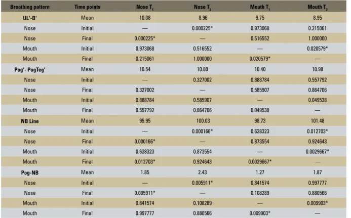
ANOVA results revealed that F was statisti-cally significant (p<0.05) for UL’-B’, Pog’-Pog Teg’, NB line and Pog-NB, and that there was a significant difference between mean values according to time point, breathing pattern, or both. The B’-Pog’ and UL’-PogTeg’ values were not statistically different (p>0.05) be-tween breathing patterns or time points. To identify which conditions (breathing pattern x time points) were different from each other, the Tukey HSD test for multiple comparisons was used (Table 1).
TABLE 1 - Multiple comparison of Tukey HSD breathing patterns (Pogonion).
*Statistically significant difference (p≤0.05).
Breathing pattern Time points Nose T1 Nose T2 Mouth T1 Mouth T2
UL'-B' Mean 10.08 8.96 9.75 8.95
Nose Initial — 0.000225* 0.973068 0.215061
Nose Final 0.000225* — 0.516552 1.000000
Mouth Initial 0.973068 0.516552 — 0.020579*
Mouth Final 0.215061 1.000000 0.020579* —
Pog'- PogTeg' Mean 10.54 10.80 10.40 10.98
Nose Initial — 0.327002 0.888784 0.557792
Nose Final 0.327002 — 0.585907 0.864706
Mouth Initial 0.888784 0.585907 — 0.049538
Mouth Final 0.557792 0.864706 0.049538 —
NB Line Mean 95.95 100.03 98.73 101.48
Nose Initial — 0.000166* 0.638323 0.012703*
Nose Final 0.000166* — 0.873554 0.924643
Mouth Initial 0.638323 0.873554 — 0.0029667*
Mouth Final 0.012703* 0.924643 0.0029667* —
Pog-NB Mean 1.85 2.43 1.27 1.87
Nose Initial — 0.005911* 0.841574 0.997777
Nose Final 0.005911* — 0.108289 0.880566
Mouth Initial 0.841574 0.108289 — 0.009903*
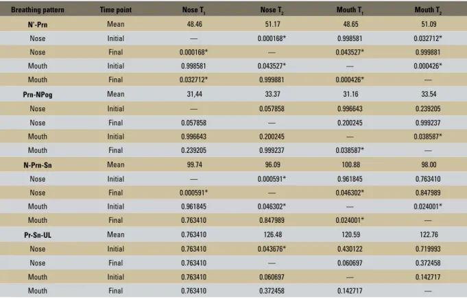
TABLE 2 - Multiple comparison of Tukey HSD breathing patterns (Nose).
*Statistically significant difference (p≤0.05).
Breathing pattern Time point Nose T1 Nose T2 Mouth T1 Mouth T2
N’-Prn Mean 48.46 51.17 48.65 51.09
Nose Initial — 0.000168* 0.998581 0.032712*
Nose Final 0.000168* — 0.043527* 0.999881
Mouth Initial 0.998581 0.043527* — 0.000426*
Mouth Final 0.032712* 0.999881 0.000426* —
Prn-NPog Mean 31,44 33.37 31.16 33.54
Nose Initial — 0.057858 0.996643 0.239205
Nose Final 0.057858 — 0.200245 0.999237
Mouth Initial 0.996643 0.200245 — 0.038587*
Mouth Final 0.239205 0.999237 0.038587* —
N-Prn-Sn Mean 99.74 96.09 100.88 98.00
Nose Initial — 0.000591* 0.961845 0.763410
Nose Final 0.000591* — 0.046302* 0.847989
Mouth Initial 0.961845 0.046302* — 0.024001*
Mouth Final 0.763410 0.847989 0.024001* —
Pr-Sn-UL Mean 0.763410 126.48 120.59 122.76
Nose Initial 0.763410 0.043676* 0.430122 0.719993
Nose Final 0.763410 — 0.060697 0.372458
Mouth Initial 0.763410 0.060697 — 0.142717
Mouth Final 0.763410 0.372458 0.142717 —
nose
The study hypothesis was tested using two-way repeated measures analysis of vari-ance (ANOVA). The two time points and two breathing patterns had a normal distribution for the variables under analysis. The Kolmogorov-Smirnov test was used to test normality, and the level of significance was set at 0.05.
The Tukey HSD test for multiple comparisons was used to detect which conditions (breathing pattern x time points) differed from each other when ANOVA results revealed a difference be-tween the mean values of N’-Prn, Prn-NPog, N-Prn-Sn and N-Prn-Sn-UL according to time point, breathing pattern, or both (Table 2).
dISCuSSIOn
Facial analysis has been used as a valuable diagnostic resource since the beginning of
or-thodontics,20 and the lips, cheeks and nose, in
particular, define the unique facial appearance of each individual.19
Skeletal growth and changes in soft tis-sues affect occlusion and facial esthetics. The amount of soft tissue over the symphysis has a fundamental role in facial harmony and in the response to skeletal changes.5 In
addi-tion, the measures obtained using cephalom-etry, although two-dimensional, provide con-crete data that make comparisons possible and complement diagnoses.16 For this reason,
cephalometry is widely used in several scien-tific studies.10,11
Changes in facial soft and hard tissues should be taken into consideration during orthodontic diagnosis and treatment planning.2,22,28 Growth
The results of the present study revealed changes in the hard and soft tissue pogonion at the two time points under analysis, 10-14 and 13-16 years of age, which are in agreement with Koch et al13 and
Mal-tagliati et al15 findings. Pogonion thickness, evaluated
according to the UL’-Pog’ measure, resulted from different bone remodeling patterns due to greater bone deposition in the lingual surface of the pogoni-on, characteristic of the growth phase, as confirmed by histological6 and implant1 studies.
In the present study, the soft tissue pogonion was directly associated with the increase of the skeletal pogonion, which differs from data re-ported by Genecov, Sinclair and Dechow,9 who
found that the growth of the soft tissue pogoni-on in patients undergoing orthodpogoni-ontic treatment was constant and relatively independent from the underlying bone.
The analysis of the B’-Pog’ showed that this measure is shorter in nasal breathers and greater in mouth breathers. This study, however, did not investigate the reason for this reduction in na-sal breathers. A smaller posterior movement of Point B might have occurred in consequence of downward and backward mandibular rotation, although the possibility of less bone deposition in the pogonion region was not ruled out.
The shorter UL’-B’ and longer Pog-NB may be explained by the posterior displacement of Point B and the bone deposition in the pogonion surface. Moreover, there were variations in the mandibular plane angle. According to Rosenstein,23 the UL’-B’
line tends to be even shorter in individuals whose treatment includes extractions.
Prahl-Andersen et al18 found that nose length,
prominence and shape are associated with the growth in height and length of the maxilla and the mandible.
The nose projection onto the posteroanterior plane had a statistically significant difference in this study, which is in agreement with findings
by Brant et al,3 who reported significant changes
in N-Prn-Sn when groups with and without pre-molar extractions were compared. According to Chanocas,4 the tip of the nose grows downward
in individuals with Class II malocclusion. The nasolabial angle (Prn-Sn-UL) had signifi-cant changes (p≤0.05) between the two time points as a result of nasal growth and upper lip retraction, which takes place during the normal development of individuals, when the nasolabial fold may also become longer.7 The results are in agreement with
those reported by Salgado et al,26 who found
sig-nificant variations in nasolabial angles of individu-als with Angle Class II and Class III malocclusion, and greater values in Class II cases. However, the results differ from those reported by Brant et al,3
who found no significant differences in the nasola-bial angle of individuals that underwent orthodon-tic treatment with or without extractions.
Nasal breathing is essential for adequate growth and development of the craniofacial complex.27 Some studies found differences in
the craniofacial development of mouth breath-ers and nasal breathbreath-ers. In mouth breathbreath-ers, there seems to be a greater inclination of the angle of the mandibular plane, and the growth pattern is vertical.14,28 This finding contradicts
the results of this study, which found no inter-action between breathing patterns and any of the variables under study.
Changes in the thickness of the soft tissues of the nose, lips and pogonion should be taken into consideration during diagnosis, treatment planning, and actual treatment of individuals during growth.
COnCLuSIOn
1. Bjork A. Variations in the growth pattern of the human mandible: longitudinal radiographic study by the implant method. J Dent Res. 1963;42(1)Pt 2:400-11.
2. Bowker W, Meredith HV. A metric analysis of the facial proile.
Angle Orthod. 1959;29(3):146-6.
3. Brant JCO, Siqueira VCV. Alterações no peril facial tegumentar,
avaliadas em jovens com Classe II, 1ª divisão, após o tratamento ortodôntico. Rev Dental Press Ortod Ortop Facial. 2006;11(2):93-102.
4. Chaconas SJ. A statistical evaluation of nasal growth. Am J Orthod. 1969;56(4):262-84.
5. Czarnecki ST, Nanda RS, Currier GF. Perceptions of a balanced
facial proile. Am J Orthod Dentofacial Orthop. 1993;104(2):180-7.
6. Enlow DH, Harris DB. A study of the postnatal growth of the human mandible. Am J Orthod. 1964;50(1):25-50. 7. Fitzgerald JP, Nanda RM, Currier GF. An evaluation of the
nasolabial angle and the relative inclinations of the nose and upper lip. Am J Orthod Dentofacial Orthop. 1992;102(4):328-34. 8. Formby WA, Nanda RS, Currier GF. Longitudinal changes
in the adult facial proile. Am J Orthod Dentofacial Orthop.
1994;105(5):464-76.
9. Genecov JS, Sinclair PM, Dechow PC. Development of the nose
and soft tissue proile. Angle Orthod. 1990;60(3):191-8.
10. Hoffelder LB, de Lima EM. Avaliação dos tecidos tegumentares da região do nariz, dos lábios e do mento em indivíduos brasileiros com Classe II esquelética. J Bras Ortodon Ortop Facial. 2006;11(61):70-82.
11. Hoffelder LB, de Lima EM, Martinelli FL, Bolognese AM. Soft-tissue changes during facial growth in skeletal Class II individuals. Am J Orthod Dentofacial Orthop. 2007;131(4):490-5.
12. Holdaway RA. The soft tissue covering of the skeletal face as related to orthodontic problems. Am J Orthod. 1964;50(6):405-20.
13. Koch R, Gonzales A, Witt E. Proile and soft tissue changes during
and after orthodontic treatment. Eur J Orthod. 1979;1(3):193-9. 14. Lessa FCR, Enoki C, Feres MFN, Valera FCP. Lima WTA,
Matsumoto MAN. Inluência do padrão respiratório na morfologia
craniofacial. Rev Bras Otorrinolaringol. 2005;71(2):156-60.
15. Maltagliati LA, Henriques JFC, Janson G. A inluência do
tratamento ortopédico nas estruturas faciais de indivíduos com má oclusão de Classe II, 1ª divisão: um estudo comparativo. J Appl Oral Sci. 2004;12(2):164-70.
16. Pereira PSG, Kajiwara JK, Grellet M. Estudo morfológico do desenvolvimento da cartilagem quadrangular do nariz e implicações nas cirurgias septoplásticas. Rev Bras Otorrinolaringol. 2002;68(2):209-17.
17. Posen JM. A longitudinal study of the growth of the nose. Am J Orthod. 1967;53(10):746-56.
18. Prahi-Andersen B, Ligthelm-Bakker AS, Wattel E, Nanda R.
Adolescent growth changes in soft tissue proile. Am J Orthod
Dentofacial Orthop. 1995;107(5):476-83.
19. Profit WR, Fields HW, Sarver DM. Contemporary orthodontics.
4th ed. St. Louis: C.V. Mosby; 2007.
20. Reis SAB, Capelozza Filho L, Cardoso MA, Scanavini MA. Características cefalométricas dos indivíduos Padrão I. Rev Dental Press Ortod Ortop Facial. 2005;10(1):67-78. 21. Ricketts RM. Planning treatment on the basis of the facial pattern
and an estimate of its growth. Angle Orthod. 1957;27(1):14-37.
22. Ricketts RM. A inluence of orthodontic treatment on facial growth
and development. Angle Orthod. 1960;30(3):103-33.
23. Rosenstein SW. A longitudinal study of anteroposterior growth of the mandibular symphysis. Angle Orthod. 1964;34(3):155-67. 24. Wieler WJ, Barros AM, Barros LA, Camargo ES, Ignácio SA,
Maruo H, et al. A combined protocol to aid diagnosis of breathing model. J Dent Clin Res. 2007;3(2):101-14. 25. Saga AY. Estudo comparativo das dimensões craniofaciais
entre respiradores predominantemente nasais e bucais na maloclusão Classe II, divisão 1 de Angle. 2002 [dissertação]. Curitiba (PR): Universidade Católica do Paraná; 2002.
26. Salgado JAP, Moraes LC, Castilho JCM, Moraes MEL. Avaliação do
ângulo nasolabial, em radiograia cefalométricas laterais, dividido
em ângulo superior e inferior, por uma linha paralela ao plano de Frankfort, em indivíduos portadores de má-oclusão Classe II e
Classe III de Angle. Ciênc Odontol Bras. 2003;6(3):40-7.
27. Spinelli MLM, Casanova PC. Respiração bucal. 2002. [Acesso 12 fev 2002]. Disponível em: www.odontologia.com.br/artigos.
28. Subtelny JD. A soft tissue growth and treatment changes. Am J Orthod. 1961;31(2):105-22.
29. Steiner CC. Cephalometrics for you and me. Am J Orthod. 1953;39(10):729-55.
REfEREnCES
Contact address Orlando Tanaka
Rua Imaculada Conceição, 1155 - Centro Zip code: 80.215-901 - Curitiba / PR, Brazil E-mail: [email protected]