Original Article
Artigo Original
Oral-motor and electromyographic
characterization of patients submitted to
open and closed reductions of mandibular
condyle fracture
Caracterização miofuncional orofacial e
eletromiográfica de pacientes submetidos
à correção da fratura condilar por redução
aberta e fechada
Amanda Pagliotto da Silva1
Fernanda Chiarion Sassi1
Claudia Regina Furquim de Andrade1
Keywords
Speech-language and Hearing Sciences Electromyography Stomatognathic System Mandibular Condyle Bone Fractures
Descritores
Fonoaudiologia
Eletromiograia
Sistema Estomatognático Côndilo Mandibular Fraturas Ósseas
Correspondence address:
Claudia Regina Furquim de Andrade Universidade de São Paulo – USP Rua Cipotânea, 51, Cidade
Universitária, São Paulo (SP), Brazil, CEP: 05360-160.
E-mail: [email protected]
Received: July 8, 2015 Study carried out at “Divisão de Fonoaudiologia, Instituto Central do Hospital das Clínicas da Faculdade de
Medicina da Universidade de São Paulo – USP” - São Paulo (SP), Brazil. 1 Universidade de São Paulo – USP - São Paulo (SP), Brazil.
Financial support: nothing to declare.
Conlict of interests: nothing to declare.
ABSTRACT
Purpose: To characterize the oral-motor system of adults with mandibular condyle facture comparing the
performance of individuals submitted to open reduction with internal ixation (ORIF) and closed reduction with mandibulomaxillary ixation (CRMMF). Methods: Study participants were 26 adults divided into three
groups: G1 – eight individuals submitted to ORIF for correction of condyle fracture; G2 – nine individuals
submitted to CRMMF for correction of condyle fracture; CG – nine healthy volunteers with no alterations of the orofacial myofunctional system. All participants underwent the same clinical protocol: assessment of the orofacial myofunctional system; evaluation of the mandibular range of motion; and surface electromyography (sEMG) of the masticatory muscles. Results: Results indicated that patients with condyle fractures from both
groups presented signiicant differences compared with those from the control group in terms of mobility of the
oral-motor organs, mastication, and deglutition. Regarding the measures obtained for mandibular movements,
participants with facial fractures from both groups showed signiicant differences compared with those from
the control group, indicating greater restrictions in mandibular motion. As for the analysis of sEMG results,
G1 patients presented more symmetrical masseter activation during the task of maximal voluntary teeth clenching. Conclusion: Patients with mandibular condyle fractures present signiicant deicits in posture, mobility, and function of the oral-motor system. The type of medical treatment does not inluence the results of muscle function during the irst six months after fracture reduction. Individuals submitted to ORIF of the condyle fracture present
more symmetrical activation of the masseter muscle.
RESUMO
Objetivo: Realizar a caracterização da performance motora orofacial de indivíduos adultos com fratura em côndilo, comparando indivíduos submetidos à redução aberta e fechada. Método: 26 adultos divididos em três grupos: G1 – composto por 8 indivíduos submetidos à redução aberta para correção da fratura em côndilo; G2 – composto por 9 indivíduos submetidos à redução fechada para correção da fratura em côndilo; GC – 9 indivíduos voluntários saudáveis, sem alterações do sistema miofuncional orofacial. Todos os participantes foram submetidos à avaliação que consistiu na aplicação de um protocolo clínico para a avaliação da motricidade orofacial, a amplitude dos
movimentos mandibulares e a avaliação da musculatura mastigatória por meio da eletromiograia de superfície
(EMGs). Resultados: Os resultados indicaram que ambos os grupos com fratura de côndilo se diferenciaram signiicantemente do grupo controle, apresentando prejuízo na mobilidade dos órgãos fonoarticulatórios e nas
funções de mastigação e deglutição. Para as medidas de amplitude mandibular, os grupos se diferenciaram do grupo controle apresentando maior restrição de movimentos. Na avaliação dos músculos mastigatórios por meio da EMGs, G2 se diferenciou de G1 e de GC, apresentando maior assimetria no funcionamento do músculo masseter.
Conclusão: Os resultados sugerem que, independentemente do tratamento adotado para correção da fratura no
INTRODUCTION
The occurrence of facial trauma cases has grown over the past four decades mostly because of the increased number of
trafic accidents and urban violence, which are the main causative
factors of these types of trauma. Studies show that interpersonal
violence, trafic accidents, and daily household and sporting
accidents have a direct relation with facial trauma, with men being the most affected by these fractures(1,2).
Mandibular fractures, especially those in the condylar and subcondylar regions, are the most common among facial fractures(2). This type of injury can alter the functioning of the
temporomandibular joint (TMJ)(1). The mandibular condyle is essential for the masticatory movements. Changes in its morphology can lead to reduced bite force and discomfort during chewing(3). Condylar fractures can cause temporomandibular
disorders, orofacial pain, joint disc displacement, malocclusion,
facial asymmetry, condylar resorption, changes in mandibular growth, ankylosis(4), and alterations in joint lubrication(5).
The literature shows a direct correlation between severity of condylar fracture and damage to the articular disc, with
higher fractures leading to retrodiscal tissue inlammation(6).
Changes in soft tissues, such as joint disc displacement, seem
to interfere with the functional outcome of treatment(7). These alterations are primarily observed on the side of the fracture, but they can also occur opposite to it(8). Patients who present medial condylar displacement after fracture are more likely to develop ankylosis, mainly due to the reduction of mandibular
mobility associated with this type of injury(3,9).
Two concepts in the mandibular condyle fracture management
are found in the current speciic literature: surgical open reduction with internal ixation (ORIF) and closed reduction with mandibulomaxillary ixation (CRMMF). There are several
possibilities to conduct CRMMF; however, nearly all approaches involve no direct reduction of the fracture, the use of soft diet for as long as two months, and placement of Erich arch bar to
guide occlusion or promote intermaxillary ixation(10). In contrast,
to conduct ORIF, it is possible to perform anatomical reduction and ixation of the factored site, allowing early functional
reestablishment and decreased incidence of problems arising from bone vicious healing(10).
Although the existing studies seek to verify and analyze
which would be the most effective and least painful treatment, results are divergent(3,11,12). A study conducted by Nogami et al. (12) clinically compared the outcomes of arthrocentesis and
conventional conservative treatment with maxillomandibular ixation for unilateral high condylar fractures. The authors concluded that the irst presented better results, with greater
mandibular mobility in mouth opening for patients undergoing this type of treatment, and also found that patients had less pain
and joint discomfort.
Success in the management of mandibular fractures, especially those of the condyle region, is directly related to the choice of
treatment for the case based on diagnosis, stability of bone ixation,
and rehabilitation prognosis. Factors such as patient’s age, type of fracture, health status, associated fractures, dentition, and the
possibility of restoring occlusion are also considered(13). Muscle rehabilitation is often mentioned when discussing mandibular condyle fractures. Regardless of the type of treatment, it is known that muscular rehabilitation is necessary to improve
and enhance the post-fracture functionality of jaw muscles(13). Boyde(14) analyzed the response of long bones to exercise
and noted that exercise led to increased osseous density and
less resorption, contributing to bone remodeling. There are few studies addressing this theme with respect to speech-language pathology therapy. Studies in the area indicate that the changes
observed during myofunctional evaluation showed signiicant
improvement after speech-language pathology therapy, especially when the mandibular range of motion was considered(1).
The main alterations related to the oral-motor system observed in patients after facial trauma are limitation in the mandibular range of motion; pain in the facial and/or neck musculature due to muscle tension caused by poor positioning of the mandible; muscle alterations regarding traction and direction of muscle
strength; changes in sensitivity; alteration in the jaw movements
compromising mastication owing to limitation and asymmetry of mandibular movements, and TMJ noise(1).
Eficient evaluation of dental occlusion and masticatory
function can bring relevant information to decide on the type of treatment to be performed/indicated(15,16). Based on what has
been previously exposed, there is the need to search, speciically,
for muscle functioning standards of the oral-motor system of patients with mandibular condyle fracture, aiming to guide the therapeutic approach and establish parameters to determine the speech-language pathology prognosis.
The objective of the present study was to characterize the
oral-motor system of adults with mandibular condyle facture comparing the performance of individuals submitted to surgical
open reduction with internal ixation and closed reduction with mandibulomaxillary ixation.
METHODS
This prospective, observational, cross-sectional study was approved by the Research Ethics Committee of the aforementioned institution under protocol no. CAPPesq 495.639. All study participants signed an Informed Consent Form (ICF) before data collection procedures began.
Participants
Data collection occurred between December 2012 and December 2014. The sample was composed of individuals of both genders, aged 18 years or older, diagnosed with facial trauma with mandibular condyle fracture. Participants were referred to the Speech-language Pathology Department of the “Instituto Central do Hospital das Clínicas” - ICHC, “Faculdade de Medicina da Universidade de São Paulo” - FM-USP for evaluation.
The participants were divided into two research groups: G1 - composed of patients undergoing surgical open reduction
G2 - composed of patients undergoing closed reduction with
mandibulomaxillary ixation (CRMMF) for correction of
condylar fracture.
Is worth mentioning that the standard protocol adopted in the institution by the Department of Plastic Surgery and Burns for the correction of mandibular condyle fractures is as follows:
a) ORIF – surgical correction of condylar fracture with rigid ixation using plate and screws; b) CRMMF – indirect reduction of condylar fracture using Erich arch bar with maxillomandibular ixation by rubber bands and use of soft diet for up to two months.
Patients from both groups only underwent speech-language pathology assessment after removal of arch bar and wires and medical release.
For comparison purposes, we recruited a group of healthy volunteers (CG - control group) with no alterations in the orofacial myofunctional system or in the scapular region, according to the literature; with complete permanent dentition
(the absence/extraction of third molars could be accepted);
with absence of severe malocclusion and without use of orthodontic appliance during the evaluation period; or with prior speech-language pathology therapy(15).
All groups were paired according to age and gender. Exclusion
criteria comprised previous history of head and neck surgery, speech-language pathology comorbidities (hearing and/or
communicative deicits or complaints), neurological disorders,
and compromised cognition or awareness that would impair understanding of the verbal information needed for the evaluation.
Clinical assessment of orofacial motricity
Participants of the three groups were submitted to clinical
evaluation of orofacial motricity. To this end, the expanded protocol of Orofacial Myofunctional Evaluation with Scores (OMES-E) was used(16). This protocol aims to assess the
components of the stomatognathic system (lips, tongue, jaw,
and cheeks) with respect to appearance/posture, mobility, and performance during deglutition and mastication. The observed
data were converted to a numerical scale, with maximum
individual score of 230 points. Data were collected by means of visual inspection during evaluation and, subsequently, by analyzing the photos and footage recorded with the use of a digital camera (Sony DSC – W120).
In order to ensure the reliability of clinical assessment results, all participants were evaluated by two speech-language
pathologists, who were independent examiners with expertise in the area. The Kappa coeficient was utilized to verify the concordance between the examiners for the overall score of the OMES-E, whose result showed high inter-rater agreement (0.89).
Mandibular range
A methodology based on the existing literature was used
to assess the mandibular range of motion(1,17). The following measures taken with the use of a digital pachymeter (Digimess Pró-Fono, Pró-Fono Produtos Especializados para Fonoaudiologia Ltda., Brasil) were obtained:
1) maximal mouth opening - distance between the incisal edges
of upper and lower incisor teeth plus the measure of vertical overbite;
2) mandibular lateralization - horizontal distance of the line between the mandibular central incisors to the line between
the maxillary central incisors after lateral slip of the jaw to the right and left. Appropriate adjustment was performed
in case of presence of midline deviation;
3) mandibular protrusion – sum of the measure of the horizontal
overlap (overjet) with the measure of the maximum horizontal sliding of the jaw.
Evaluation of masticatory muscles - Surface
electromyography (sEMG)
All surface electromyography (sEMG) exams were conducted
by the same speech-language pathologist under the same environmental conditions. Assessment of the electric activity
of masticatory muscles of the participants was based on speciic
methodology(18). To this end, the sEMG was performed using a 4-channel Miotool 400 electromyography device with all channels
calibrated as follows: at 500 microvolts (μV) with a bandpass ilter (20-500 Hz), a notch ilter (60 Hz), and 100x gain, with low noise level (<5 μV RMS). Miograph 2.0 software (Miotec
Biomedical Equipment) was used to capture and process the
sEMG exam. This software program performs online acquisition,
storage, and processing of signals and runs on the Windows XP (Microsoft) operating system. The electrical activity signals of the muscle movements were captured with disposable, bipolar
surface Ag/AgCl electrodes, model SDS500, double, ixed with
Transpore tape (3M).
Prior to data collection, the facial skin of participants was cleaned using gauze soaked in 70% alcohol and local trichotomy was performed to ensure good impedance during
the exam. Electrodes were placed at the midpoint of the central
muscle, in the longitudinal direction of the muscle bundle, at the mesodistal position, so that simultaneous electrical activities of the temporalis and masseter muscles were evaluated in both hemifaces. The resulting signals were analyzed by root mean
square (RMS) and expressed in microvolts (V). The ground
cable was connected to the electrode and set on the right wrist.
The electric activity of the muscles was veriied according to
the situations described ahead:
• at rest – recording time of 30 seconds; three collections were performed to obtain the mean electrical activity;
• at maximal voluntary dental clenching with maximal
intercuspal position (MIC) – participants were requested
to apply maximum bite force possible for ive seconds.
Three consecutive recordings were performed with a 5-second interval between trials;
• at maximal voluntary dental clenching with cotton rolls
between the teeth (Al) - a cotton roll was placed bilaterally
requested to apply maximum bite force possible for ive
seconds. Three consecutive recordings were performed with a 5-second interval between trials.
Analysis of the electromyograms
Temporal domain analysis was conducted for the outcomes
of the sEMG exams. At rest, the values obtained represent
the mean (RMS) of the electromyographic activity observed over 30 seconds. The amplitude of muscle activity during the voluntary dental clenching tasks (Al and MIC) was obtained by selection of the representative period of muscle activation (i.e., on and off situations). The “on” situation was determined by the onset of muscle contraction above baseline values, whereas the “off” situation was determined by the return of the muscle to its baseline value. The mean values of the EMG amplitude for the Al and MIC tasks were normalized in relation to the activity at rest.
Reliability of the sEMG data was also analyzed. To this end, 15 electromyographic samples representative of the MIC and Al dental clenching tasks were randomly selected from a total of 156. These samples underwent blind and independent analysis
by two researchers with experience in the ield. The correlation coeficient was found to be high for all comparisons (95% conidence interval; [CI] = 0.8873-0.9533), indicating high
inter-rater consistency.
Data analysis
Data were statistically analyzed using the SPSS, 22 version, software program. Descriptive analysis was performed including
mean, standard deviation, minimum and maximum values, median,
and 1st and 3rd quartiles. Because data distribution did not comply with normality, comparison between the performances of all groups was conducted using the Kruskal-Wallis nonparametric test, whereas the post hoc analyses of pairs were conducted by
the Dunn test. The level of signiicance adopted for all statistical
analyses was 5% (p<0.05).
The muscle asymmetry index for the maximal dental
intercuspation measures with and without cotton rolls was calculated by dividing the side with lower muscle activation
by the side with greater activation. This index was calculated
for each participant separately.
RESULTS
After the implementation of all phases of the research previously described in the methodology, the study groups were characterized as follows: G1 was composed of eight individuals (one woman and seven men) with mean age of 35.5 years (±12.20). Regarding the location of factures, G1 included two patients with bilateral condyle fracture, three patients with left condylar fracture, and three patients with right condylar fracture. All patients presented associated fractures: seven with fractures of the mandibular body, four with mandibular ramus
fractures, four with maxillary fractures, three with zygomatic
fractures, four with orbital fractures, and two with fractures of the nasal bone.
G2 was composed of nine individuals (two woman and seven men) with mean age of 30.7 years (±11.61). Concerning the location of factures, G2 included two patients with bilateral condyle fracture, three patients with left condylar fracture, and four patients with right condylar fracture. As in the previous
group, all patients presented associated fractures: ive with
fractures of the mandibular body, one with parasymphyseal fracture, one with symphyseal fracture, one with mandibular
ramus fracture, six with maxillary fractures, one with zygomatic
fracture, three with orbital fractures, and two with fractures of the nasal bone.
The control group (CG) was composed of nine individuals (one woman and eight men) with mean age of 33.1 years (±12.45). The total study sample comprised 26 participants.
Statistical analysis indicated signiicant difference between the
ages of the groups (p=0.713). With regard to the time elapsed (in days) between fracture reduction surgery and speech-language pathology clinical assessment in the case of G1, and between
maxillomandibular ixation and clinical assessment in the case of G2, statistical analysis also indicated signiicant difference
between the groups (G1 – 48.5±25 and G2 – 38.8±27; p=0.678). Comparative analyses between the groups for the categories of orofacial motricity clinical assessment, according to the
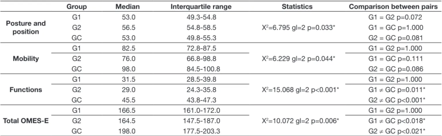
expanded protocol of Orofacial Myofunctional Evaluation with Scores (OMES-E), are described in Table 1. As expected,
the study groups (G1 and G2) differed signiicantly from the control group (CG) for all items and total of the OMES-E
protocol, but not between themselves. Although the initial
analysis indicated signiicant differences between the groups
for the item functions, post hoc analysis showed no difference between the groups. The median indicated that the group that
underwent open reduction with internal ixation (ORIF) presented
higher scores than the group that underwent closed reduction
with mandibulomaxillary ixation (CRMMF) with respect to
the items mandibular mobility, functions, and total score of the
protocol. Overall, the results indicated that, regardless of the
type of treatment used for fracture reduction, the two groups showed similar behavior in the oral-motor evaluation.
Comparison between the groups for the measures of mandibular range of motion is shown in Table 2. Signiicant difference between the study groups and the CG was observed
for all measures taken, except for mandibular protrusion. No signiicant difference was observed between the ORIF group
and the CRMMF group. Considering the medians obtained, it was possible to observe that G2 presented higher values than G1,
mainly with regard to the measure of maximal mouth opening.
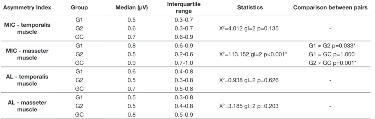
For comparison purposes of muscle sEMG between groups,
the asymmetry index was calculated as described in the analysis
compared with those of G2 and CG. G1 also presented asymmetry in muscle function, but it was closer to that of CG.
Comparative analysis of the data (Table 4) indicated that
the groups differed only with respect to the task of maximal
voluntary dental clenching with cotton rolls between the teeth
(Al). Signiicant statistical difference was found between G1 and G2, and between G2 and CG regarding maximal voluntary dental clenching with maximal intercuspal position (MIC) for the masseter muscle. G2 exhibited more asymmetrical muscle
sEMG compared with those of the other two groups.
Table 1. Comparison between groups according to the results of the categories of the OMES-E protocol
Group Median Interquartile range Statistics Comparison between pairs
Posture and position
G1 53.0 49.3-54.8
X2=6.795 gl=2 p=0.033*
G1 = G2 p=0.072
G2 56.5 54.8-58.5 G1 = GC p=1.000
GC 53.0 49.8-55.3 G2 = GC p=0.081
Mobility
G1 82.5 72.8-87.5
X2=6.229 gl=2 p=0.044*
G1 = G2 p=1.000
G2 76.0 66.8-98.8 G1 = GC p=0.111
GC 98.0 84.5-100.8 G2 = GC p=0.086
Functions
G1 31.5 28.5-39.8
X2=15.068 gl=2 p<0.001*
G1 = G2 p=1.000
G2 29.0 24.3-35.8 G1 ≠ GC p=0.011*
GC 45.5 43.8-47.3 G2 ≠ GC p<0.001*
Total OMES-E
G1 166.5 161.0-172.0
X2=10.072 gl=2 p=0.006*
G1 = G2 p=1.000
G2 164.5 147.5-187.0 G1 ≠ GC p<0.018*
GC 198.0 177.5-203.3 G2 ≠ GC p<0.021*
*Statistically significant result (p<0.05); Kruskal-Wallis and Dunn post hoc tests
Caption: OMES-E = Orofacial myofunctional evaluation with expanded scores; G1 = undergoing surgical open reduction with internal fixation (ORIF); G2 = undergoing closed reduction with mandibulomaxillary fixation (CRMMF); CG = control group
Table 2. Comparison between groups for the medians of mandibular range of motion
Group Median (mm) Interquartile range Statistics Comparison between pairs
Maximal mouth opening
G1 20.6 13.6-29.1
X2=16.692 gl=2 p<0.001*
G1 = G2 p=0.614
G2 33.3 26.0-42.5 G1 ≠ GC p<0.001*
GC 55.3 44.1-59.4 G2 ≠ GC p<0.014*
Lateralization to the right
G1 2.7 1.3-5.2
X2=12.205 gl=2 p=0.002*
G1 = G2 p=1.000
G2 4.6 2.6-6.2 G1 ≠ GC p<0.001*
GC 8.2 6.4-9.1 G2 ≠ GC p=0.004*
Lateralization to the left
G1 4.2 1.6-6.2
X2=10.990 gl=2 p=0.004*
G1 = G2 p=1.000
G2 4.2 2.2-5.2 G1 ≠ GC p=0.024*
GC 7.9 7.2-8.7 G2 ≠ GC p=0.008*
Mandibular protrusion
G1 3.7 1.6-7.8
X2=5.521 gl=2 p=0.063
-G2 4.4 2.6-6.9
GC 7.0 6.0-7.4
*Statistically significant result (p<0.05); Kruskal-Wallis and Dunn post hoc tests
Caption: mm = millimeters; G1 = undergoing surgical open reduction with internal fixation (ORIF); G2 = undergoing closed reduction with mandibulomaxillary fixation (CRMMF); CG = control group
Table 3. Descriptive analysis of the asymmetry indices of temporal and masseter muscles
Asymmetry Index
Minimum Maximum Median 1st quartile 3rd quartile
G1
temporalis - MIC 0.17 0.86 0.53 0.33 0.69
masseter - MIC 0.22 0.92 0.75 0.61 0.87
temporalis - AL 0.19 0.88 0.61 0.40 0.81
masseter - AL 0.24 0.90 0.48 0.31 0.79
G2
temporalis - MIC 0.07 0.95 0.59 0.31 0.73
masseter - MIC 0.07 0.64 0.48 0.23 0.55
temporalis - AL 0.03 0.87 0.50 0.33 0.81
masseter - AL 0.05 0.97 0.54 0.42 0.75
GC
temporalis - MIC 0.31 0.95 0.71 0.62 0.86
masseter – MIC 0.44 0.99 0.85 0.66 0.97
temporalis - AL 0.29 0.92 0.67 0.50 0.80
masseter - AL 0.42 0.99 0.75 0.51 0.90
DISCUSSION
Overall, the results indicated that for clinical evaluation
of the orofacial myofunctional system, both study groups (G1 and G2) differed from the control group (GC), presenting impairment of mastication, deglutition, and mobility of the phonoarticulatory organs. Both study groups did not differ with respect to this assessment item regardless of the type of treatment adopted for fracture reduction, suggesting that the oral-motor performance remains the same. As for the measures of mandibular range of motion, the study groups also differed from the CG, showing greater restriction to mandibular mobility. Qualitatively, the group submitted to closed reduction
with mandibulomaxillary ixation (CRMMF) showed greater
mandibular range of motion compared with that of the group
undergoing surgical open reduction with internal ixation (ORIF). Regarding the surface electromyography (sEMG)
of the masticatory muscles, the CRMMF group differed from
the ORIF and control groups, presenting greater asymmetry in the sEMG of the masseter muscle at the maximal intercuspal
position (MIC).
As previously presented, the medical procedures used for treating facial fractures can be surgical or not. The dentist
may choose to manage the fracture using CRMMF or ORIF.
According to the literature, the choice of technique is directly related to the fracture location and characteristics, displacement of fragments, and condition of the teeth(3,10). In the ORIF technique, there is an array of surgical approaches to be used,
as well as different types of materials to perform the ixation of
fractured bone fragments(3). The CRMMF technique is usually
adopted when the fracture presents signiicantly disfavored
displacements, that is, displacement of fractured bone fragments is likely to become worse through muscular action(3,11), being therefore chosen in more severe fractures.
Data on orofacial myofunctional evaluation in patients
with facial trauma are scarce in the speciic literature(1,2).
The studies that more closely resemble the cases herein described address the postoperative performance of patients undergoing orthognathic surgery. In these cases, because there is surgical preparation and planning, it is possible to conduct an assessment preliminary to the surgery for determination of the facial pattern and comparison with postoperative outcomes(19-22). In the case of condylar trauma, because it is not possible to assess the patient before the fracture, it is also not possible to know whether changes of occlusion or dental deformities
already existed, leading to alterations in the orofacial motricity.
Post-surgery intramuscular factors indicative of atrophy are observed in studies on dental deformities and orthognathic surgery. For patients who underwent mandibular distraction, this characteristic remains for up to 6 months after surgery, probably due to muscle strain caused by the surgical procedure, which
leads to a decrease in the regeneration of muscle ibers(19). It is known that the musculature produces its own growth factors,
which regulate iber hypertrophy and muscle volume. These
growth factors are still not known for the masticatory muscles in humans(20). Most likely, some types of fracture may lead to muscular strain, initiating a process of muscle atrophy and
altering the formation of the masseter muscle ibers. A study that investigated the type of muscle iber in individuals with
dentofacial deformities showed that when occlusion is smaller,
muscle activity is reduced and fewer muscle ibers are recruited,
leading to reduced muscle volume(21). Qualitatively, when the
OMES-E scores of the groups were considered, analysis of
the results raised the hypothesis that the surgical treatment of fractures allows for better and early muscle activation, which
justiies why participants of the ORIF group (G1) presented
better orofacial myofunctional performance in the short term. As for participants of the CRMMF group (G2), the muscle strains may occur after the fracture, without prompt correction,
justifying the worse performance of orofacial functions and
mobility.
Table 4. Comparison of the asymmetry index of temporal and masseter muscles between groups
Asymmetry Index Group Median (µV) Interquartile
range Statistics Comparison between pairs
MIC - temporalis muscle
G1 0.5 0.3-0.7
X2=4.012 gl=2 p=0.135
-G2 0.6 0.3-0.7
GC 0.7 0.6-0.9
MIC - masseter muscle
G1 0.8 0.6-0.9
X2=113.152 gl=2 p<0.001*
G1 ≠ G2 p=0.033*
G2 0.5 0.2-0.6 G1 = GC p=1.000
GC 0.9 0.7-1.0 G2 ≠ GC p=0.001*
AL - temporalis muscle
G1 0.6 0.4-0.8
X2=0.938 gl=2 p=0.626
-G2 0.5 0.3-0.8
GC 0.7 0.5-0.8
AL - masseter muscle
G1 0.5 0.3-0.8
X2=3.185 gl=2 p=0.203
-G2 0.5 0.4-0.8
GC 0.8 0.5-0.9
*Statistically significant result (p<0.05); Kruskal-Wallis and Dunn post hoc tests
Regarding mandibular mobility, there are more studies with different outcomes for patients with facial trauma, comparing various treatments, such as open and closed reduction. Two studies reported that the group of patients
submitted to ORIF presented lower occurrence of clicking
and pain, better condylar regeneration, and better mandibular range of motion compared with those of the group of patients submitted to CRMMF(22,23). When the functional results of the CRMMF technique are compared with those of intra-articular irrigation for the treatment of mandible condyle fractures, the latter showed better outcomes, with these patients presenting mean mouth opening of 40 mm after three months of follow
up, whereas patients of the irst group only reached this measure after six months(11). However, these studies did not
perform analysis of the masticatory function, only veriied
the mandibular mobility.
For the treatment with ORIF, studies show varied mouth
opening, from 32 to 64 mm, presence of mandibular deviation,
and pain on maximal mouth opening(3,10,12,22,23). Dificulties with sample size and heterogeneity of individuals are factors reported by most studies. Studies that investigated
patients undergoing CRMMF showed that, six months after
the procedure, the displaced disc is deformed by reduced thickening of the posterior band and a decrease in the mass
of the anterior band of the central area, leading to a biconvex
disc. The higher the condylar fracture, the worse the damage to the retrodiscal tissue(6,8).
CRMMF treatment of the fracture followed by functional rehabilitation of the muscles is considered safe; however, the
main advantage of the ORIF treatment is the reduction of the
displaced fragment to its most anatomical form possible(10). As a
drawback, the ORIF treatment is considered an invasive treatment
that can cause damage to nerves and blood vessels during the surgical procedure, as well as post-operative complications such as infections(13). In the present study, no statistically signiicant differences were found between the groups with open and closed reductions of fractures with respect to mouth opening, laterality,
and jaw protrusion. Again, qualitative analysis shows that measures were smaller for the ORIF group (G1) compared with those of the other groups. This information can be justiied both
by the small study sample size and the reduced time between the medical procedure and the speech-language pathology clinical assessment - less than three months for all individuals.
When compared with the control group, signiicant differences were observed for all movements, except for mandibular
protrusion. Mandibular motion results in changes in the intraoral space, affecting the functions of chewing, swallowing and speaking, because it enables motion of the intraoral structures(24). Maximal mouth opening is traditionally mentioned
as the major task for the assessment of temporomandibular joint (TMJ) function(25). According to Schneider(23), mouth opening should be considered a less sensitive component compared with the other movements, because a rotational
component can compensate for a deiciency in the translation
of the mandibular condyle in the glenoid fossa. Protrusion has
been suggested as a more sensitive marker for evaluating the translation movement of the condyle(23).
Regarding the surface electromyographic data (sEMG), studies on the muscle rehabilitation of patients undergoing orthognathic surgery are also found. In general, they describe patients presenting wide variation in bite force measures and sEMG values, with alteration in the occlusal plane angle related
to major changes in muscle function data (decreased) owing to
verticalization of the direction of the muscle activation vector by increasing the occlusal plane(26). For trauma patients, it is not possible to determine the occlusal plane prior to fracture.
However, it can be considered the existence of this change
in direction of the muscle activation vector after surgery for fracture reduction or, yet, that this change occurs during bone regeneration. Further studies should be conducted to investigate this aspect.
In relation to condyle fractures, as observed in the results of the present study, possible muscular strain and postoperative occlusal changes may have led to a worse symmetry with respect to the activation of the masseter muscle for the CRMMF group
(G2). For patients of the ORIF group (G1), reapproximation of
the bone bases probably enabled better muscle performance, even considering surgical manipulation, which can, in turn, lead to the occurrence of edema, interference with the healing process, and consequent deterioration of muscle performance(27). A study with patients undergoing orthognathic surgery that
also used the asymmetry index(28) describes improvement in muscle balance after surgery and after correction of dentofacial deformities.
With respect to the monitoring of patients postoperatively to orthognathic surgery, studies have shown alteration in the sEMG values in the early postoperative period, with improvement after 6 months(26,29). These studies showed that, with muscle rehabilitation, recovery of muscle function was faster, but after 6 months, the performance of individuals with and without treatment was similar. Another study suggests that, for patients undergoing orthognathic surgery, recovery of the masticatory function precedes physiological muscle changes(26). Evaluation after speech-language pathology therapy is needed for patients with condyle trauma so that possible muscle disorders can be observed in the long term.
In general, it is known that compensation may occur during muscle rehabilitation (hyperfunction of the masseter, temporalis and sternocleidomastoid muscles)(4) resulting from changes in
bone and muscle structure and in joint function. These offsets are, at irst, necessary for functional viability, considering
that structural impairment prevents normal physiology and
requires the use of adjacent muscles(4,26,28). Nevertheless, this compensation should be carefully assessed during rehabilitation sessions of the muscular and orofacial functions(1) aiming to
minimize muscular atrophy, changes in muscle ibers, condylar
CONCLUSION
Overall, the results of the present study suggest that, regardless
of the treatment used to reduce the fracture in a period of up to 6 months after correction, the oral-motor system and mandibular range of motion remain the same for patients undergoing both open or closed reduction of mandibular condyle fractures. The group of patients undergoing surgical open reduction with
internal ixation of the fracture showed better symmetry in the
activation of the masseter muscle when compared with that of
the group treated with closed reduction with mandibulomaxillary ixation.
REFERENCES
1. Bianchini EMG, Mangilli LD, Marzotto SR, Nazário D. Pacientes acometidos por trauma da face: caracterização, aplicabilidade e resultados do tratamento fonoaudiológico específico. Rev CEFAC. 2004;6(4):388-95.
2. Mello FV Fo, Fernandes C. Epidemiologia em traumas de face. In: Felicio
CM, Trawitzki LVV, editores. Interfaces da medicina, odontologia e fonoaudiologia no complexo cérvico-craniofacial. Barueri: Pró-Fono;
2009. Capítulo 16. p. 315-332.
3. Jensen T, Jensen J, Norholt E, Dahl M, Lenk-Hansen L, Svensson P. Open
reduction and rigid internal fixation of mandibular condylar fractures by an intraoral approach: a long-term follow-upstudy of 15 patients. J Oral Maxillofac Surg. 2006;64(12):1771-9. PMid:17113444. http://dx.doi.
org/10.1016/j.joms.2005.12.069.
4. Choi BH, Yi CK, Yoo JH. MRI examination of the TMJ after surgical
treatment of condylar fractures. Int J Oral Maxillofac Surg.
2001;30(4):296-9. PMid:11518351. http://dx.doi.org/10.1054/ijom.2001.0054.
5. Hattori IK, Watari I, Takei M, Ishida Y, Yonemitsu I, Ono T. Effect of functional shift of the mandible on lubrication of the temporomandibular
joint. Arch Oral Biol. 2012;57(7):987-94. PMid:22325029. http://dx.doi.
org/10.1016/j.archoralbio.2012.01.006.
6. Dwivedi AND, Tripathi R, Gupta PK, Tripathi S, Garg S. Magnetic
resonance imaging evaluation of temporomandibular joint and associated soft tissue changes following acute condylar injury. J Oral Maxillofac
Surg. 2012;70(12):2829-34. PMid:23141983. http://dx.doi.org/10.1016/j.
joms.2012.08.026.
7. Gallo LM. Movements of the temporomandibular joint disk. Semin Orthod. 2012;18(1):92-8. http://dx.doi.org/10.1053/j.sodo.2011.10.005. 8. Yu YH, Wang MH, Zhang SY, Fang YM, Zhu XH, Pan LL, et al. Magnetic
resonance imaging assessment of temporomandibular joint soft tissue injuries of intracapsular condylar fracture. Br J Oral Maxillofac Surg.
2013;51(2):133-7. PMid:22560788. http://dx.doi.org/10.1016/j.bjoms.2012.03.019. 9. Benaglia MB, Gaetti-Jardim EC, Oliveira JGP, Mendonça JCG. Bilateral
temporomandibular joint ankylosis as sequel of bilateral fracture of the mandibular condyle and symphysis. Oral Maxillofac Surg.
2014;18(1):39-42. PMid:23306946. http://dx.doi.org/10.1007/s10006-012-0384-z. 10. Choi KY, Yang JD, Chung HY, Cho BC. Current concepts in the mandibular
condyle fracture management part II: open reduction versus closed reduction. Arch Plast Surg. 2012;39(4):301-8. PMid:22872831. http://
dx.doi.org/10.5999/aps.2012.39.4.301.
11. Colletti G, Battista VMA, Allevi F, Giovanditto F, Rabbiosi D, Biglioli F.
Extraoral approach to mandibular condylar fractures: Our experience with 100 cases. J Craniomaxillofac Surg. 2014;42(5):186-94. PMid:24099654. http://dx.doi.org/10.1016/j.jcms.2013.08.005.
12. Nogami S, Yamauchi K, Kataoka Y, Takano H, Yamashita Y, Takahashi T. Clinical comparison between arthrocentesis and conventional conservative
treatment with maxillomandibular fixation for unilateral high condylar fractures. J Oral Rehabil. 2014;41(2):141-7. PMid:24372314. http://dx.doi.
org/10.1111/joor.12124.
13. Kang DH. Surgical management of a mandible subcondylar fracture. Arch Plast Surg. 2012;39(4):284-90. PMid:22872829. http://dx.doi.org/10.5999/ aps.2012.39.4.284.
14. Boyde A. The real response of bone to exercise. J Anat. 2003;203(2):173-89. PMid:12924818. http://dx.doi.org/10.1046/j.1469-7580.2003.00213.x. 15. Proffit WR, Fields HW Jr, Moray LJ. Prevalence of malocclusion and orthodontic treatment need in the United States: estimates from the NHANES III survey. Int J Adult Orthodon Orthognath Surg. 1998;13(2):97-106. PMid:9743642.
16. Felício CM, Folha GA, Ferreira CL, Medeiros AP. Expanded protocol of orofacial myofunctional evaluation with scores: validity and reability. Int
J Pediatr Otorhinolaryngol. 2010;74(11):1230-9. PMid:20800294. http://
dx.doi.org/10.1016/j.ijporl.2010.07.021.
17. Magnani DM, Sassi FC, Vana LPM, Alonso N, Andrade CRF. Evaluation of oral-motor movements and facial mimic in patients with head and neck burns by a public service in Brazil. Clinics. 2015;70(5):339-45. PMid:26039950. http://dx.doi.org/10.6061/clinics/2015(05)06. 18. Mangilli LD, Sassi FC, Sernik RA, Tanaka C, Andrade CRF. Caracterização
eletromiográfica e ultrassonográfica da função mastigatória em indivíduos com oclusão normal. J Soc Bras Fonoaudiol. 2012;24(3):211-7. PMid:23128168.
http://dx.doi.org/10.1590/S2179-64912012000300005.
19. Sciote JJ, Horton MJ, Rowlerson AM, Ferri J, Close JM, Raoul G. Human masseter muscle fiber type properties, skeletal malocclusions, and muscle
growth factor expression. J Oral Maxillofac Surg. 2012;70(2):440-8.
PMid:21821327. http://dx.doi.org/10.1016/j.joms.2011.04.007. 20. Park MK, Cho SM, Yun KI, Park JU. Change in bite force and electromyographic
activity of masticatory muscle in accordance with change of occlusal plane.
J Oral Maxillofac Surg. 2012;70(8):1960-7. PMid:21982694. http://dx.doi.
org/10.1016/j.joms.2011.07.022.
21. Frongia G, Ramieri G, Corrado D. Changes in electric activity of masseter and anterior temporalis muscles before and after orthognathic surgery in
skeletal class III patients. Oral Surg Oral Med Oral Pathol Oral Radiol.
2013;116(4):398-401. PMid:24035106. http://dx.doi.org/10.1016/j. oooo.2013.06.008.
22. Breuel W, Krause M, Schneider M, Harzer W. Genetic stretching factors
in masseter muscle after orthognathic surgery. Br J Oral Maxillofac
Surg. 2013;51(6):530-5. PMid:23280152. http://dx.doi.org/10.1016/j.
bjoms.2012.11.009.
23. Schneider M, Erasmus F, Gerlach KL, Kuhlisch E, Loukota RA, Rasse M, et al. Open reduction and internal fixation versus closed treatment and
mandibulomaxillary fixation of fractures of the mandibular condylar process:
a randomized, prospective, multicenter study with special evaluation of
fracture level. J Oral Maxillofac Surg. 2008;66(12):2537-44. PMid:19022134.
24. Szentpétery A. Clinical utility of mandibular movements ranges. J Orofac Pain. 1993;7(2):163-8. PMid:8358362.
25. Lötters FJB, Zwijnenburg AJ, Megens CCEJ, Naeije M. Relationship
between condylar and incisor point displacement during habitual maximum open-close movements. J Oral Rehabil. 1996;23(8):548-54. PMid:8866268.
http://dx.doi.org/10.1111/j.1365-2842.1996.tb00894.x.
26. Ko EWC, Huang CS, Lo LJ, Chen YR. Alteration of masticatory electromyographic activity and stability of orthognathic surgery in patients
with skeletal class III malocclusion. J Oral Maxillofac Surg.
2013;71(7):1249-60. PMid:23562358. http://dx.doi.org/10.1016/j.joms.2013.01.002. 27. Le Bell Y, Lehtinen R, Peltomäki T, Peltola J. Function of masticatory system
after surgical-orthodontic correction of maxilomandibular discrepancies.
Proc Finn Dent Soc. 1993;89(3-4):101-7. PMid:8134329.
28. Frongia G, Ramieri G, De Biase C, Bracco P, Piancino MG. Changes in electric activity of masseter and anterior temporalis muscles before and
after orthognathic surgery in skeletal class III patients. Oral Surg Oral Med Oral Pathol Oral Radiol. 2013;116(4):398-401. PMid:24035106. http://
dx.doi.org/10.1016/j.oooo.2013.06.008.
29. Ko EWC, Teng TTY, Huang CS, Chen YR. The effect of early physiotherapy on the recovery of mandibular function after orthognathic surgery for class III correction. Part II: electromyographic activity of masticatory muscles. J
Craniomaxillofac Surg. 2015;43(1):138-43. PMid:25439089. http://dx.doi.
org/10.1016/j.jcms.2014.10.028.
Author contributions
APS contributed to the literature search, data collection, and writing of the manuscript; FCS participated in the organization, analysis, and interpretation of data and writing of the manuscript; CRFA was responsible for the design,