1133
CLINICS 2009;64(11):1133-5
LETTER TO THE EDITOR
Trakya University, Faculty of Medicine, Department of Anatomy, Balkan Campus - Edirne, Turkey.
Email: [email protected] Tel: +90/284/2355935
AN ANOMALY OF FLEXOR MUSCLES OF THE FIFTH
(LITTLE) FINGER OF THE HAND: AN ANATOMICAL
CASE REPORT
doi: 10.1590/S1807-59322009001100016
Ali Yılmaz, Cüneyt Bozer, Enis Uluçam, Oğuz Taşkınalp
INTRODUCTION
Various anomalies of the lexor digitorum supericialis (FDS) and lexor digitorum profundus (FDP) muscles of the little inger have been reported in the literature. These include many variations of the FDS and FDP, such as absence of the little inger FDS tendon, aplasia of the FDP to the little inger, an accessory FDS to the little inger, doubling of the tendon, attachments to other supericialis tendons or to the profundus tendon, tendon insertion anomalies, and additional muscle slips from the FDS of the ring inger to the FDS of the little inger.1-16
Variations of the FDS have been termed retrogressive or progressive according to the presence of remnants of the connections between two sheets of muscles or the occasional separation (up to the origin point) of individual muscle bellies, respectively.10,14
Furthermore, single-origin (either antebrachial or palmar) and dual-origin (both antebrachial and palmar) hypotheses have been proposed for the origin of the FDS, based on the variations seen, the innervations present, and ontogeny and phylogeny.7,17
A general survey of the literature failed to reveal any case reports similar to ours. Therefore, the purpose of this case study is to describe a muscular variant, which to our knowledge has not previously been identiied.
CASE REPORT
A routine dissection course on the anterior aspect of
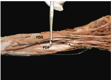
the left forearm of a 32-year-old female cadaver revealed a double FDS tendon, with one tendon acting as the FDP tendon to the little inger. In addition, there was a connection between this anomalous tendon and the FDP tendon to the ring inger. The FDP tendon originated from the FDS muscle at the forearm and had a normal distal attachment. The FDS was innervated by the median nerve. The blood supply of the tendon acting as the FDP tendon to the little inger was from a muscular branch of the ulnar artery. It was innervated by a branch from the median nerve. There were no muscular anomalies in the right arm or hand (Figs. 1-3).
DISCUSSION
Absence of the FDP of the little inger was recently reported by Furrer et al. Their case report showed the presence of the FDS and the absence of the FDP by ultrasound and magnetic resonance imaging in the right little inger of a nine-year-old boy.13 However, unlike the present
case, there was no muscle or tendon substituting for the FDP tendon to the little inger.
In three specimens, Austin et al. demonstrated tendinous slips from the FDS to the FDP of the little inger. These slips were well deined and very narrow and prevented FDS
1134
CLINICS 2009;64(11):1133-5 Anomalous lexor of the little inger
Yılmaz A et al.
function independently of FDP function to the little inger.15
A review of FDS and FDP muscle variations by Bergman et al. mentioned that there has also been a report of a slip from the ulnar side of the FDP running to join the FDS tendon of little inger.14 In addition, the FDP was fused with the FDS
in 11% of 205 arms examined; however, the FDS and FDP muscle bellies were not fused in the present case. The FDP
tendon to the little inger originated separately from the FDS muscle.14
The review by Bergman et al. also reported that occasionally a tendon separated from the FDS joins one of the tendons of the FDP muscle (accessorius profundus digitorum).14 However, in the present case, there was no
tendon to the little inger from the FDP muscle, so the FDS tendon to the little inger could not be described as accessory. Wahba et al. reported an accessory belly of the FDP that originated from the intercompartmental septum on the medial side of the forearm just proximal to the wrist joint and inserted on the proximal phalanx of the little inger. Despite this anomaly, the FDS and FDP muscles and tendons were completely intact, with no abnormal variations. This muscle was deined as a combination of an accessory FDP muscle belly acting on the metacarpophalangeal joint of the little inger, and Wahba et al. have named this variant an accessory lexor digiti minimi profundus muscle.16 Kisner
found a double FDS tendon to the little inger with absence of the profundus on the left hand of a 15-year-old patient during surgery; however, unlike the present case, there was a single ifth FDS tendon with normal appearance dividing into FDP and FDS tendons at the level of the distal palmar crease.6
The FDS tendon of the little finger is frequently interconnected with the FDS of the ring finger. In the present case, the anomalous FDP tendon to the little inger was attached to the FDP tendon to the ring inger. The close functional link between the ring inger FDS and the little inger FDS explains how an injury to either the ring or little inger can cause malfunction of the adjacent inger. The FDS has been used as a motor for a wide variety of tendon transfer operations in the hand. Most of these transfers are on the volar side of the hand and are used to restore synergistic actions (such as opposition of the thumb) or as intrinsic replacements.1,9,18 Recognition of these anomalies is
important during surgical repair of tendon lacerations. The treating surgeon should consider both digits when dealing with an injury to either inger.5,15,19
Figure 3 - Anterior aspect of the left forearm. FDS: l exor digitorum super-FDS: lexor digitorum super-icialis, FDP: lexor digitorum profundus. Black arrow indicates the FDP tendon originating from the FDS muscle
Figure 2 - Close view of the connection between the anomalous tendon and the FDP tendon to the ring inger. The white arrow indicates the FDP tendon originating from the FDS muscle, and the white asterix indicates the intertendinous connection
REFERENCES
1. Beasley RW. Beasley’s Surgery of the Hand. New York: Thieme Medical Publishers Inc; 2003.
2. Johnson D, Ellis H. Pectoral Girdle and Upper Limb: Forearm. In: Standring S, editor. Gray’s Anatomy The Anatomical Basis of Clinical Practice. 39th ed. Philadelphia: Elsevier Ltd; 2005. pp. 876-7.
3. Cassell MD, Bergman RA. Palmaris longus muscle substituting for the ring inger slip of lexor digitorum supericialis. Anat Anz. 1990;171:201-4.
4. Elliot D, Khandwala AR, Kulkarni M. Anomalies of the lexor digitorum supericialis muscle. J Hand Surg [Br]. 1999;24:570-4.
5. Gonzalez MH, Whittum J, Kogan M, Weinzweig N. Variations of the lexor digitorum supericialis tendon of the little inger. J Hand Surg. 1997;22:277-80.
1135
CLINICS 2009;64(11):1133-5 Anomalous lexor of the little inger
Yılmaz A et al.
7. Kobayashi N, Saito S, Wakisaka H, Matsuda S. Anomalous lexor of the little inger. Clin Anat. 2003;16:40-3.
8. Kostakoglu N, Borman H, Kecik A. Anomalous flexor digitorum supericialis muscle belly: an unusual case of mass in the palm. Br J Plast Surg. 1997;50:654-6.
9. Nayak SR, Pai MM, Krishnamurthy A, Kumar MSJ, Vadgaonkar R, Prabhu LV. An unusual lexor of the little inger and ulnar nerve entrapment: a case report. Neuroanatomy. 2007;6:30-1.
10. Nayak SR, Ramanathan L, Prabhu LV, Raju S. Additional lexor muscles of the forearm: case report and clinical signiicance. Singapore Med J. 2007;48:231-3.
11. Robinson SC. An anomalous lexor digitorum supericialis muscle-tendon unit associated with ulnar neuropathy. A case report. Clin Orthop Relat Res. 1985;194: 169-71.
12. Tountas CP, Halikman LA. An anomalous lexor digitorum sublimis muscle. A case report. Clin Orthop Relat Res. 1976;121:230-3. 13. Furrer M, Schweizer A, Meuli-Simmen C. Aplasia of the lexor digitorum
profundus tendon of the small inger. J Hand Surg Eur Vol. 2007;32:111-2.
14. Bergman RA, Afifi AK, Miyauchi R. Illustrated Encyclopedia of Human Anatomic Variation: Opus I: Muscular System: Muscles of the upper limb: Forearm and hand [cited 2009 June 2]. Available from: http://www.anatomyatlases.org/AnatomicVariants/MuscularSystem/ Text/F/17Flexor.shtml
15. Austin GJ, Leslie BM, Ruby LK. Variations of the lexor digitorum supericialis of the small inger. J Hand Surg. 1989;14:262-7. 16. Wahba MY, Singh GD, Lozanoff S. An anomalous accessory lexor
digiti minimi profundus muscle: a case study. Clin Anat. 1998;11:55-9. 17. Haines RW. The lexor muscles of the forearm and hand in lizards and
mammals. J Anat. 1950;84:13-29.
18. Agee J, McCarroll HR, Hollister A. The anatomy of the lexor digitorum supericialis relevant to tendon transfers. J Hand Surg. 1991;16:68-9. 19. Bowman P, Johnson L, Chiapetta A, Mitchell A, Belusko E. The clinical