RESEARCH ARTICLE Pub. 1160
ISSN 1679-9216
Received: 9 July 2013 Accepted: 17 October 2013 Published: 12 November 2013
Setor de Obstetrícia Veterinária, Departamento de Medicina Veterinária Preventiva e Reprodução Animal, Faculdade de Ciências Agrárias e Veterinárias - UNESP, Jaboticabal, São Paulo, Brazil. CORRESPONDENCE: M.A.R. Feliciano [marcusfeliciano@yahoo.com.br - Tel.: +55 (16) 32092626]. FCAV/ UNESP, Via de Acesso Prof. Dr. Paulo Donato Castellani, Bairro Distrito Industrial, s/n. CEP 14.884-900 Jaboticabal, SP, Brazil.
B-mode Ultrasound and Doppler Mode for Early-stage
Pregnancy Diagnosis in Shi-Tzu Bitches
Marcus Antonio Rossi Feliciano, Anelise Carvalho Nepomuceno, Diogo José Cardilli, Leandro Nassar Coutinho, Maria Emília Franco Oliveira, Vívian Tavares de Almeida,
Fernanda Gonçalves Canello & Wilter Ricardo Russiano Vicente
ABSTRACT
Background: The very fi rst sign to confi rm pregnancy in bitches is the gestacional sac detection. In this primary moment, it is very small with only few millimeters. The average time for gestational sacs visualization is approximately 20 days after mating (2 mm) surrounded by a thin hyperechoic wall (trophoblasts). The Doppler is a new method for pregnancy diagnosis in bitches. The measurement of velocity peak and vascular resistance index of the corpus luteum are used for such purpose. The precocity on detection pregnancy in bitches is an important tool for differential diagnosis between physi-ologic uterine alterations and uterine diseases. The aim of the current study was to assess the effi ciency of the B-mode and Triplex Doppler ultrasound for early-stage pregnancy diagnosis in Shi-Tzu bitches, using ecobiometry of the gestational sac and corpus luteum vascular index.
Materials, Methods & Results: Ten healthy Shi-Tzu bitches were evaluated. Pregnancy diagnosis was carried out using the B-mode. The ecobiometry of the gestational sac were characterized by the variables the outer (OLL) and inner (ILL) latero-lateral diameters, and the outer (ODV) and inner (IDV) dorso-ventral diameters. The corpus luteum vascularization was studied by the sistolic velocity peak (PVS), end-diastolic velocity (EDV), vascular resistance (RI) and pulsativity indexes (PI) using the Triplex Doppler. The ultrasound pregnancy diagnosis was carried out on days 14.2 ± 2.04 post mat-ing. The earliest pregnancy diagnosis occurred on day 12. Positive correlation on linear regression was observed between ILL and IDV and the date of the pregnancy diagnosis (r2 = 0.7 and P = 0.0027; r2 = 0.7 e P = 0.0025, respectively). On
corpus luteum Triplex Doopler assessment, PVS was 11.67 ± 2.53 cm/s, EDV was 5.52 ± 1.6 cm/s, PI was 1.04 ± 0.45 and RI was 0.61 ± 0.1.
Discussion: Early diagnosis observed in the present study for canine pregnancy is an important differential in veterinary obstetrics. Gestational sac was detected in the uterine horn on the 20th day post LH peak, 18 days following ovulation in a Beagle bitch and the ultrasound diagnosis of pregnancy was performed 18-24 days following the fi rst and last mating, respectively, in Yorkshire dogs. Those results highlighted the difference between the dates for early-stage pregnancy diag-nosis among different canine breeds. The precocity for the ultrasound pregnancy diagdiag-nosis depends on the visualization of the gestational sac, which is directly related to embryogenesis. The assessment of the ecobiometry of the gestational sacs may aid on the early-stage ultrasound pregnancy diagnosis in bitches. The values of corpus luteum vascular index in Shi-Tzu bitches in the current study were different from those found in other trial in bitches and in women. Such values may be justifi ed by the differences on the corpus luteum structures between the canine and human species. Regarding to Triplex Doppler in pregnant bitches, the values of PI and EDV could be determined, which hadn’t been reported before in veterinary medicine for the study of corpus luteum blood fl ow. In conclusion, the detection of pregnancy on early-stage using ultrasound examination is feasible in Shi-Tzu bitches, by the 12th day post conception. It was also possible to de-terminate important ecobiometry values of the gestacional sac and corpus luteum vascular index using Triplex Doppler.
INTRODUCTION
The sign that confi rms pregnancy in bitches is the gestacional sac detection, a mature blastocyst which is responsible for the embryonic development. In this moment, the gestational sac is very small and has only few millimeters in diameter. The average time for gestational sacs visualization is approximately 20 days after mating, appearing as anecoic structures (fi rst chorionic fl uid) with 2 mm, surrounded by a thin hyperechoic wall (trophoblasts) [15].
The blood fl ow assessment of the corpus lu-teum and ovary using Doppler is a mean of diagnosing pregnancy and predicting fetal viability. The measure-ment of peak velocity and corpus luteum vascular re-sistance index are used for such purpose [1]. There are few reports of Doppler use for pregnancy monitoring and uterine arteries fl ow characteristics of the of preg-nant and no pregpreg-nant in small animal practice [9,13]. The precocity on pregnancy detection in bitches is an important tool for differential diagnosis between physiologic uterine alterations and uterine diseases. Insi-de a gestation period lasting approximately 60 days [18] the diagnosis of diseases should be as fast as possible to allow interventions by the responsible clinician.
Therefore, the aim of the current study was to assess the effi ciency of the B-mode and Doppler ultrasound examination for early-stage gestational diagnosis in Shi-Tzu bitches and determination of ecobiometric parameters of the gestational sacs and
corpus luteum vascular index.
MATERIALS AND METHODS
Ten no pregnant multiparous Shi-Tzu bitches weighting 4-7 kg, aged 4-6 years old, from a private kennel were evaluated. The animals were previously submitted to physical and obstetric examination and only patients considered healthy were admitted in the current study.
The detection of proestro phase beginning was performed by the kennel owner, who was advice to contact the research team in order to confi rm the phase of the estral cycle. The estrus signs (i.e., accep-tance of matting) and vaginal cytology carried out the confi rmation of the optimal fertility period. Following confi rmation, the female were put along with a male for three days or artifi cial insemination was performed. The early-stage gestational diagnosis was performed two weeks following the fi rst mating or ar-tifi cial insemination, analogically to studies performed
on human obstetrics [5], according to methodology previously described [11].
The B-mode ultrasound and Doppler were performed using the MyLabTM30 VET machine1 and a
7-12MHz linear transducer. A single examiner carried out the ultrasound.
Prior to the procedures, the abdomen of the bi-tches were widely clipped. The animals were put in dorsal recumbency, which was changed to right or left lateral recumbency as required for optimal ultrasound scanning.
The presence of gestational sac was determined using the B-mode ultrasound examination. The eco-biometry of the gestational sac was determined using the measurement of the outer (OLL) and inner (ILL) latero-lateral diameters, and outer (ODV) and inner (IDV) dorso-ventral diameters.
The corpus luteum vascularization, assessed by pulsed Doppler, was used to determine the peak systolic velocity (PSV), end-diastolic velocity (EDV), resistance index (RI = [PSV - EDV] / PSV) and pul-satility index (PI = [PSV - EDV] / mean velocity) [6]. Three evaluation per animal were performed and a mean value was determined for each index.
Pulse Doppler was employed to determine sample volume, using the uniform insonation method. The cursor was positioned in the area of a vessel within the ovarian parenchyma to measure the trace of spectral curve fl ow and vascular index, which were obtained automatically following software identifi cation of the ultrasonic scanner for each waveform [7].
The power Doppler function was employed to increase the sensitivity of the blood fl ow measurements and to transform the examination from angle-indepen-dent to insonation or inciangle-indepen-dent angle [4].
Statistical analysis
The current study followed a randomized design. The data were previously tested for normality and homogeneity of the variance (F-test). The PROC MEANS-SAS® and GraphPad Prisma 4 were used. The signifi cance level adopted in the current study was 5%.
RESULTS
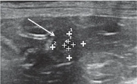
The mean pregnancy period in 10 Shi-Tzu bitches was 58 days and the number of puppies per pregnancy, on average, was four. The ultrasound pregnancy diagnosis was accomplished at 14.2 ± 2.04 days following mating, requiring only a single ultra-sound examination to detect pregnancy. The earliest pregnancy was detected on the 12th day post mating (Figure 1) and the mean number of gestational vesicles detected per animal was four.
The mean values of the ecobiometry of the gestational sac (EGS) found in the current study and
the correlation between the date of pregnancy diag-nosis (DPD) in Shi-Tzu bitches are shown in Table 1 and Figure 2.
Regarding the Triplex Doppler mode, peak systolic velocity was 11.67 ± 2.53 cm/s, end-diastolic velocity 5.52 ± 1.6 cm/s, pulsativity index was 1.04 ± 0.45 and vascular resistance index was 0.61 ± 0.1 for the left ovary. Statistical values were not possible to be measured on the right ovary due to its anatomic positioning, to the presence of artifacts and to reduced sampling. However, it was possible to detect the pre-sence of corpus luteum on those structures.
Figure 1. Ultrasound imaging of the early-stage and ecobiometry of the gestational sac in Shi-Tzu bitches.
Table 1. Mean values of OLL, ILL, ODV and IDV of the gestational sacs and linear regression between the date of pregnancy diagnosis and the ecobi-ometry of the gestational sacs in Shi-Tzu bitches.
OLL ILL ODV IDV
Mean values 0.83 ± 0.22 0.31 ± 0.18 0.73 ± 0.19 0.26 ± 0.14
Regression equation Y = 0.069x - 0.15 Y = 0.077x - 0.78 Y = 0.069x - 0.25 Y = 0.058x - 0.57
r2 0.42 0,7 0.56 0.7
Slope 0.069 ± 0.028 0.077 ± 0.018 0.069 ± 0.021 0.058 ± 0.013
P (5%) 0.0426* 0.0027* 0.0128* 0.0025*
*P < 0.005. Outer latero-lateral diameter (OLL); inner latero-lateral diameter (ILL); outer dorso-ventral diameter (ODV); inner dorso-ventral diameter
(IDV); r2: coeffi cient of determination.
DISCUSSION
Early diagnosis observed in the present study for canine pregnancy is an important differential in veterinary obstetrics for several reasons including: (1) confi rmation of pregnancy as soon as possible for the owners anxious; (2) provide subsidies to the vet for interference in cases of accidental and unscheduled mating [14]; promote obste-tric planning management for the pregnant female as well as an effective health strategy [10]; and (3) assist in the diagnosis of gestational alterations that may compromise this physiological process in bitches [21].
Even if the ultrasound is considered a non-in-vasive diagnostic technique [14], the stress resulting from the immobilization of the animal [12] or even atypical environment [15] may have a negative effect on the reproduction of the animals [19]. In pregnant bitches, hormonal, metabolic, and neural alterations caused by stress can promote embryonic absorption, fetal mortality and problems during parturition [3]. Therefore, in this study we adopted strategies to minimize possible stress to the animals, such as: the constant presence of the owner; the prior presentation and interaction of the animal with the sonographer; and a peaceful and quiet environment with pleasant temperatures and humidity.
Gestacional sacs can be early viewed between 19th and 20th days of pregnancy in bitches but such small fl uid structures can be overlapped by intestinal gas at the beginning of pregnancy. B-mode ultrasound presented precision of 94, 98 and 99% for de diagnosis of pregnancy in bitches following 24, 25 and 28 days of pregnancy, respectively [17]. Other studies reported the visualization of gestational sacs during B-mode ultrasound examination between 15 and 20 days of pregnancy [20]. Regarding the date of the gestacional
sac detection reported in those studies, it is suggested that the current trial showed early-stage ultrasound pregnancy diagnosis in Shi-Tzu bitches.
A gestational sac was detected in the uterine horn on the 20th day post LH peak, 18 days following ovulation in a Beagle bitch [17]. The ultrasound diag-nosis of pregnancy was performed 18-24 days follo-wing the fi rst and last mating, respectively, in Yorkshire dogs [8]. Those results highlighted the difference between the dates for early-stage pregnancy diagnosis among different canine breeds. Such fact reveals the importance of studies on other specifi c canine breeds for better understanding of the early-stage ultrasound pregnancy diagnosis, as performed in the current trial. The precocity for the ultrasound pregnancy diagnosis depends on the visualization of the gesta-tional sac, which is directly related to embryogenesis. The morula is formed 8 days following fertilization and consequently blastulation occurs (organization of the blastocyst, blastocele e trophoblast) [16]. The ultrasound visualization of anecogenic content (which aids on the differentiation of the gestational sac from the uterine tissue in bitches), may be associated to the formation of the blastocele. Therefore, the assessment of the ecobiometry of the gestational sacs may aid on the early-stage ultrasound pregnancy diagnosis in bitches.
The inner latero-lateral and dorso-ventral dia-meters may be associated to the accumulation of fl uid (blastocele) and the visualization of the gestational sac. Such proposal is in accordance with the positive cor-relation for the values of the linear regression between the date of pregnancy diagnosis and the ecobiometry of the gestational sac in the current study (ILL, r2 = 0.7
Higher values of diameter of gestational sacs were found in bitches [13] and queens [2] in com-parison with the results obtained in Shi-Tzu in the current study. The lateral diameter was also found higher than the dorso-ventral diameter in other trials [2,13] as reported in our study. Such fact occurs due to the direction of the fetal development, which occurs cranio-caudaly.
The values of corpus luteum vascular index in Shi-Tzu bitches in the current study were different from those found in other trial in bitches [11] and in women [5] which were (left ovary: PVS = 30.31 cm/s, IR = 0.52; right ovary: PVS = 20.46 cm/s, RI = 0.42) and (PVS = 27 cm/s, RI = 0.41 - 0.57), respectively. Such values may be justifi ed by the differences on the corpus luteum structures between the canine and human species.
Regarding the Triplex Doppler in pregnant bitches, the values of PI and EDV could be determi-ned, which hadn’t been reported before in veterinary medicine for the study of corpus luteum blood fl ow.
CONCLUSION
In conclusion, the detection of pregnancy on early-stage using ultrasound examination is feasible in Shi-Tzu bitches, by the 12th day post conception. It was also possible to determinate important values of ecobiometry of the gestational sac and corpus luteum
vascular index using Triplex Doppler.
SOURCE AND MANUFACTURER 1MyLabTM30 VET - ESAOTE, Genoa, Italy.
Acknowledgements. The authors would like to thank FAPESP
for the fi nancial support to the current research and for post-doctor scholarship support (Processes 2010/16913-7 and 2011/06011-9).
Ethical approval. The trial was conducted following approval
by the Animal Welfare and Ethics Committee of the School of Agrarian and Veterinary Sciences of the São Paulo State Uni-versity (FCAV/UNESP Jaboticabal) [process no 017314/10].
Declaration of interest. The authors report no confl icts of
interest. The authors alone are responsible for the content and writing of the paper.
REFERENCES
1 Alcazar J.L., Acosta M.J., Laparte C. & Ruiz M.L. 1996. Assessment of luteal blood fl ow in normal early pregnancy. Journal Ultrasound in Medicine. 15(1): 53-56.
2 Blass H.G., Eik-Nes S.H. & Berg S. 2000. Three-dimensional fetal ultrasound. Best Practices Research Clinical Obstetrics Gynaecology. 14(1): 611-627.
3 Brito A.B., Miranda S.A., Ruas, M.R., Santos R.R. & Domingues S.F.S. 2011. Assessment of feline fetal viability by conceptus echobiometry and triplex Doppler ultrasonography of uterine and umbilical arteries. Animal Reproduction Science. 122(1): 276-281.
4 Dar-Bello A.C., Riul T.R. & De Oliveira L.M. 2005. Desnutrição e estresse na gestação: medidas comportamentais das mães e dos fi lhotes durante a lactação. Temas em Psicologia. 13(1): 34-44.
5 Di Salvo P., Bocci F., Zelli R. & Polisca A. 2006. Doppler evaluation of maternal and fetal vessels during normal gestation in the bitch. Research Veterinary Science. 81(1): 322-388.
6 Drost W.T. 2007. Basic ultrasound physics. In: Trhall D.E. (Ed). Veterinary Diagnostic Radiology. 5th edn. St Louis: Saunders Elsevier, pp.38-49.
7 Durfee S.M. & Frates M.C. 1999. Sonographic spectrum of corpus luteum in early pregnancy: gray-scale, color, and pulsed Doppler appearance. Journal of Clinical Ultrasound. 27(2): 55-59.
8 Feliciano M.A.R., Muzzi L.A.L., Leite C.A.L. & Junqueira M.A. 2007. Two-dimensional conventional, high resolu-tion two-dimensional and three-dimensional ultrasonography in the evaluaresolu-tion of pregnant bitch. Arquivo Brasileiro de Medicina Veterinária e Zootecnia. 59(1): 1333-1337.
9 Feliciano M.A.R., Nepomuceno A.C., Crivelaro R.M., Oliveira M.E.F., Coutinho L.N. & Vicente W.R.R. 2013.
Foetal echoencephalography and Doppler ultrasonography of the middle cerebral artery in canine fetuses. Journal of Small Animal Practice. 54(1): 149-152.
10 Feliciano M.A.R., Vicente W.R.R., Leite C.A.L. & Muzzi L.A.L. 2008. Vascular resistance index and systolic velocity peak of the corpus luteum in pregnant bitch: a case report. Revista Brasileira de Reprodução Animal. 32(1): 191-196.
www.ufrgs.br/actavet
1160
12 Freitas J.G. & Silva A.R. 2008. Diagnóstico de gestação em cadelas. Revista Brasileira de Reprodução Animal. 32(1): 58-66.
13 Jabin V.C.P., Finardi J.C., Mendes F.C.C., Weiss R.R., Kozicki L.E. & Moraes R. 2007. Use of ultrasonography exams to determinate the parturition day by Yorkshire canine breed. Archives of Veterinary Science. 12(1): 63-70.
14 Mesquita S.F.P., Nocolielo M., Barbieri M.F., Michiyori B.T., Silva E.R., Klein M.O., Rosa J.L., Cavariani M.M. & Camargo I.C.C. 2009. Efeitos do estresse induzido por imobilização na prenhez da rata. Revista Brasileira de Zootecnia. 11(3): 277-282.
15 Miranda S.A. & Domingues S.F.S. 2010. Conceptus ecobiometry and triplex Doppler ultrasonography of uterine and umbilical arteries for assessment of fetal viability in dogs. Theriogenology. 74(1): 608-617.
16 Monteiro C.L.B., Madeira V.L.H. & Silva L.D.M. 2011. Diagnóstico da gestação em gatas. Revista Brasileira de Reprodução Animal. 35(4): 385-392.
17 Muir W.W. 1990. The equine stress response to anesthesia. Equine Veterinary Journal. 22(5): 302-303.
18 Noden D.N. & Lahunta A. 1991.Embriologia de los animales Domésticos. Zaragoza: Acribia, 399p.
19 Nyland G.T. & Mattoon J.S. 2002. Ultrasonography of the genital system. In: Nyland G.T. & Mattoon J.S. (Eds). Veterinary Diagnostic Ultrasound. Philadelphia: W.B. Saunders, pp.141-151.
20 Prestes N.C. & Landim-Alvarenga F.C. 2012.Obstetrícia Veterinária. Rio de Janeiro: Guanabara Koogan, 272p.
21 Rivier C. & Rivest S. 1991. Effect of Stress on the Activity of the Hypothalamic-Pituitary-Gonadal Axis: Peripheral and Central Mechanisms. Biology of Reproduction. 45(1): 523-532.
22 Shille V.M. & Gontarek J. 1985. The use ultrasonography for pregnancy diagnosis in the bitch. Journal of the American Veterinary Medical Association. 187(1): 1021-1025.