D isse m inate d Lange rhans’ ce ll
histio cyto sis and m assive
pro te in-lo sing e nte ro pathy
Instituto da Criança, Hospital das Clínicas,
Faculdade de Medicina, Universidade de São Paulo, São Paulo, SP, Brasil
T.M. Santos-Machado, L.M. Cristófani, M.T.A. Almeida, P.T. Maluf, P.A. Costa, M.A. Pereira, J.L.B.C. Brito and V. O done-Filho
Abstract
Symptomatic involvement of the gastrointestinal (GI) tract as a promi-nent symptom in Langerhans cell histiocytosis (LCH) is uncommon, occurring in less than 1 to 5% of all cases, even when the disease is in its disseminated form. Up to now, there have been reports of 18 cases of LCH with GI manifestations, including our 2 cases, with diarrhea (77.7%), protein-losing enteropathy (33.3%) and bloody stool being the most frequent findings. The authors present two patients with severe diarrhea and refractory hypoalbuminemia, and with the pro-tein-losing enteropathy documented by Cr51-labeled albumin studies. A review of the literature indicated that the presence of GI symptoms is often associated with systemic disease as well as with poor progno-sis, mainly under 2 years of age. Radioisotopes are useful for docu-menting protein loss in several diseases with high specificity and sensitivity, and their utilization in the cases reviewed here permitted diagnoses in 6 children, as well as improved therapeutic management. Co rre spo nde nce
V. O done Filho Instituto da Criança Hospital das Clínicas, FM, USP Av. Dr. Eneas C. Aguiar, 647 05403-900 São Paulo, SP Brasil
Fax: + 55-11-853-2602 Research partially supported by the Centro de Estudos Prof. Pedro de Alcântara and Fundação Maksoud para o Desenvolvimento da Cirurgia Pediátrica. Publication supported by FAPESP.
Received March 16, 1999 Accepted July 12, 1999
Ke y wo rds
·Langerhans’ cell histiocytosis ·Histiocytosis X ·Gastrointestinal
involvement
·Protein-losing enteropathy ·Hypoalbuminemia ·Cr51-labeled albumin test
Intro ductio n
In 1953, Lichtenstein (1) introduced the term histiocytosis X to integrate Hand-Schüller-Christian syndrome, Letterer-Siwe disease, and eosinophilic granuloma of the bone, a group of diseases with similar histo-pathological characteristics. In 1973, histio-cytosis X was described as local prolifera-tion as well as disseminaprolifera-tion of Langerhans histiocytes (2). Since Langerhans histiocytes are considered to be the main lesion cells in this disorder, in 1983, Risdall et al. (3) sug-gested naming the disease Langerhans cell
histiocytosis (LCH).
follow-ing characteristics: immunohistochemical confirmation with staining for S-100 pro-tein, ATPase, alpha-D-mannosidase, or pea-nut lectin binding. A definite diagnosis requires the presence of Birbeck granules in the lesion cells upon electron microscopic examination or demonstration of T6 (CD1A) antigenic determinants on the surface of the lesion cells.
Although the literature on LCH is exten-sive, this disorder in combination with gas-trointestinal (GI) involvement is rarely de-scribed (5-7). Even in these few cases, GI complications seldom appear as prominent clinical manifestations. The main clinical findings regarding intestinal disease include diarrhea (sometimes bloody) and protein-losing enteropathy (8).
In this study, we report two patients with disseminated LCH admitted to the Instituto da Criança (ICr). Both cases involved mas-sive protein-losing enteropathy demonstrated by tests performed with Cr51-labeled albu-min. Based on our experience and on a re-view of the existing literature on GI involve-ment in LCH, we discuss the role of enteral protein loss in the maintenance of hypoalbu-minemia as an aggravating factor in patients with disseminated LCH.
Case re po rts
Case 1
A 2-year-old white girl with a three-month history of diarrhea, daily fever, anasarca, oliguria, an ulcerated lesion of the scalp, disseminated petechiae, hepatomegaly and splenomegaly, as well as a tumor on the left frontal region was examined in our service. Initial laboratory findings revealed: he-moglobin level = 3.7 g/dl; hematocrit value = 12%; white blood cell count = 1,900/mm3; platelet count <5,000/mm3; blood urea nitro-gen level = 137 mg/dl (considered as dehy-dration); creatinine clearance level = 0.5 mg/ dl; serum albumin level = 1.1 g/dl; total
bilirubin level = 4.6 mg/dl (or 3.5 mg/dl of direct bilirubin); prothrombin activity = 24.4%; skin and bone marrow biopsy results consistent with LCH.
The patient continued with anasarca, per-sistent hypoalbuminemia, and massive diar-rhea, in spite of several blood and albumin transfusions, as well as administration of parenteral nutritional support. Enteral pro-tein loss was investigated by Cr51-labeled albumin tests. The dose administered was equivalent to 0.5 microcurie/kg. Results re-vealed an enteral loss equivalent to 81.5 ml of plasma per day (normal value = 14.6 ± 9 ml/day).
The patient did not respond to support with daily albumin. Her hypoalbuminemia persisted in spite of treatment with vinblas-tine, methotrexate, prednisone, and etopo-side. She died two months after diagnosis due to respiratory and cardiac failure, diges-tive hemorrhage, and hemorrhagic ascites.
Case 2
A 2-year-old white boy with a six-month history of jaundice, fever, pallor, diarrhea, loss of weight, hepatomegaly, ascites, skin and mucosal lesions, and diabetes insipidus was examined in our service.
Laboratory findings revealed: hemoglo-bin level = 11.9 g/dl; white blood cell count = 22,200/mm3; platelet count = 310,000/ mm3; blood urea nitrogen level = 43 mg/dl (considered as dehydration); creatinine clear-ance level = 0.6 mg/dl; total bilirubin level = 10.3 mg/dl (or 9.7 mg/dl of direct bilirubin); prothrombin activity = 38%; serum albumin level = 2.1 g/dl; biopsy results of a palate lesion consistent with LCH.
enteral loss equivalent to 75 ml of plasma per day.
Despite daily albumin administration, di-arrhea and hypoalbuminemia persisted. Treatment with prednisone, cyclophospha-mide, chlorambucil, and hydroxyurea was unsuccessful. The patient developed severe thrombocytopenia and died five months af-ter diagnosis.
The families of both children refused permission for further studies at autopsies.
D iscussio n
LCH with GI tract involvement is rare and is usually recognized only at post mortem examination of patients with multi-organ le-sions (9-13). Although most studies that have included large series of patients with dis-seminated LCH do not report GI symptoms (6,7,14), two children admitted to our serv-ice had diarrhea, representing 4.6% of all patients with LCH seen at our institution between 1976 and 1991.
In spite of the difficulty in assessing the frequency of GI involvement, Hyams et al. (15) reported GI symptoms in less than 1% of all patients, based on a review of other series that included 145 patients (12,16-18). Skopnic et al. (19) showed an estimated incidence of 5% of GI tract involvement in disseminated LCH. Idliki and Hamoudi (20) discussed a case with primary LCH of the bowel at the time of diagnosis.
Virtually all children described in the literature had multi-systemic disease, but GI disorders were rarely a prominent feature. Typically, LCH occurs either at sites in the skin or lymph nodes, where Langerhans cells or their precursors thrive, or in organs such as the lung or liver, as a result of certain pathological conditions (21,22). Although it would seem that GI tract invasion would be related to multi-systemic, widespread dis-ease, Keeling and Harries (11) have demon-strated that histological involvement of the GI tract in LCH may be more frequent than
the classical textbook descriptions of this disease suggest.
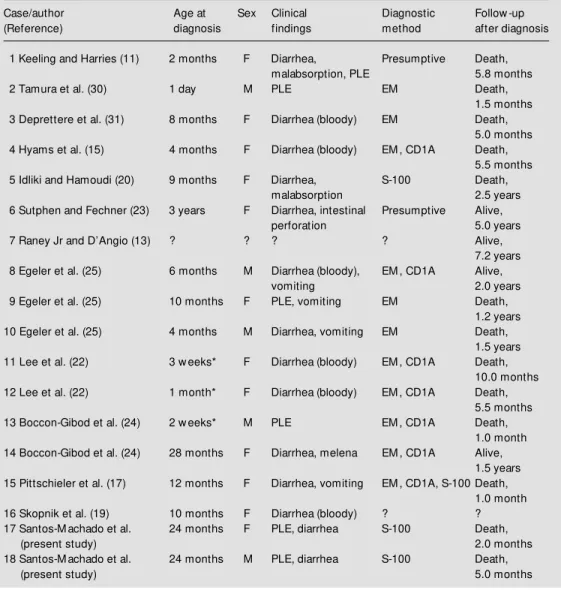
A summary of 18 reported cases, includ-ing ours, is presented in Table 1. Patients ages ranged from the neonatal period (con-genital form) to 3 years, with a median age of 4 months. Two thirds of the affected chil-dren were females. Table 2 outlines the most frequent clinical manifestations of LCH with GI complications, including diarrhea (some-times bloody), vomiting, and protein-losing enteropathy. Malabsorption and intestinal ulceration with melena and perforation were rarely described (11,23-25). As Table 2 il-lustrates, LCH patients with intestinal dis-ease present a high frequency (33.3%) of protein-losing enteropathy.
Even though GI tract dysfunction has not been identified as critical for prognosis (26), most of the patients affected by it have fared poorly, probably due to multi-systemic dis-semination of the disease. According to Jaffe et al. (8), the definitive diagnosis of LCH based on the Histiocyte Society criteria (4) is usually made beforehand at another site. Although histologic findings confirm the presence of S-100 protein, clinical findings are often sufficient to document GI involve-ment.
After confirming our diagnoses by posi-tive stains of S-100 protein from skin biop-sies, we documented the protein loss with Cr51-labeled albumin tests, a method that is widely used to identify enteric protein loss (27).
albu-Table 1 - Clinical manifestations of GI tract involvement in LCH in 18 children, including our 2 cases.
* Congenital; EM = electron microscopy; PLE = protein-losing enteropathy; ? = not reported.
Case/author Age at Sex Clinical Diagnostic Follow -up
(Reference) diagnosis findings method after diagnosis
1 Keeling and Harries (11) 2 months F Diarrhea, Presumptive Death,
malabsorption, PLE 5.8 months
2 Tamura et al. (30) 1 day M PLE EM Death,
1.5 months
3 Deprettere et al. (31) 8 months F Diarrhea (bloody) EM Death,
5.0 months
4 Hyams et al. (15) 4 months F Diarrhea (bloody) EM , CD1A Death,
5.5 months
5 Idliki and Hamoudi (20) 9 months F Diarrhea, S-100 Death,
malabsorption 2.5 years
6 Sutphen and Fechner (23) 3 years F Diarrhea, intestinal Presumptive Alive,
perforation 5.0 years
7 Raney Jr and D’Angio (13) ? ? ? ? Alive,
7.2 years
8 Egeler et al. (25) 6 months M Diarrhea (bloody), EM , CD1A Alive,
vomiting 2.0 years
9 Egeler et al. (25) 10 months F PLE, vomiting EM Death,
1.2 years
10 Egeler et al. (25) 4 months M Diarrhea, vomiting EM Death,
1.5 years
11 Lee et al. (22) 3 w eeks* F Diarrhea (bloody) EM , CD1A Death,
10.0 months
12 Lee et al. (22) 1 month* F Diarrhea (bloody) EM , CD1A Death,
5.5 months
13 Boccon-Gibod et al. (24) 2 w eeks* M PLE EM , CD1A Death,
1.0 month
14 Boccon-Gibod et al. (24) 28 months F Diarrhea, melena EM , CD1A Alive,
1.5 years 15 Pittschieler et al. (17) 12 months F Diarrhea, vomiting EM , CD1A, S-100 Death,
1.0 month
16 Skopnik et al. (19) 10 months F Diarrhea (bloody) ? ?
17 Santos-M achado et al. 24 months F PLE, diarrhea S-100 Death,
(present study) 2.0 months
18 Santos-M achado et al. 24 months M PLE, diarrhea S-100 Death,
(present study) 5.0 months
min (27). Our patients presented massive protein losses of 81.5 ml/day, while the nor-mal value is 14.6 ± 9 ml/day.
In six cases reviewed in the literature in which protein-losing enteropathy was pres-ent, all patients (100%) died. Deaths oc-curred 1 month to 1.5 years after diagnosis. Patients who did not experience protein loss lived longer, possibly due to the absence of refractory hypoalbuminemia. Tamura et al. (30) believe that loss of serum protein through the bowel is another contributing factor to the poor prognosis of patients with multi-Table 2 - Clinical and laboratory findings (% ) in 18
LCH patients w ith GI tract symptoms, including our 2 cases.
Symptoms Number %
of cases
Diarrhea 14 77.7
Protein-losing enteropathy 6 33.3
Bloody stool 6 33.3
Vomiting 4 22.2
M alabsorption 2 11.1
M elena 1 5.5
Perforation 1 5.5
Re fe re nce s
1. Lichtenstein L (1953). Histiocytosis X: In-tegration of eosinophilic granuloma of the bone, “ Let t erer-Siw e disease” , and “ Schüller-Christian disease” as related manifestations of a single nosologic en-tity. Archives of Pathology, 56: 84-102. 2. Nezelof C, Basset F & Rousseau M F
(1973). Histiocytosis X: Histogenetic argu-ments for a Langerhans’ cell origin. Bio-medicine, 18: 365-371.
3. Risdall RJ, Dehner LP, Duray P, Kobrinsky N, Robison L & Nesbit M E (1983). Histio-cytosis X (Langerhans’ cell histioHistio-cytosis). Archives of Pathology and Laboratory M edicine, 107: 59-63.
4. Writing Group of the Histiocyte Society (1987). Histiocytosis syndromes in chil-dren. Lancet, 1: 208-209.
5. Lahey M E (1962). Prognosis in reticuloen-dotheliosis in children. Journal of Pediat-rics, 60: 664-671.
6. Nesbit M E (1986). Current concepts and treatment of histiocytosis X (Langerhans’ cell histiocytosis). In: Voûte PA, Barret A, Bloom HJ, Lemerle J & Neidhardt M K (Editors), Cancer in Children - Clinical M an-agement. Vol. 1. Springer-Verlag, Berlin, 176-184.
7. Nezelof C, Frileux-Herbert F & Cronier-Sachot J (1979). Disseminated histiocyto-sis X. Analyhistiocyto-sis of prognostic factors based on a retrospective study of 50 cases. Can-cer, 44: 1824-1834.
8. Jaf f e R, W ollm an M R, Kocoshis S, Penchansky L & Gilbert-Barness E (1993). Pathological cases of the month. Langer-hans’ cell histiocytosis w ith gastrointesti-nal involvement. American Journal of Dis-eases in Children, 147: 79-80.
9. Daneshbod K & Kissane JM (1978). Idio-pathic differentiated histiocytosis. Ameri-can Journal of Clinical Pathology, 70: 381 -389.
10. Favara BE & Jaffe R (1987). Pathology of Langerhans’ cell histiocytosis.
Hematol-ogy/Oncology Clinics of North America, 1: 75-97.
11. Keeling JW & Harries JT (1973). Intestinal malabsorption in infants w ith histiocyto-sis X. Archives of Disease in Childhood, 48: 350-354.
12. Oberman HA (1961). Idiopathic histiocy-tosis: A clinicopathologic study of 40 cases and review of the literature on eosi-nophilic granuloma of the bone, Hand-Schüller-Christian disease and Letterer-Siw e disease. Pediatrics, 28: 307-327. 13. Raney Jr RB & D’Angio GJ (1989).
Lang-erhans’ cell histiocytosis (histiocytosis X). Experience at the Children’s Hospital of Philadelphia 1970-1984. M edical and Pe-diatric Oncology, 17: 20-28.
14. Lahey M E (1975). Histiocytosis X - an a-nalysis of prognostic factors. Journal of Pediatrics, 87: 184-189.
15. Hyams JS, Hasw ell JE, Gerber M A & Berman M M (1985). Colonic ulceration in histiocytosis X. Journal of Pediatric Gas-troenterology and Nutrition, 4: 286-290. 16. Lucaya J (1971). Histiocytosis X.
Ameri-can Journal of Diseases in Children, 121: 289-295.
17. Pittschieler K, Radetti G, Egarter E & M engarda G (1989). Chronischer Durchfall als Hauptsymptom einer Histiozytose X. Helvetica PaediatricaActa, 43: 467-471. 18. Talbot M L (1974). Histiocytosis X.
Ameri-can Surgeon, 40: 89-96.
19. Skopnik H, Greven P, Füzesi L, M ertens R & Heiman G (1991). Gastrointestinale Symptome bei disseminierten Langer-hans-Zell-Histiozytosen. Kasuistik and Lit erat urüberchit . M onat sschrif t f ür Kinderheilkunde, 139: 239-243. 20. Idliki O & Hamoudi AB (1984). Primary
histiocytosis X of bow el. Pediatric Pathol-ogy, 2: 492 (Abstract).
21. Basset F, Soler P & Hance AJ (1986). The Langerhans’ cell in human pathology. An-nals of the New York Academy of
Sci-ences, 465: 324-329.
22. Lee RG, Braziel RM & Stenzel P (1990). Gastrointestinal involvement in Langer-hans’ cell histiocytosis (histiocytosis X): Diagnosis by rectal biopsy. M odern Pa-thology, 3: 154-157.
23. Sutphen JL & Fechner RE (1986). Chronic gastroenteritis in a patient w ith histiocy-tosis X. Journal of Pediatric Gastroenter-ology and Nutrition, 5: 324-328. 24. Boccon-Gibod LA, Krichen HA,
Carlier-M ercier LCarlier-M B, Salaun JF, Fontaine JL & Leuerger AR (1992). Digestive tract in-volvement w ith exudative enteropathy in Langerhans’ cell histiocytosis. Pediatric Pathology, 12: 515-524.
25. Egeler RM , Schipper M E & Heymas HS (1990). Gastrointestinal involvement in Langerhans’ cell histiocytosis (histiocyto-sis X): A clinical report of three cases. European Journal of Pediatrics, 149: 325-329.
26. Lahey M E (1981). Prognostic factors in histiocytosis X. American Journal of Pedi-atric Hematology/Oncology, 3: 57-60. 27. Waldman TA (1966). Protein-losing
enter-opathy. Gastroenterology, 50: 422-443. 28. Waldman TA (1961). Gastrointestinal
pro-tein loss demonstrated by Cr51-labeled albumin. Lancet, 2: 121-123.
29. Waldman TA & Wochner RD (1963). The use of Cr51-labeled albumin in the study of protein-losing enteropathy. Protides of the Biological Fluids, Proceedings of the Colloquium, 11: 224-226.
30. Tam ura T, Um et su M , M ot oya H & Yokoyama S (1980). Congenital Letterer-Siw e disease associated w ith protein-los-ing enteropathy. European Journal of Pe-diatrics, 136: 77-80.
31. Deprettere A, Aevoet G, Van Acker KJ & Dockx P (1983). Intractable diarrhea in his-tiocytosis X. Helvetica PaediatricaActa, 38: 291-294.
systemic LCH lesions.
We suggest the use of Cr51-labeled albu-min to investigate protein-losing enteropa-thy in patients with disseminated LCH, with or without important GI symptoms. The