Inte ractio ns be twe e n intrace llular
Ca
2+
sto re s: Ca
2+
re le ase d fro m
the NAAD P po o l po te ntiate s cAD
PR-induce d Ca
2+
re le ase
Departments of Anesthesia and Internal Medicine, Mayo Clinic and Foundation, Rochester, MN, USA E.N. Chini
Abstract
Cells possess multiple intracellular Ca2+-releasing systems. Sea urchin egg homogenates are a well-established model to study intracellular Ca2+ release. In the present study the mechanism of interaction be-tween three intracellular Ca2+ pools, namely the nicotinic acid adenine dinucleotide phosphate (NAADP), the cyclic ADP-ribose (cADPR) and the inositol 1',4',5'-trisphosphate (IP3)-regulated Ca2+ stores, is explored. The data indicate that the NAADP Ca2+ pool could be used to sensitize the cADPR system. In contrast, the IP3 pool was not affected by the Ca2+ released by NAADP. The mechanism of potentia-tion of the cADPR-induced Ca2+ release, promoted by Ca2+ released from the NAADP pool, is mediated by the mechanism of Ca2+-induced Ca2+ release. These data raise the possibility that the NAADP Ca2+ store may have a role as a regulator of the cellular sensitivity to cADPR.
Co rre spo nde nce
E.N. Chini
Department of Anesthesiology Mayo Clinic and Foundation 200 First Street,
Rochester, MN 55905 USA
Fax: + 1-507-255-7300 E-mail: chini.eduardo@ mayo.edu Research supported by the Mayo Foundation.
Received December 5, 2001 Accepted March 5, 2002
Ke y wo rds
·cADPR
·NAADP
·Calcium
·Sea urchin eggs
·Fertilization
·IP3
Intro ductio n
The release of Ca2+ from intracellular
stores is a widespread component of several signaling pathways (1-3). Nicotinic acid ade-nine dinucleotide phosphate (NAADP) is a recently discovered nucleotide with
intracel-lular Ca2+-releasing properties (4-11).
NAADP-induced Ca2+ release was first
de-scribed in sea urchin egg homogenates (5).
The Ca2+ release mechanism elicited by
NAADP differs in many ways from the Ca2+
release controlled by cyclic ADP-ribose (cADPR) and inositol 1',4',5'-trisphosphate
(IP3) (2,4-17). Properties of this Ca2+
-releas-ing molecule include: i) absence of
regula-tion by the intracellular divalent caregula-tions Mg2+
and Ca2+ (6-8,15); ii) NAADP-induced Ca2+
release is fully inactivated by exposure to low concentrations of NAADP (14), and iii)
Ca2+ release induced by NAADP appears to
be insensitive to changes of pH over a wide range (8,17). These characteristics make
NAADP a unique trigger of intracellular Ca2+
(2,9,10). In addition to the NAADP-induced
Ca2+ release system, cells also possess other
intracellular Ca2+ messengers such as cADPR
and IP3 (5). The exact physiological role of
three different intracellular Ca2+-releasing
systems in cells is not known. However, it is
possible that these different Ca2+ pools may
intra-cellular Ca2+ oscillation (9,12,13,16,18). In
the present study I explored in vitro the
mechanisms by which NAADP could
modu-late the Ca2+ release elicited by cADPR. It
was found that NAADP could potentiate the
cADPR-induced Ca2+ release by
sensitiza-tion of the ryanodine receptor by a
mechan-ism similar to the Ca2+-induced Ca2+ release.
This result indicates that crosstalk between
intracellular Ca2+ pools may modulate the
complex mechanism of intracellular Ca2+
mobilization.
Mate rial and Me tho ds
Se a urchin e gg ho m o ge nate s
Homogenates from Lytechinus pictus egg
were prepared as described previously (5). Frozen homogenates were thawed in a 17ºC water bath and diluted to 1.25% with an intracellular medium containing 250 mM N-methyl glutamine, 250 mM potassium glu-conate, 20 mM HEPES buffer, pH 7.2, 1 mM
MgCl2, 2 U/ml creatine kinase, 4 mM
phos-phocreatine, 1 mM ATP, 3 µg/ml oligomy-cin, and 3 µg/ml antimycin. After incubation at 17ºC for 3 h, 3 µM fluo-3 was added. Fluo-3 fluorescence was monitored at 490 nm excitation and 535 nm emission in a 250-µl cuvette, held at 17ºC with a circulating water bath and continuously mixed with a mag-netic stirring bar, in a Hitachi spectrofluo-rometer (F-2000).
45Ca uptake and release were measured
by a filtration method using glass-fiber fil-ters as described in Ref. 6. The remaining
intravesicular 45Ca was determined by
filtra-tion of 0.2 ml of a 1.25% (v/v) egg homoge-nate through a prewashed GF/C glass filter (Whatman) under vacuum, followed by rapid washing three times with 1 ml of an ice-cold
intracellular medium containing 3 mM LaCl3.
The radioactivity retained on the filter was determined by standard scintillation count-ing.
Mate rial
L. pictus and Aplysia californica were obtained from Marinus Inc., Long Beach, CA, USA. Fluo-3 was purchased from
Mo-lecular Probes, Eugene, OR, USA, and IP3,
ryanodine, oligomycin and antimycin were from Calbiochem, San Diego, CA, USA. All other reagents, of the highest purity grade available, were supplied by Sigma Co., St. Louis, MO, USA. NAADP and cADPR were synthesized as described before (5).
The reported experiments were repeated at least three to six times.
Re sults and D iscussio n
NAAD P and cAD PR induce Ca2+ re le ase fro m
diffe re nt Ca2+ po o ls
First we investigated the mechanisms of
Ca2+ uptake in sea urchin egg homogenates,
which were found to have both
thapsigargin-sensitive and -inthapsigargin-sensitive Ca2+ uptake
sys-tems. These data indicate that egg homoge-nates have both a sarcoplasmic-endoplasmic
reticulum Ca2+ ATPase (SERCA)-like pool
and a second different mechanism of Ca2+
uptake that is not mediated by a SERCA-like enzyme. As shown in Figure 1, the thapsi-gargin-insensitive system is slower.
How-ever, the maximum amount of Ca2+ uptake
was identical in the presence or absence of thapsigargin (Figure 1). Next we determined
whether the intracellular Ca2+-releasing
C
a
2
+ u
p
ta
k
e
(
n
m
o
l)3.0
2.0
1.0
0.0
0 10 20 30 80
Time (min)
Figure 1. Ca2+ uptake in sea
ur-chin egg homogenates. The
de-termination of Ca2+ uptake w as
performed using 45Ca as
de-scribed in M aterial and M ethods. Sea urchin egg homogenates w ere incubated in the presence (open circles) or absence (filled circles) of 10 µM thapsigargin (a
agents cADPR, IP3, and NAADP could
acti-vate Ca2+ efflux in both
thapsigargin-sensitive and -inthapsigargin-sensitive pools (Figure 2). In agreement with data previously reported by Genazzani and Galione (15), the results
indicated that cADPR and IP3 promoted Ca2+
release only through the thapsigargin-sensi-tive pools (Figure 2). In contrast, NAADP
was able to induce Ca2+ release from both
thapsigargin-sensitive and -insensitive pools (Figure 2), indicating that the NAADP and
cADPR Ca2+ pools in sea urchin egg
homo-genates are at least partially independent.
Po te ntiatio n o f the Ca2+-induce d Ca2+ re le ase
by Ca2+ re le ase d fro m the NAAD P po o l
It has been previously reported that
extravesicular Ca2+ can not only potentiate
but is also necessary for the Ca2+ release
induced by ryanodine receptor agonists such as cADPR and ryanodine (6,19). In contrast,
the NAADP-induced Ca2+ release does not
behave like a Ca2+-induced Ca2+ release
(6,15). It has been proposed that the Ca2+
released by NAADP could modulate the Ca2+
-induced Ca2+ release system activated by
cADPR (18,20,21). However, no direct evi-dence for this action has been reported to
date. Here we demonstrate that Ca2+ release
from the NAADP pool could potentiate the
Ca2+ release induced by ryanodine and
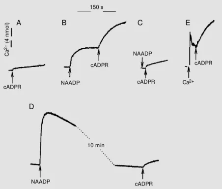
cADPR. As shown in Figure 3, after the addition of 12 nM NAADP a small amount
of Ca2+ was released from the vesicles, and
the addition of subthreshold concentrations of cADPR at the peak (steady state) of the
Ca2+ release led to a significant potentiation
of the cADPR-induced Ca2+ release (Figure
3). This effect was not mediated by NAADP itself but by the increase in extravesicular
Ca2+, since when the Ca2+ release induced by
NAADP was abolished by previous desensi-tization of the NAADP receptor the
cADPR-induced Ca2+ release was not enhanced by
NAADP (Figure 3C). The increase of
extravesicular Ca2+ induced by NAADP
in-Figure 2. Ca2+ release induced by nicotinic acid adenine dinucleotide phosphate (NAADP)
from the thapsigargin-insensitive pool. The sea urchin egg homogenates w ere loaded w ith
45Ca as described in Figure 1. After 3 h of Ca2+ uptake, Ca2+ release w as initiated by
addition of 1 µM IP3, 100 nM cyclic ADP-ribose (cADPR) or 100 nM NAADP. The Ca2+
release w as performed in homogenates loaded in the absence (open circles) or the presence (filled circles) of 10 µM thapsigargin.
In
tr
a
v
e
s
ic
u
la
r
C
a
2
+ (
%
)
120
IP3 cADPR NAADP
30 s
100
80
60
40
20
0
C
a
2
+(4
n
m
o
l)
150 s
cADPR NAADP
cADPR
NAADP
cADPR
cADPR Ca2+
10 min
cADPR NAADP
A B C E
D
Figure 3. Potentiation of the cyclic ADP-ribose (cADPR)-induced Ca2+ release by Ca2+
released from the nicotinic acid adenine dinucleotide phosphate (NAADP) pool. Free Ca2+
concentrations w ere measured as described in M aterial and M ethods using fluo-3. The
arrow indicates the sequential addition of different Ca2+ channel agonists. In A the arrow
indicates the addition of 16 nM cADPR that by itself does not promote Ca2+ release. In B
the homogenate w as first treated w ith 12 nM NAADP and 16 nM cADPR w as added at the
peak (steady state) of the Ca2+ release induced by NAADP. In C the homogenate w as
pretreated w ith 2 nM NAADP for 20 min (not show n) to promote self-desensitization of the NAADP receptor. After that the homogenate w as treated w ith 12 nM NAADP and 16 nM cADPR. In D homogenates w ere treated w ith a saturating concentration of 60 nM NAADP
and then, after the Ca2+ released by NAADP w as taken up again, the homogenate w as
treated w ith 16 nM cADPR. In E the homogenate w as treated w ith 4 nmol Ca2+ prior to the
addition of 16 nM cADPR. The data are representative of 12 different experiments done w ith three different preparations of sea urchin egg homogenates.
. . . .
... ... ..
pool can sensitize the ryanodine receptor to cADPR. In contrast, we found no effect of
NAADP on the Ca2+ release induced by IP
3.
Furthermore, Ca2+ released from the IP
3 pool
was not consistently able to sensitize the
cADPR-induced Ca2+ release (data not
shown). This is probably due to the fact that
cADPR and IP3 induce Ca2+ release from the
same Ca2+ pool in sea urchin egg
homoge-nates (15).
A second mechanism for NAADP
modu-lation of the cADPR-induced Ca2+ release
has been described by Churchill and Galione (12), who reported that in intact sea urchin
eggs NAADP-induced Ca2+ oscillations were
mediated via a two-pool mechanism that
primed the cADPR- and the IP3-sensitive
Ca2+ stores (12). In fact, priming the Ca2+
pools with Ca2+ (13) can increase the
appar-ent affinity for cADPR and IP3.
The precise role of NAADP-modulated
Ca2+ release is not known. However, it has
been proposed that in pancreatic acinar cells
NAADP could be the trigger of Ca2+
oscilla-tions induced by cholecystokinin (20,21).
The cited investigators proposed that Ca2+
released by NAADP in response to
chole-cystokinin may activate the Ca2+-induced
Ca2+ release mediated by cADPR, leading to
amplification of the Ca2+ signaling and
gen-eration of the Ca2+ oscillation (20,21). A
similar role for NAADP has been proposed
for the mobilization of Ca2+ in starfish
oo-cytes (18). The present study is the first to
demonstrate a direct effect of the Ca2+
re-leased by NAADP on the apparent affinity of the ryanodine receptor for cADPR (Fig-ure 4). This further indicates that NAADP may have an important role in the complex
mechanism of intracellular Ca2+
mobiliza-tion in several vertebrate and invertebrate
cells (4,5,16-18,20,21). In fact, the Ca2+
re-leased from the NAADP pool can modulate
the intracellular Ca2+ release by at least two
different mechanisms: a) by priming the
in-tracellular Ca2+ pools (16) and b) by direct
sensitization of the Ca2+-induced Ca2+ release.
C
a
2
+ r
e le a s e ( n m o l/ m in ) 4 3 2 1 0 10 8 6 4 2 0 C a 2
+ r
e le a s e ( n m o l/ m in )
0 200 400 600 800
Ryanodine (µM )
0 10 20 30 50
cADPR (nM ) 40
Figure 4. Effect of Ca2+ released
by NAADP on the apparent af-finity of the ryanodine receptor for ryanodine and cyclic
ADP-ribose (cADPR). Homogenates
w ere treated w ith no addition (filled circles), or w ith the addi-tion of 12 nM NAADP (open circles) as show n in Figure 3B. The dose-response dependence for ryanodine (A) and cADPR (B) w as determined by the addi-tion of different concentraaddi-tions
of t he Ca2+releasing com
-pounds as show n in the figure. The addition of ryanodine and cADPR w as perf orm ed af t er
NAADP-induced Ca2+ release
w as at its plateau level (see
Fig-ure 3B). The Ca2+ released by
NAADP potentiates the effect of bot h ryanodine and cADPR about 2.5 to 3 times. The data represent the mean ± SEM of four experiments. 7 6 5 4 3 0 C a 2
+ r
e le a s e ( n m o l/ m in ) 2 1
0 1 2 3 4 5 6
Extravesicular Ca2+ above baseline (nmol)
Figure 5. Effect of extravesicular
Ca2+ on cADPR-induced Ca2+
re-lease. Ca2+ release w as
moni-tored as described in M aterial and M ethods. The figure
indi-cates the Ca2+ released by 16
nM cADPR under different
lev-els of extravesicular Ca2+ above
baseline. The Ca2+ released
un-der ambient extravesicular Ca2+
is indicated by a triangle. The
ext ravesicular Ca2+ w as
in-creased by the addition of differ-ent concdiffer-entrations of NAADP
(squares) or Ca2+ (circles). The
addit ion of cADPR w as per-formed at the plateau level of
Ca2+ induced by NAADP or Ca2+
itself, as show n in Figure 1. The data are the mean ± SEM of three independent experiments.
creased the apparent affinity of the ryano-dine receptor for cADPR and ryanoryano-dine
(Fig-ure 4). Increasing the extravesicular Ca2+
could reproduce the effect of NAADP on the
Ca2+ release mediated by cADPR by the
addition of Ca2+ itself to the sea urchin egg
homogenates (Figures 3E and 5). In fact, when normalized for the increase in
extravesicular Ca2+ upon the potentiation of
the cADPR-induced Ca2+ release, the effects
of NAADP and of addition of Ca2+ itself
were near identical (Figure 5). These data
indicate that Ca2+ released from the NAADP
A
Multiple intracellular Ca2+ stores are
pres-ent in many cells (1,4-6,20,21) and may play a role in several physiological processes in-cluding muscle contraction, exocrine and endocrine secretion, fertilization, neuronal activation and immune cell function (1,2,9,
13,16-18,20). Exactly how Ca2+ exerts its
intracellular effects is not completely under-stood. The answer may lie in the complex interaction between intracellular and
extra-cellular Ca2+ pools to generate specific
spa-tial-temporal intracellular Ca2+ signals. In
this regard, the present results describing the direct interactions between NAADP (a
non-Ca2+-induced Ca2+ release) and cADPR (a
Ca2+-induced Ca2+ release) Ca2+ stores may
be of broad physiological importance. In fact, the determination of the specific role of
different Ca2+ stores in several cellular
func-tions deserves further investigation.
Re fe re nce s
1. Berridge M J (1993). A tale of tw o mes-sengers. Nature, 365: 388-389.
2. Dousa TP, Chini EN & Beers KW (1996). Adenine nucleotide diphosphate: emerg-ing second messengers actemerg-ing via
intra-cellular Ca2+ release. American Journal of
Physiology, 271: C1007-C1024.
3. Galione A & White A (1994). Ca2+ release
induced by cyclic-ADP-ribose. Trends in Cell Biology, 4: 431-436.
4. Cheng J, Yusufi ANK, Thompson M A, Chini EN & Grande JP (2001). Nicotinic acid adenine dinucleot ide phosphat e
(NAADP), a new Ca2+ releasing agent, in
kidney. Journal of the American Society of Nephrology, 12: 54-60.
5. Chini EN, Beers KW & Dousa TP (1995). Nicotinate-adenine dinucleotide
phos-phate (NAADP) triggers a specific Ca2+
release in sea urchin eggs. Journal of Bio-logical Chemistry, 270: 3216-3223. 6. Chini EN & Dousa TP (1996).
Nicotinate-adenine dinucleotide phosphate-induced
Ca2+ release does not behave as a Ca2+
-induced Ca2+ release system.
Biochemi-cal Journal, 316: 709-711.
7. Chini EN & Dousa TP (1996).
Palmitoyl-CoA potentiates the Ca2+ release elicited
by cyclic ADP-ribose. American Journal of Physiology, 270: C530-C537.
8. Chini EN, Liang M & Dousa TP (1998). Differential effect of pH upon cyclic-ADP-ribose and nicotinate-adenine dinucleotide
phosphate-induced Ca2+ release systems.
Biochemical Journal, 335: 499-504. 9. Galione A, Patel S & Churchill GC (2000).
NAADP-induced calcium release in sea urchin eggs. Biology of the Cell, 92: 197-204.
10. Genazzani AA & Galione A (1996). A Ca2+
release mechanism gated by the novel pyridine nucleotide, NAADP. Trends in Pharmacological Sciences, 18: 108-110. 11. Lee HC & Aarhus R (1995). A derivative of
NADP mobilizes calcium stores insensi-tive to inositol trisphosphate and cyclic ADP-ribose. Journal of Biological Chemis-try, 270: 2152-2157.
12. Churchill GC & Galione A (2001).
NAADP-induces Ca2+-oscillations via a tw o-pool
mechanism by priming IP3- and
cADPR-sensitive Ca2+ stores. EM BO Journal, 20:
2666-2671.
13. Galione A, M cDougall A, Busa W B, Willmott N, Gillot J & Whitaker M (1993). Redundant m echanism s of calcium -induced calcium release underlying cal-cium w aves during fertilization of sea-ur-chin eggs. Science, 261: 348-352. 14. Genazzani AA, Empson RM & Galione A
(1996). Unique inactivation properties of
NAADP-sensitive Ca2+ release. Journal of
Biological Chemistry, 271: 11599-11602. 15. Genazzani AA & Galione A (1996).
Nico-tinic acid-adenine dinucleotide phosphate
mobilizes Ca2+ from a
thapsigargin-insen-sitive pool. Biochemical Journal, 315: 721-725.
16. Perez-Terzic CM , Chini EN, Shen SS,
Dousa TP & Clapham DE (1995). Ca2+
release triggered by nicotinate adenine dinucleotide phosphate in intact sea ur-chin eggs. Biochemical Journal, 312: 955-959.
17. Yusufi ANK, Cheng J, Thompson M A, Chini EN & Grande JP (2001). NAADP
elicits specific microsomal Ca2+ release
from mammalian cells. Biochemical Jour-nal, 353: 531-536.
18. Santella L, Kyozuka K, Genazzani AA, De Riso L & Carafoli E (2000). Nicotinic acid adenine dinucleotide phosphate-induced
Ca2+ release: interaction among distinct
Ca2+ mobilizing mechanisms in starfish
oocytes. Journal of Biological Chemistry, 275: 8301-8306.
19. Lee HC (1993). Potentiation of calcium-and caffeine-induced calcium release by cyclic ADP-ribose. Journal of Biological Chemistry, 268: 293-299.
20. Cancela JM , Churchill GC & Galione A (1999). Coordination of agonist-induced
Ca2+-signalling patterns by NAADP in
pan-creatic acinar cells. Nature, 398: 74-76. 21. Cancela JM , Gerasimenko OV,
Gerasi-menko JV, Tepikin AV & Petersen OH (2000). Tw o different but converging
mes-senger pathw ays to intracellular Ca(2+)