Polyethylene glycols (PEG) of various molecular weights are low toxic biocompatible polymers widely used in biology, medicine and pharmaceuticals [1]. Typically, PEG is used as a precipitating agent for the isolation and purification of proteins, nucleic acids and viruses. Fusogenic properties of the polymer provide a preparation of hybridoma cell cultures by the fusion of the two cell types. Surface modification by covalently attached PEG is the basis of the camouflage biotechnology, designed to reduce the immunogenicity of molecules and cells in order to extend their survival in vivo. The process of pegylation enhances the solubility of macromolecules in water and improves biocompatibility, that is important for drug development. In combination with other components the PEG is used to form hydrogels, which allow a controlled release of therapeutic agents, moreover they are also applied in regenerative medicine and some medical devices.
PEG has also demonstrated cryoprotective properties in respect of certain cell types,
providing strong protection against effects of extreme environmental factors associated with freezing and thawing processes [2, 3]. Such factors include [4]: the formation of crystals, the growth of salt concentration in the supercooled liquid, increasing the osmotic pressure, phase transitions and lateral separation of lipids in membrane systems, dehydration of macromolecules. PEG of m.w. 1500 (PEG-1500) does not cross the plasma membrane, that is referred to the exocellular type of cryoprotectants, which cause considerable interest among researchers [2–6], because on their base washing-free methods of cell cryopreservation can be developed. PEG-1500 provides effective protection of the erythrocytes during freezing down to ultralow temperatures (–196 C) [2]. However, the transfer of cryopreserved erythrocytes to physiological conditions reveals latent membrane injuries that may be caused by a modification of the structural and functional state of the individual components of the membrane under the influence of PEG-1500. Taking into account the prospects of PEG-1500 UDС 576.314.6:577.322.23:576.524:57.043 https://doi.org/10.15407/biotech9.05.054
MODIFICATION OF ERYTHROCYTE MEMBRANE
PROTEINS BY POLYETHYLENE GLYCOL 1500
Key words: Ca2+-ATPase, CD44, polyethylene glycol 1500, erythrocyte.
N. G. Zemlianskykh Institute for Problems of Cryobiology and Cryomedicine
L. A. Babijchuk of the National Academy of Sciences of Ukraine, Kharkiv
E-mail: [email protected]
Received 12.07.2016
using for erythrocyte cryopreservation and establishment of blood banks, the research of membrane modification in the cryoprotectant presence may be the key condition to understanding and leveling of the adverse effects of the polymer interaction with cells by adjusting the processing conditions of cells with cryoprotective medium or introducing additional components into it.
It is known that the stability of erythrocytes largely depends on the state of membrane proteins associated by point contacts to cytoskeletal components into an integrated protein network. One of these integral proteins involved in forming connections with the cytoskeleton is a surface marker CD44 [7]. It can be assumed that the effect of PEG-1500, communicating only with the outer surface of the membrane, on integral proteins is stipulated by disturbance of their interactions with cytoskeleton proteins. Application protein-linking reagents, in particular diamide, would assist in clarifying the involvement of cytoskeletal proteins to a change in the integral protein CD44 expression under the influence of PEG-1500. It is known that the diamide causes oxidation of sulfhydryl groups and the formation of disulfide bridges between proteins [8]. In erythrocytes the diamide induces crosslinking of mainly spectrin polypeptides, which are the main component of the erythrocyte cytoskeleton, and, to a lesser extent, the main component of integral membrane proteins — band 3 [9]. Restraining the dynamics of protein-protein interactions in membrane-cytoskeleton complex in such a manner, it is possible to evaluate the role of this structure in changing of the СD44 characteristics in erythrocytes under the influence of the cryoprotectant. It should also consider the fact that protein-protein interactions in membrane-cytoskeleton complex of erythrocytes are substantially dependent on intracellular Ca2+ level, which is regulated by the only one element of active transport of this cation in erythrocytes: plasma membrane Ca2+-ATPase.
The aim of the work was to study the effect of PEG-1500 on Ca2+-ATPase
activity and an integral membrane protein CD44, involved in forming of contacts with erythrocyte cytoskeleton. Application of diamide, restraining the dynamics of protein-protein interactions, will help to clarify the role of cytoskeletal proteins in the alteration of CD44 expression in PEG-1500 presence.
Materials and Methods
In the study the following reagents were used: disodium ATP, Tris, Hepes, EGTA, diamide (Sigma, USA), CaCl2, MgCl2, PEG-1500 (Fluka, USA), bovine serum albumin (BSA) (PAA Laboratories GmbH, Austria), KCl, NaCl, glucose, other reagents of Russian and Ukrainian production (chemically pure or extra-pure grades) and CD44-FITC (BD Biosciences) — monoclonal fluorophore-conjugated antibodies having the name, which is one-to-one to the identified surface marker.
The objects of the study were donor’s blood erythrocytes, obtained from the Blood Center (Kharkiv), with a shelf life not more than 5 days at 3–5 С. Erythrocytes were washed from plasma and leukocyte components with salt medium A (150 mM NaCl, 10 mM Tris-HCl, pH 7,4), as described previously [10].
The erythrocyte membranes (ghosts) were isolated by the method [11]. The ghosts were sealed in the medium, used for determining of an enzymatic activity of Ca2+-ATPase [12], which included 135 mM KCl, 10 mM Tris, 10 mM HEPES (pH 7.4), 0.037 mM MgCl2, 4 mM ATP, 1 mM EGTA, 1.5 mM PMSF, and necessary amounts of CaCl2, added at the rate of free [Cai2+] at the level of 2–410–6 M (the medium B). For the calculation of free Ca2+
concentration the software MAXChelator (http://www.standford.edu/~cpatton/maxc. html) was used. The produced ghosts divided into two equal parts and incubated in media with or without CaCl2 (only in the presence of EGTA) at 37 С. After sealing stage the restored cells were pelleted and washed with a saline medium A. Before the beginning of the experiment the aliquots of ghosts from each sample were taken to determine the base level of inorganic phosphate (Pi) and protein concentration. Restored cells (100 μl) were incubated in solutions of PEG-1500 [5–30% (0,033–0,2 M)], prepared on the basis of the medium A, for 20 min at 22 С. The medium A was used as a control when assessing the Ca2+-ATPase activity. The changes in
the Ca2+-ATPase activity were assessed by the difference in the accumulation of Pi in Са2+-containing and Са2+-free media after
Effect of PEG-1500 on the erythrocytes membrane surface marker CD44 was evaluated similarly to the previously described procedures [10], by incubating the cells for 20 hrs in solution of 30% PEG-1500, prepared on the base of Ringer medium with adding 5 mM glucose, 125 mM NaCl, 5 mM KCl, 1 mM MgCl2, 1 mM CaCl2, 32 mM HEPES (pH 7.4). As a control in this series of experiments the Ringer-glucose medium was used. A part of the samples before exposure to PEG-1500 was incubated in the presence of 2.5 mM diamide at 37 С for 1 hr in Ringer medium. The erythrocyte CD44 parameters were determined by flow cytometry (FACS Calibur, Becton Dickenson, USA). In each measurement 30,000 events were counted. Data were analyzed using WinMDI 2.8 software.
Our findings were statistically analyzed using the software package Statgraphics plus 2.1. Data are presented as M ± SE (mean value ± standard error). Statistical significance of differences between experimental groups was evaluated using Fisher’s rank test for multiple comparison of sample with the least significant difference. Each series comprised at least 8 experiments.
Results and Discussion
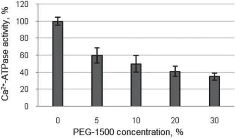
Assessment of Ca2+-ATPase activity in erythrocytes in the presence of PEG-1500 showed that the adding even a small amount of the cryoprotectant into a medium led to inhibition of Ca2+-ATPase activity (Fig. 1).
In the presence of significant concentra-tions of polymer (20–30%), which are capable of exerting a pronounced cryoprotective effect, there was observed a decrease in the rate of Pi accumulation approximately 3 times. Evidently, the inhibition of Ca2+-ATPase activity in erythrocytes, exposed to hypertonic medium of PEG-1500, will entail an increase in intracellular Ca2+ and can affect the protein-protein interactions in membrane-cytoskeleton complex.
In the study of changes in surface marker CD44 in erythrocytes under the influence of the cryoprotectant by flow cytometry we assessed two parameters: composition of cell suspensions characterized by the ratio of CD44+- and CD44–-erythrocytes (for convenience the percentage of only CD44+ -erythrocytes was assessed), and level of surface marker expression for CD44+-erythrocytes, i.e. the amount or density of CD44 molecules in membranes of individual cells.
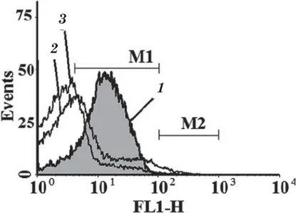
The characteristic of the marker CD44 expression in cells is the median of the distribution histograms showing a value, relative to which, cells in the suspension are divided into two equal size parts. Since only a part of erythrocytes in suspension binds the label CD44-FITC, it was necessary to assess the distribution of native erythrocytes, which were not incubated with CD44-FITC, within FL1 fluorescence channel to clearly identify the area of СD44–-erythrocytes (negative control). Cutting off СD44–-cells (Fig. 2, curve 1), we
identified the marker margins, corresponding to CD44+-erythrocyte area (Fig. 2, curve 2).
In general, in native suspension about 80% (79,3±4,9%) of the erythrocytes were СD44-positive with an average median of the histogram distribution of 13.3±1.3 arbitrary units (a. u.) of fluorescence intensity (FI).
In the samples of native erythrocytes the restraining of dynamics of protein-protein interactions in membrane-cytoskeleton complex, caused by diamide-induced cross-links, entails a decrease in the expression level of СD44 (9.6 ± 0.5 a. u. IF) without significant changes (75,9 ± 4,1) in the amount of CDD44+ -cells (Fig. 2, curve 3).
Exposure of erythrocytes in the medium containing 30% PEG-1500 (Fig. 3, curve 2) was accompanied by a decrease in CD44 expression (9.1 ± 1.6 a. u. IF) in membranes and reducing of CD44+-erythrocyte amount (41.9 ± 4.8%) in the range of the marker standard margins M1 with simultaneous appearance of a small amount of cells (2.3 ± 1.0%) with CD44 expression above the control values (median value 157.3 ± 15.1 a.u. IF) corresponding to the margins M2 in histograms. Pretreatment of cells with diamide exerted more significant impact on CD44 in erythrocytes, exposed to 30% cryoprotectant solution (Fig. 3, curve 3) as compared to the control. Amount of CD44+-cells decreased to 22.5 ± 5.3, and the surface marker expression level was only 5.7 ± 0.4 a. u. IF. However, pretreatment of cells with diamide prevented the development of processes in membrane, resulting in the formation of the erythrocyte subpopulation with higher expression level of CD44, corresponding to the margins M2.
Thus, an exposure of erythrocytes in 30% PEG-1500 causes a change in the Ca2+-АТРase
confirms, as exemplified by СD44, the ability of PEG-1500 to change the structural and functional properties of membranes via modifying of regulatory effects of the cytoskeletal proteins.
A model of reconstructed erythrocytes provides the possibility to assess the Са2+
-ATPase activity affected by exocellular compound under the conditions allowing it to prevent a direct contact with cytoplasmic domains of an enzyme. Effects of organic compounds, many of those as know to exhibit cryoprotective properties, on the Са2+-ATPase
activity were investigated in various biological models. In particular, the stimulation of enzymatic activity of isolated Са2+-ATPase
derived from erythrocyte membranes was demonstrated in presence of PEG, dimethyl sulfoxide (DMSO), glycerol, ethylene glycol [15]. However, the observed effect is rather associated with the characteristics of model system than reflects the influence of these substances on the Са2+-ATPase activity in a cell, because the stimulation of its activity was due to the recovery of hydrophobic interactions within membrane domains of isolated enzyme under the influence of amphiphilic substances [15]. It was shown that in various experimental models trends of changing in the Са2+-ATPase
activity under the influence of organic solvents could be disagreed. In particular, despite the fact that DMSO stimulates the Са2+-ATPase activity of isolated purified enzyme [15], it does not change the basic Са2+ -ATPase activity in membranes of erythrocyte ghosts and inhibits calmodulin-stimulated enzyme activity [16]. The effect of glycerol on the Са2+-ATPase in the sealed ghosts and
saponin-permeabilized erythrocytes were also different [17]. Since the ability of Са2+ -pump to transport ions Са2+ is primarily determined by its catalytic activity, one can expect an increase in the level of intracellular Са2+ in the erythrocytes incubated in PEG-containing solutions. Growing of Са2+ level
in the PEG presence is promoted via water loss by cells under the influence of osmotic gradient due to a high concentration of the non-penetrating across membrane polymer. Ions Са2+, functioning as a second messenger, undoubtedly play a role in cell survival under stress conditions.
Increasing in the Са2+ level, which has been observed even in the presence of an isotonic solution of PEG-1500 [18], should affect the structural state of membrane components, and changes in a surface marker expression can be resulted from occurring structural rearrangements caused by stress factors. Whereas the cryoprotective agent is referred as “compatible cosolvents” and it is capable to maintain the structure of protein macromolecules, membranes and cells in a state close to native without causing denaturation or impaired structural order [19–21], but it has been still able to induce structural and functional changes in erythrocytes, which during a prolonged exposure resulted in lowering of CD44+-cells amount and marker expression level. The most likely cause of the mentioned changes in erythrocytes in cryoprotectant presence is the loss of membrane material in the form of vesicles with the capture of molecules of the surface marker CD44 in their composition. For erythrocyte membranes the vesiculation is a recognized
Fig. 1. Change in Ca2+-ATPase activity in the sealed erythrocytes depending on the PEG-1500 concentration
Fig 2. Distribution histogram of native erythrocytes labeled with CD44-FITC after exposure to the Ringer medium:
1 (here and in Fig. 3) — distribution histogram of the native erythrocytes labeled with CD44-FITC (M1 specifies the area of CD44+-erythrocytes);
2 — distribution histogram of the native erythrocytes not labeled with CD44-FITC (negative control); 3 — distribution histogram of the native erythrocytes, pretreated with diamide
The data are of a typical experiment. The X axis is value of fluorescence intensity of cells in FL-1 channel (a.u. IF FL1), represented by logarithmic scale values. The Y axis is the number of calculable events in normalized form
Fig. 3. Distribution histogram of erythrocytes labeled with CD44-FITC after exposure to 30% solution of PEG-1500:
2 — distribution histogram of the erythrocytes exposed in presence of 30% PEG-1500;
3 — distribution histogram of the erythrocytes exposed in presence of 30% PEG-1500, which have been pretreated with diamide
The X axis is value of fluorescence intensity of cells in FL-1 channel (a.u. IF FL1) represented by logarithmic scale values. The Y axis is the number of calculable events in normalized form (the data of a typical experiment)
1
3
2
3
2
mechanism of cell response to either stress or aging [22–24]. The changes, affecting CD44, may depend on structural modification of lipid bilayer and proteins of the membrane-cytoskeleton complex. In particular, the lipid packing mismatch in neighboring areas, caused for example by phase transitions in specific membrane areas, affected by dehydration processes, are able to induce the formation of vesicles [25]. Furthermore, the CD44 molecules are known to exist in the membrane as individual molecules or a part of the intricate protein complexes. In particular, CD44 may be associated with the cytoskeleton network via ankyrin and protein 4.1 [26]. These links are dynamic and may vary in different parts of membrane. Non-associated with the cytoskeleton the CD44 molecules can be captured easier into the vesicles forming on the erythrocyte membranes under stress. It should be noted that changes in the intracellular Ca2+ level play a key role in initiation of these processes, since Ca2+ and calmodulin decrease the affinity of interaction between the protein 4.1 and CD44 [26], whereby the latter becomes a free membrane protein or gets able to form a link with ankyrin. Expected redistribution of interaction within network of membrane-cytoskeleton protein complex involving CD44, ankyrin, protein 4.1 and band 3 in the presence of cryoprotectants can affect the physical properties of membrane.
Important information to understand the cryoprotectant mechanism actions on erythrocyte membrane can be provided by an evidence on the specificity of changes in the marker CD44 expression under the effect of a 30% PEG-1500 solution, which is characterized by the appearance of cells with higher expression of the marker CD44 (M2). This subpopulation emergence upon the absence of capability for protein synthesis in erythrocytes can be associated with structural changes in membranes wherein there is partial or complete fusion of membrane systems, that leads to the enrichment of individual cells with the marker. It is known that PEG contributes to an aggregation and fusion of cells [27, 28]. However, PEG-1500 has been previously shown [29] does not cause a fusion of erythrocytes. For this purpose, PEG-6000 is used at a concentration of about 40%, furthermore cell fusion requires additional conditions, one of those is cellular rehydration [30], not presented in our experiments. Nevertheless, in the
erythrocyte membranes on the contact with high PEG-1500 concentrations the structural changes can be initiated that eventually lead to the formation of sufficient area with a modified structure that allows fusion of individual membranes. In the described experimental conditions, fusion of vesicles, carrying the marker, with the cells seems the most probable.
per monomer unit [33] that determines its dehydrating effect against various macromolecules and membranes. Further, the PEG molecules can be distributed at the air-water interface forming stable m o n o l a y e r s [ 3 5 , 3 6 ] . S u r f a c e - a c t i v e properties of the polymer, its dehydrating action and changing in the polarity in the presence of PEG may have some influence on membrane surface [37] that may cause a decrease in membrane fluidity [38]. It has been shown that the adding of PEG modifies the surface potential of lipid monolayers [33] and causes a shift of temperature of the phase lipid transition [39]. It has also been found that PEG molecules induce dehydration of multilamellar lipid vesicles that is accompanied by a reduction in effective size of the polar headgroups of the membrane phospholipids and enhanced van der Waals interactions between the acyl chains of the matrix lipid [40, 41]. These structural changes are typically compensated by an increased thickness of the membrane and decrease in the mobility of lipid molecules that leads to segregation of different types of membrane lipids. Probably similar changes may affect some areas of erythrocyte membranes and exert a certain negative impact on the stability of membranes in the presence of PEG-1500 under stressful conditions of cryopreservation.
The observed changes in Ca2+-ATPase activity in erythrocytes under the influence of PEG-1500 does not give a comprehensive view on its mechanism of inhibition and requires further assessment of its functioning characteristics, in particular the analysis of Ca2+-transporting capacity in presence of PEG-1500 in the medium. In future such a study could potentially give a clearer insight in the state and role of Ca2+-regulating constituents in changes of structural and functional state of the cells at using of exocellular cryoprotectants and low temperature effects. Especially important issue is the assessment of changes in the level of intracellular Ca2+, which can be both a regulator of the structural state of membrane and trigger of apoptosis under stressful conditions. Effect of PEG-1500 on the surface marker C D 4 4 a p p a r e n t l y i s i m p l e m e n t e d i n
various ways, and the change in the level of intracellular Ca2+, which determines t h e m o d i f i c a t i o n o f p r o t e i n - p r o t e i n interactions in the membrane-cytoskeleton complex, plays definitely an important role. The consequence of the modification of the structural state of the membrane components in the presence of PEG-1500 may be resulted in changing the erythrocyte stability under stress conditions.
Thus, the experiments revealed a decrease in the Ca2+-ATPase activity, as
well as a reduction in the expression of surface markers CD44 and CD44+-cell amount on the exposure of erythrocytes in the presence of PEG-1500. A decrease in the Ca2+-ATPase activity may be caused by a modification of the membrane structure under the influence of physical and chemical properties of 30% PEG-1500 solution. The consequence of reducing of the Ca2+
REFERENCES
1. Hutanu D., Frishberg M. D., Guo L., Darie C. C. Recent applications of polyethylene glycols (PEGs) and PEG derivatives. Mod. Chem. Appl. 2014, 2 (2), 1–6. doi: 10.4172/2329-6798.1000132.
2. Babijchuk L. A., Zemlianskykh N. G. Optimization and advantages of washing-out method of erythrocyte cryopreservation with PEO-1500. Problems for Cryobiology. 2001, V. 1, P. 35–44. (In Russian).
3. Feuerecker M., Kaufmann I., Salam A. P., Choukèr A. Effects of cryopreservation with polyethylene glycol on the expression of CD11b and CD62L on the surface of polymorphonuclear leukocytes. Cryo Lett. 2012, 33 (2), 151–160.
4. Gao D., Critser J. K. Mechanisms of cryoinjury in living cells. ILAR J. 2000, V. 41, P. 187–196. doi.org/10.1093/ilar.41.4.187
5. Stoll C., Holovati J. L., Acker J. P., Wolkers W. F. Synergistic effects of liposomes, trehalose, and hydroxyethyl starch for cryopreservation of human erythrocytes. Biotechnol. Prog. 2012, 28 (2), 364–371. doi: 10.1002/btpr.1519. 6. El-Shewy H. M., Kendall W. F. Jr., Darrabie M.,
Collins B. H., Opara E. C. Polyvinyl pyrrolidone: a novel cryoprotectant in islet cell cryopreservation. Cell Transplant. 2004, 13 (3), 237–243. doi. org/10.3727/000000004783983927
7. Borland G., Ross J. A., Guy K. Forms and functions of CD44. Immunology. 1998, 93 (2), 139–148. doi: 10.1046/j.1365– 2567.1998.00431.x
8. Fischer T. M., Haest C. W., Stohr M., Kamp D., Deuticke B. Selective alteration of erythrocyte deformability by SH–reagents: evident for an involvement of spectrin in membrane shear elasticity. Biochem. Biophys. Acta. 1978, 510 (2), 270–282.
9. Haest C. W. Kamp D., Deuticke B. Topology of membrane sulfhydryl groups in the human erythrocyte. Demonstration of a non-reactive population in intrinsic proteins. Biochem. Biophys. Acta. 1981, 643 (2), 319–326. doi. org/10.1016/0005-2736(81)90077-8
10. Zemlianskykh N. G., Babijchuk L. A. Effect of cryopreservation stresses on erythrocyte surface marker CD44. Biol. Membr. 2014, 31 (6), 416–426. (In Russian). doi: 10.7868/ S0233475514050107
11. Fairbanks G., Steck T. L., Wallach D. F. Electrophoretic analysis of the major polypeptides of the human erythrocyte membrane. Biochemistry. 1971, 10 (13), 2606–2617. doi: 10.1021/bi00789a030 12. Pokudin N. I., Petruniaka V. V., Orlov S. N. Does
calmodulin participate in the regulation of the
Ca-pump of erythrocytes in vivo? Biokhimia. 1988, 53 (5), 753–758. (In Russian).
13. Rathbun W., Betlach V. Estimation of enzymically produced orthophosphate in the presence of cysteine and adenosine triphosphate. Anal. Biochem. 1969, 28 (1–3), 436–445.
14. Bredford M. M. A rapid and sensitive method for the quantitation of microgram quantities of protein utilizing the principle of protein-dye binding. Anal. Biochem. 1976, 72 (7), 248–254.
15. Benaim G., de Meis L. Activation of the purified erythrocyte plasma membrane Ca2+ -ATPase by organic solvents. FEBS Lett. 1989, 244 (2), 484–486. doi.org/10.1016/0014-5793(89)80589-7
16. McConnell E. J., Wagoner M. J., Keenan C. E., Raess B. U. Inhibition of calmodulin-stimulated (Ca2+ + Mg2+)-ATPase activity by dimethyl sulfoxide. Biochem. Pharmacol. 1999, 57 (1), 39–44. doi.org/10.1016/S0006-2952(98)00259-7
17. Zemlyanskikh N. G., Kofanova O. A. Modulation of human erythrocyte Ca2+– ATPase activity by glycerol: the role of calmodulin. Biochemistry (Mosc). 2006, 71 (8), 900–905. doi: 10.1134/ S0006297906080128
18. Kucherenko Y. V., Bernhardt I. The study of Ca2+ influx in human erythrocytes in isotonic polyethylene (glycol) 1500 (PEG–1500) and sucrose media. Ukr. Biokhim. Zh. 2006, 78 (6), 46–52.
19. Timasheff S. N. Solvent stabilization of protein structure. Meth. Mol. Biol. 1995, V. 40, P. 253–269. doi: 10.1385/0-89603-301-5:253
20. Carpenter J. F., Crowe J. H. The mechanism of cryoprotection of proteins by solutes. Cryobiology. 1988, 25 (3), 244–255. doi.org/10.1016/0011-2240(88)90032-6 21. Konov K. B., Isaev N. P., Dzuba S. A.
Low-temperature molecular motions in lipid bilayers in the presence of sugars: insights into cryoprotective mechanisms. J. Phys. Chem. B. 2014, 118 (43), 12478–12485. doi: 10.1021/jp508312n
22. Holovati J. L., Wong K. A., Webster J. M., Acker J. P. The effects of cryopreservation on red blood cell microvesiculation, phosphatidylserine externalization, and CD47 expression. Transfusion. 2008, 48 (8), 1658–1668. doi: 10.1111/j.1537-2995.2008.01735.x.
in activated neutrophils. Sci. World J. 2011, V. 11, P. 173–185. doi: 10.1100/ tsw.2011.25.
24. Bosman G. J., Lasonder E., Groenen-Döpp Y. A., Willekens F. L., Werre J. M. The proteome of erythrocyte — derived microparticles from plasma: new clues for erythrocyte aging and vesiculation. J. Proteomics. 2012, 76 (Spec. N), 203–210. doi: 10.1016/j. jprot.2012.05.031.
25. Balasubramanian S. K., Wolkers W. F., Bischof J. C. Membrane hydration correlates to cellular biophysics during freezing in mammalian cells. Biochim. Biophys. Acta. 2009, 1788 (5), 945–953. doi: 10.1016/j. bbamem.2009.02.009.
26. Nunomura W., Takakuwa Y., Tokimitsu R., Krauss S. W., Kawashima M., Mohandas N. Regulation of CD44 — protein 4.1 interaction by Ca2+ and calmodulin. Implications for modulation of CD44 — ankyrin interaction. J. Biol. Chem. 1997, 272 (48), 30322–30328. doi: 10.1074/jbc.272.48.30322
27. Zhao W. Y., Xiong H. Y., Yuan Q., Zeng L., Wang L. M., Zhu Y. H. In vitro effects of polyethylene glycol in University of Wisconsin preservation solution on human red blood cell aggregation and hemorheology. Clin. Hemorheol. Microcirc. 2011, 47 (3), 177–185. doi: 10.3233/CH-2010-1379. 28. Lentz B. R. PEG as a tool to gain insight into
membrane fusion. Eur. Biophys. J. 2007, 36 (4–5), 315–326. doi:10.1007/s00249-006-0097-z
29. Maggio B., Ahkong Q. F., Lucy J. A. Poly(ethylene glycol), surface potential and cell fusion. Biochem. J. 1976, 158 (3), 647–650. doi: 10.1042/bj1580647
30. Ahkong Q. F., Desmazes J. P., Georgescauld D., Lucy J. A. Movements of fluorescent probes in the mechanism of cell fusion induced by poly(ethylene glycol). J. Cell Sci. 1987, 88 (3), 389–398.
31. Winterhalter M., Burner H., Marzinka S., Benz R., Kasianowiczt J. J. Interaction of poly(ethylene-glycols) with air-water interfaces and lipid monolayers: investigations on surface pressure and surface potential. Biophys. J. 1995,
69 (4), 1372–1381. doi: 10.1016/S0006-3495(95)80006-8
32. Arnold K., Herrmann A., Pratsch L., Gawrisch K. The dielectric properties of aqueous solutions of poly(ethylene glycol) and their influence on membrane structure. Biochim. Biophys. Acta. 1985, 815 (3), 515–518.
33. Bailey F. E., Koleske J. V. Poly(ethylene Oxide). New York: Academic Press. 1976, 173 р.
34. Chen J., Spear S. K., Huddleston J. G., Rogers R. D. Polyethylene glycol and solutions of polyethylene glycol as green reaction media. Green Chem. 2005, 7 (2), 64–82. doi. org/10.1016/j.jchromb.2004.01.047
35. Kawaguchi M., Tohyama M., Mutoh Y., Takahashi A. Ellipsometric study of polymer monolayers spread at the air/water interface. Langmuir. 1988, 4 (2), 407–410.
36. Sauer B. B., Yu H., Yazdanian M., Zografi G., Kim M. W. An elipsometric study of polymer monolayers at the air/water interface. Macromolecules. 1989, 22 (5), 2332–2337. 37. Arnold K., Pratsch L., Gawrisch K. Effect
of poly(ethylene glycol) on phospholipid hydration and polarity of the external phase. Biochim. Biophys. Acta. 1983, 728 (1), 1 2 1 – 1 2 8 . d o i . o r g / 1 0 . 1 0 1 6 / 0 0 0 5 -2736(83)90444-3
38. Herrmann A., Pratsch L., Arnold K., Lassmann G. Effect of poly(ethylene glycol) on the polarity of aqueous solutions and on the structure of vesicle membranes. Biochim. Biophys. Acta. 1983, 733 (1), 87–94. doi. org/10.1016/0005-2736(83)90093-7
39. Tilcock C. P. S., Fisher D. Interaction of phospholipid membranes with PEGs. Biochim. Biophys. Acta. 1979, 577 (1), 53–56. 40. Lehtonen J. Y. A., Kinnunen P. K. J. Changes
in the lipid dynamics of liposomal membranes induced by PEG. Biophys. J. 1994, 66 (6), 1981–1990. doi: 10.1016/S0006-3495(94)80991-9
МОДИФІКАЦІЯ МЕМБРАННИХ ПРОТЕЇНІВ ЕРИТРОЦИТІВ ПОЛІЕТИЛЕНГЛІКОЛЕМ 1500
Н. Г. Землянських Л. О. Бабійчук
Інститут проблем кріобіології і кріомедицини НАН України, Харків
E-mail: [email protected]
Метою роботи було вивчення впливу полі-етиленгліколю (ПЕГ-1500) на активність
Са2+-АТРази і зміну експресії поверхневого маркера CD44 у мембранах еритроцитів лю-дини. Визначення активності Са2+-АТРази виконували в замкнутих тінях еритроци-тів за рівнем накопичення неорганічного фосфору. Зміну експресії CD44 і кількості CD44+-еритроцитів оцінювали методом про-точної цитометрії. Встановлено інгібування активності Са2+-АТРази, а також зниження рівня експресії СD44 і зменшення кількості СD44+-клітин, що відображає складні пере-будови у мембранно-цитоскелетному комп-лексі еритроцитів під впливом ПЕГ-1500. Вплив ПЕГ-1500 на поверхневий маркер CD44 може бути опосередкований модифі-кацією протеїнів мембранно-цитоскелетного комплексу, про що свідчить посилення втра-ти CD44 мембранами в еритроцитах після за-стосування протеїнзшивного реагента діамі-ду. Зниження активності Са2+-АТРази може сприяти підвищенню рівня внутрішньо-клітинного Са2+ і призвести до модифікації взаємодій інтегральних протеїнів з компо-нентами цитоскелета, унаслідок чого може відбуватись везикуляція мембран і знижен-ня експресії маркера СD44, який динамічно пов’язаний із цитоскелетом.
Ключові слова: Са2+-АТРаза, CD44, полі
ети-лен гліколь, еритроцити.
МОДИФИКАЦИЯ МЕМБРАННЫХ ПРОТЕИНОВ ЭРИТРОЦИТОВ ПОЛИЭТИЛЕНГЛИКОЛЕМ 1500
Н. Г. Землянских Л. А. Бабийчук
Институт проблем криобиологии и криомедицины НАН Украины, Харьков
E-mail: [email protected]
Целью работы было изучение влияния по-лиэтиленгликоля (ПЭГ-1500) на активность Са2+-АТРазы и изменение экспрессии по-верхностного маркера CD44 в мембранах эри-троцитов человека. Определение активности Са2+-АТРазы проводили в замкнутых тенях эритроцитов по уровню накопления неоргани-ческого фосфора. Изменение экспрессии CD44 и количества CD44+-эритроцитов оценивали методом проточной цитометрии. Установле-но ингибирование активУстановле-ности Са2+-АТРазы, а также снижение уровня экспрессии СD44 и уменьшение количества СD44+-клеток, что отражает сложные перестройки в мемб ранно-цитоскелетном комплексе эритроцитов под влиянием ПЭГ-1500. Влияние ПЭГ-1500 на поверхностный маркер CD44 может быть опо-средовано модификацией протеинов мемб-ранно-цитоскелетного комплекса, о чем свиде-тельствует усиление потери CD44 мемб ранами в эритроцитах после применения протеин-сшивающего реагента диамида. Снижение ак-тивности Са2+-АТРазы может способствовать повышению уровня внутриклеточного Са2+ и привести к модификации взаимодействий ин-тегральных протеинов с компонентами цито-скелета, вследствие чего может происходить везикуляция мембран и снижение экспрессии маркера СD44, который динамически связан с цитоскелетом.
Ключевые слова: Са2+-АТРаза, CD44,