A model of chronic IgE-mediated food
allergy in ovalbumin-sensitized mice
Departamentos de 1Patologia Geral and 2Bioquímica e Imunologia,
Instituto de Ciências Biológicas, Universidade Federal de Minas Gerais, Belo Horizonte, MG, Brasil
J.C.S. Saldanha1*,
D.L. Gargiulo1*, S.S. Silva1,
F.H. Carmo-Pinto1,
M.C. Andrade2,
J.I. Alvarez-Leite2,
M.M. Teixeira2
and D.C. Cara1
Abstract
Food allergy is most frequently the result of IgE-mediated hypersensi-tivity reactions. Here, we describe a chronic model in which some of the intestinal and systemic consequences of continuous egg white solution ingestion by ovalbumin-sensitized eight-week-old BALB/c mice, 6 animals per group, of both sexes, were investigated. There was a 20% loss of body weight that began one week after antigen exposure and persisted throughout the experiment (3 weeks). The sensitization procedure induced the production of anti-ovalbumin IgG1 and IgE, which were enhanced by oral antigen exposure (129% for IgG1 and 164% for IgE, compared to sensitization values). Intestinal changes were determined by jejunum edema at 6 h (45% Evans blue extravasa-tion) and by a significant eosinophil infiltration with a peak at 48 h. By day 21 of continuous antigen exposure, histological findings were mild, with mast cell hyperplasia (100%) and increased mucus produc-tion (483%). Altogether, our data clearly demonstrate that, although immune stimulation was persistently occurring in response to continu-ous oral antigen exposure, regulatory mechanisms were occurring in the intestinal mucosa, preventing overt pathology. The experimental model described here reproduces the clinical and pathological changes of mild chronic food allergy and may be useful for mechanistic studies of this common clinical condition.
Correspondence D.C. Cara
Av. Antônio Carlos, 6627 Departamento de Patologia Geral ICB, UFMG
31270-901 Belo Horizonte, MG Brasil
Research supported by FAPEMIG, CAPES and CNPq.
*These authors contributed equally to this study.
Received June 2, 2003 Accepted February 10, 2004
Key words
•Food allergy •Ovalbumin •Murine model •IgE
•Hypersensitivity
Introduction
The physiological penetration of dietary macromolecules into the organism through the gut mucosa has several recognized im-munological consequences. One of these is the development of food allergy, which is defined as an adverse immunological (hy-persensitivity) response to food and can be
IgE dependent or not (1). IgE-dependent food-allergic reactions may affect one or more target organs: the skin (urticaria, angio-edema), the respiratory tract (rhinitis, asthma), the gastrointestinal tract (pain, emesis, diar-rhea), and the cardiovascular system (ana-phylactic shock) (2).
height and weight (3). Food allergy and other types of allergies, such as allergic rhinitis and asthma, are reaching epidemic propor-tions in both the developed and developing countries (4). The reasons for the changes in prevalence of allergic disorders are not known and several hypotheses have been raised to explain them, including the “hygiene hypo-thesis”, which states that a reduction in in-fections in early infancy predisposes to aller-gic responses (5,6). The understanding of regulatory mechanisms controlling allergic inflammation may provide useful insights into disease pathophysiology and aid in the development of novel therapeutic strategies. We have reported that mice previously sensitized against ovalbumin (Ova) avoided the ingestion of a sweetened solution of egg white, choosing water instead (7,8), and this aversion was related to IgE antibodies (9). Indeed, elimination diet is still the mainstay of IgE-dependent food allergy therapy (2,3). However, with the advent of manufactured food, trace amounts of allergens can still be present in food and some atopic patients appear to be continuously exposed to these allergens. In these atopic patients, continu-ous unwilling exposure to a food allergen may induce a mild and persistent allergic condition. To try mimicking the latter clini-cal situation, we have developed an experi-mental model in which sensitized mice are given egg white solution (EWS) as the only liquid source.
Material and Methods
Animals
Young adult BALB/c mice of both sexes were used. All mice received standard (Purina) mouse chow throughout the experi-ment. All investigations were in agreement with the Ethical Principles in Animal Ex-perimentation, adopted by the Ethics Com-mittee in Animal Experimentation of our Institution (CETEA/UFMG).
Sensitization protocol
Mice received (day 0) a subcutaneous (sc) injection of 0.2 ml saline containing 10
µg Ova (five times crystallized hen’s egg albumin; Sigma, St. Louis, MO, USA) plus 1 mg Al(OH)3 as adjuvant. Secondary
sensiti-zation consisted of a sc injection of 10 µg
soluble Ova 14 days after the primary sensi-tization (day 14). Although it has been shown that intraperitoneal injection of alum induces higher antibody production (10), we chose the sc route to avoid peritoneal
inflamma-tion. Seven days after the secondary sensiti-zation (i.e., on day 21), the bottle containing tap water was replaced with gauze filtered 20% EWS for a period of 3 weeks (from day 21 to day 42). This solution contains about 10 mg Ova/ml. The following control groups were studied in parallel for comparison: non-sensitized or non-sensitized mice that were given tap water throughout the experimental pro-cedure and non-sensitized mice that were given EWS from day 21 to day 42.
Nutritional status
In order to evaluate the nutritional condi-tion of all animals, liquid and food consump-tion and body weight were determined each week. After 21 days of continuous EWS ingestion, hematocrit levels were determined and serum albumin levels were measured using an Albumin Doles Kit (Goiania, GO, Brazil)in order to assess the possible pres-ence of dehydration and/or malnutrition.
Serum antibodies
USA). The reactions were developed with the streptavidin-peroxidase conjugate (Ex-traAvidin; Sigma), o-phenylene-diamine and H2O2. The plates were read at 492 nm on an
automated ELISA reader (EL800, Bio-Tek Instruments, Inc., Winooski, VT, USA). Anti-Ova IgE antibodies were measured by cap-ture-ELISA using plates coated with rat anti-mouse IgE and 50 µl total serum and biotin-ylated Ova, as previously described (11). The results are reported as arbitrary units using a positive reference serum assigned to be 1000 units.
Intestinal edema
Vascular permeability changes were evaluated by Evans blue dye extravasation (12). Twenty-one days after the primary sen-sitization with 10 µg Ova + 1 mg Al(OH)3,
the animals were exposed to a restricted and voluntary intake of EWS for 6, 24 and 48 h and then injected with Evans blue (20 mg/ kg) into the tail vein; 10 min later, the ani-mals were killed and the small intestine was dissected and weighed. The intestine was divided into four segments (duodenum, proxi-mal jejunum, distal jejunum, and ileum) and each segment was further divided into two parts. One was soaked in formamide (4 ml/g wet weight tissue at 20ºC for 24 h) to extract the Evans blue dye, and the other was dried at 60ºC for 24 h. The concentration of Evans blue was determined by spectrophotometry at 630 nm using an ELISA reader (EL800, Bio-Tek Instruments) and 96-well plates. Results were plotted on a standard curve for Evans blue (0.15-20 µg/ml). The Evans blue content of each sample is reported as mg/g dry weight tissue.
Histology and morphometry
Changes in the intestinal mucosa were evaluated at 6, 24 and 48 h, and at 7, 14 and 21 days of the experiment. The mice were sacrificed by cervical dislocation and the
small intestine was taken for histological analysis. The intestine was divided into four segments (duodenum, proximal jejunum, dis-tal jejunum, and ileum), fixed in 10% forma-lin in PBS, embedded in paraffin and cut into 3-5-µm thick sections. The sections were stained with hematoxylin-eosin for general analysis, with Toluidine blue for mast cell evaluation, with periodic acid Schiff for mucus analysis, or with 1% carbol chromo-trope 2R and hematoxylin (Sigma) for eosin-ophil granule identification.
As described elsewhere, the intestinal mucosa was submitted to morphometric anal-ysis using an image analanal-ysis program run-ning on an IBM computer (13). Images were obtained with a JVC TK-1270/RGB micro-camera and the KS300 software built in a Kontron Eletronick/Carl Zeiss image ana-lyzer. Ten fields from hematoxylin-eosin-stained sections were randomly chosen at 10X (871.392 µm2/field) for villus height
and crypt depth measurement. Ten fields were chosen randomly at 40X (53.333 µm2/
field) from chromotrope and Toluidine blue sections in order to count the number of eosinophils and mast cells, respectively, and the data are reported as number of cells/ field. For the determination of goblet cell volume, all pixels with green hues were se-lected for the creation of a binary image and subsequent calculation of the total area, and the data are reported as µm2 mucus/field.
Statistical analysis
Data were analyzed statistically by one-way analysis of variance (ANOVA) or by the Student t-test, when appropriate, with the
level of significance set at P < 0.05. If ANOVA revealed differences, the groups were compared by the Student t-test with
Results
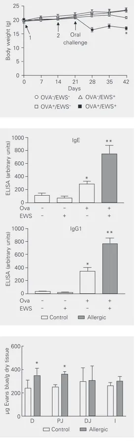
The mean daily tap water consumption was similar for sensitized (4.7 ± 0.2 ml) and non-sensitized (4.2 ± 0.2 ml) mice, whereas the mean daily EWS consumption was higher for non-sensitized mice (8.3 ± 0.8 ml) com-pared to sensitized mice (4.1 ± 0.4 ml). All groups showed the same food consumption (data not shown). One week after the oral challenge with EWS, sensitized, but not non-sensitized, mice had lost about 20% of their body weight (Figure 1). The loss of body weight was maintained throughout the ob-servation period and was not observed in animals who ingested tap water (Figure 1). The decreased body weight appeared not to be related to dehydration since there were no significant differences in hematocrit levels among groups (48-54%). Likewise, serum albumin levels were similar among groups before and after oral exposure to EWS or water (3.4-3.7 g/dl).
The sensitization procedure by itself in-duced the production of both anti-Ova IgG1 and IgE (Figure 2). Remarkably, the concen-tration of both of these antibodies more than doubled in sensitized mice that were given EWS solution, but not in sensitized mice given tap water (Figure 2). EWS consumption did not affect circulating levels of immunoglobu-lins in non-sensitized mice (Figure 2).
Twenty-one days after Ova sensitization, mice were exposed to restricted and voluntary EWS for 6 h. During this time, they ingested about 0.8 ml of the solution (8 mg Ova) and developed an increase in the vascular perme-ability of the proximal segments of the small intestine (Figure 3), as shown by the extrava-sation of Evans blue dye. There were no statis-tical differences in dye extravasation among the control groups at 6 h, or among all groups at later time points (data not shown). How-ever, the early increase in vascular permeabil-ity generated villus swelling 24 h after EWS ingestion in sensitized mice (Figure 4B).
Inflammatory cell infiltration, with an
Figure 1. Body weight of mice up to 42 days after the first sen-sitization with ovalbumin (Ova). Data are reported as means ± SEM in g for 6 mice in each group. BALB/c mice received (Ova+) or not (Ova-) 10 µg Ova
plus Al(OH)3 on day 0 and 10 µg
Ova on day 14. From day 21 to the end, some Ova+ and Ova
-animals received filtered 20% (w/v) egg white solution in their drinking bottle (EWS+). *P <
0.05 compared to Ova-/EWS+
(ANOVA-Student t-test).
µg Evans blue/g dry tissue
600
400
200
0
D PJ DJ I
*
*
Control Allergic
Body weight (g)
25
1 2 Oral
challenge * 20 15 10 5 0
0 7 14 21 28 35 42
Days
OVA-/EWS- OVA-/EWS+
OVA+/EWS- OVA+/EWS+
ELISA (arbitrary units)
1000 800 600 400 200 0 Ova EWS -+ + + + ** * IgE ** * Ova EWS -+ + + +
ELISA (arbitrary units)
1000 800 600 400 200 0 IgG1 Control Allergic Figure 2. Anti-ovalbumin (Ova)
IgG1 and IgE levels of mice sen-sitized with Ova. Serum anti-body responses of BALB/c mice sensitized with 10 µg Ova plus Al(OH)3 on day 0 and with 10 µg
Ova on day 14. From day 21 to the end, some Ova+ and Ova
-animals received filtered 20% egg white solution in their drink-ing bottle (EWS+). Data are
re-ported as means ± SEM of IgG1 and IgE expressed as arbitrary ELISA units for 6 animals per group. *P < 0.05 compared to Ova-/EWS-, and **P < 0.05
com-pared to Ova+/EWS-
(ANOVA-Student t-test).
Figure 3. Intestinal vascular per-meability. Extravasation of Evans blue dye in the small intestine (D - duodenum, PJ - proximal jejunum, DJ distal jejunum, I -ileum) triggered by oral chal-lenge with egg white solution (EWS) during a period of 6 h by sensitized (Ova+) (closed bars)
and non-sensitized (Ova-) (open
bars) mice. Animals were sensi-tized 21 days before the chal-lenge by an injection of 10 µg OVA plus 1 mg Al(OH)3. Data are
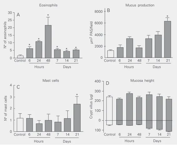
evident presence of eosinophils (Figure 4C), was seen as early as 6 h after antigen expo-sure. However, the number of eosinophils in the jejunal mucosa peaked at 48 h after antigen exposure and dropped thereafter (Fig-ure 5A). Nevertheless, eosinophils were still present at significantly higher levels than in control animals until the end of the observa-tion period (day 21).
With the continuous exposure to antigen (about 47 mg Ova/day), the pathological changes markedly improved and, at 21 days after oral antigen exposure, the intestine showed no edema and little infiltration of inflammatory cells other than eosinophils. However, there was increased mucus pro-duction by goblet cells (Figures 4D and 5B) and mast cell hyperplasia (Figures 4E and 5C) in the jejunum. The total mucosa height did not differ from control (Figure 5D).
No morphological differences were seen
Figure 4. Representa-tive histological sec-tions of the proximal jejunum from non-sen-sitized mice drinking water (A), and from sensitized mice after 24 h (B), 48 h (C) and 21 days (D, E) of con-tinuous ingestion of a filtered 20% egg white solution. A, B and D, hematoxylin and eosin staining; C, chromo-trope 2R staining; E, Toluidine blue staining. The bars are 50 µm in A, B, D and E, and the bar in C is 25 µm. The large arrow in C indicates an eosinophil, the small white arrow in E indicates a mast cell, and the white arrowhead in D indicates a goblet cell.
N
º
of eosinophils
30
25
20
15
10
5
0
Control 6 24 48 7 14 21
Hours Days
*
* *
* * *
Control 6 24 48 7 14 21
Hours Days
µm
2 PAS/field
8000
6000
4000
2000
0
Control 6 24 48 7 14 21
Hours Days
Control 6 24 48 7 14 21
Hours Days
N
º
of mast cells
4
3
2
1
0
Crypt villus (µg)
400
300
200
100
0
100 C
A B
D
Eosinophils Mucus production
Mast cells Mucosa height
Figure 5. Eosinophils (A), mucus production (B), mast cells (C), and mucosa height (D) in the jejunum of BALB/c mice sensi-tized with 10 µg ovalbumin (OVA) plus 1 mg Al(OH)3 after
continuous ingestion of an egg white solution (EWS). The open bars indicate the control group (Ova-/EWS-) and the closed bars
represent the allergic group (Ova+/EWS+) who ingested the
EWS. Data are reported as means ± SEM of number of cells (A, C), mucus production (B; PAS = periodic acid Schiff), or mucosa height (D) for 6 mice in each group *P < 0.05 com-pared to control Ova-/EWS
-(ANOVA-Student t-test).
*
in any segment of the small intestine of non-sensitized mice given water or EWS or of sensitized mice given water when compared to the intestine of naive mice.
Discussion
There is much experimental evidence showing that the introduction of antigens via the gastrointestinal route induces oral toler-ance, defined as the inhibition of specific antibody formation after subsequent paren-teral immunizations with the same antigen (14). However, our results suggest that, when mice are sensitized to OVA, the subsequent long-term, low-level exposure to this same antigen via the oral route serves as an immu-nological booster leading to high levels of IgE and IgG1 production. Indirectly, these experiments suggest that a small portion of the ingested antigen is indeed capable of transposing the gut barrier and stimulating primed lymphocytes. Usually, IgE-depend-ent food allergy reactions affect one or more target organs such as skin, respiratory tract, gastrointestinal tract, and cardiovascular sys-tem (2). An immediate syssys-temic reaction mediated by IgE is called anaphylaxis irre-spective of severity (15), but colloquially the term anaphylaxis is applied to severe, poten-tially fatal allergic reactions (2). Mainly in children, chronic food allergic disorders (IgE-associated) typically affect the gastrointesti-nal tract with different degrees of eosino-philic inflammation, edema and weight loss (16).
Sensitized mice showed the same con-sumption of egg white and tap water, but the consumption of egg white by non-sensitized mice was higher than that by sensitized ani-mals. The latter result suggests that there was an aversion to EWS by sensitized mice. This immunologically taste aversion was documented before (7,8) and was related to IgE responses (9). Recently, Basso and co-workers (17) have shown that after an oral Ova challenge, allergic mice presented higher levels of anxiety, increased Fos expression in emotionality related brain areas, and also aversion to Ova-containing solution. In our model, after prolonged egg white consump-tion, sensitized mice also showed a marked
loss in body weight. The mechanisms in-volved in this phenomenon are not clear, although this event has also been observed in rat models (18,19). Although the hematocrit and serum albumin levels were normal, sug-gesting that dehydration and/or malnutrition did not occur in mice with food allergy, we cannot exclude the possibility that a decreased absorption of other diet components or an increased energy consumption may have occurred. It is possible that the mechanisms involved in body weight loss are related to food aversion. By day 21, body weight was still low and circulating IgE and IgG1 levels were highest. These responses were related to the genetic background, since when an-other lineage was used (C57BL6), sensitiza-tion induced the producsensitiza-tion of IgE, but at lower levels when compared to BALB/c mice and there was no loss in body weight (data not shown). Also, our preliminary data have shown that IL-4 receptor alpha-deficient BALB/c mice do not produce IgE when im-munized, nor do they lose body weight when challenged. These results indicate that this model is strongly related to Th2 cytokines.
After 6 h of antigen intake, vascular per-meability was increased, as detected histo-logically at 24 h. This acute inflammatory change may be a result of local mast cell degranulation in response to Ova challenge and of the subsequent increase of histamine levels in the gut, as has observed elsewhere in sensitized mice after Ova challenge (20). In chronic allergic diseases such as asthma, during continuous antigen exposure eosino-phils are primed by IL-5 and attracted by chemokines, infiltrating the tissue (21). These cells are responsible for the late phase of the immediate allergic reaction, producing basic protein which is toxic to the epithelium (21). Particularly in the jejunum, we observed that an eosinophil infiltrate was present as early as 6 h after exposure, with a peak at 48 h followed by low but significant levels until the end of the experiment.
pathological changes were marked by goblet cell and mast cell hyperplasia. Mast cell numbers in tissues are relatively constant, even though mast cell hyperplasia is ob-served in both the inflammatory and in the repair/remodeling stage of various inflam-matory/fibrotic disorders (22). The functional significance of the accumulation of mast cells in these processes is largely unknown. In allergy, apart from their classical role in eliciting the early phase, mast cells also have an important function in late and chronic stages (23). In these stages they may interact with and be activated by infiltrated inflam-matory cells such as eosinophils and lym-phocytes and by resident structural cells such as epithelial and smooth muscle cells and fibroblasts.
Mucus hypersecretion is another charac-teristic of allergic inflammation and has been shown to be IL-13 dependent (24). It has been proposed that mucus protects the
intes-tinal wall by limiting the absorption of anti-gens (25).
Taken together, our data clearly demon-strate that, although immune stimulation was persistently occurring in response to con-tinuous oral antigen exposure, regulatory mechanisms in the intestinal mucosa pre-vented overt pathology. The mechanisms underlying the regulation of intestinal pa-thology in the model were not investigated here but clearly deserve further study. More-over, it will be important to determine whether regulatory events triggered by chronic oral antigen exposure are antigen specific and restricted to the intestinal mu-cosa. The model of food allergy described here can be important to answer the ques-tions raised above and may permit further understanding of the functional relevance of regulatory events triggered by chronic oral antigen exposure in allergic individuals.
References
1. Sampson HA (1999). Food allergy. Part 1: immunopathogenesis and clinical disorders. Journal of Allergy and Clinical Immunology, 103: 717-728.
2. Sicherer SH (2002). Food allergy. Lancet, 360: 701-710.
3. Christie L, Hine RJ, Parker JG & Burks W (2002). Food allergies in children affect nutrient intake and growth. Journal of the American Dietetic Association, 102: 1648-1651.
4. Holgate ST (1999). The epidemic of allergy and asthma. Nature, 402: B2-B4.
5. Yazdanbakhsh M, Kremsner PG & van Ree R (2002). Allergy, para-sites, and the hygiene hypothesis. Science, 296: 490-494. 6. Helm RM & Burks AW (2000). Mechanisms of food allergy. Current
Opinion in Immunology, 12: 647-653.
7. Cara DC, Conde AA & Vaz NM (1994). Immunological induction of flavor aversion in mice. Brazilian Journal of Medical and Biological Research, 27: 1331-1341.
8. Cara DC, Conde AA & Vaz NM (1997). Immunological induction of flavour aversion in mice. II. Passive/adoptive transfer and pharma-cological inhibition. Scandinavian Journal of Immunology, 45: 16-20. 9. Andrade MC (1999). Participação do processo anafilático na aversão à ingestão de clara de ovo por animais imunizados com ovalbumina. Master’s thesis, Departamento de Bioquímica e Imunologia, Uni-versidade Federal de Minas Gerais, Belo Horizonte, MG, Brazil. 10. Faquim-Mauro EL & Macedo MS (2000). Induction of
IL-4-depend-ent, anaphylactic-type and IL-4-independIL-4-depend-ent, non-anaphylactic-type IgG1 antibodies is modulated by adjuvants. International Immunol-ogy, 12: 1733-1740.
11. Russo M, Nahori MA, Lefort J et al. (2000). Suppression of asthma-like responses in different mouse strains by oral tolerance. Ameri-can Journal of Respiratory Cell and Molecular Biology, 24: 518-526. 12. Jancar S, Sirois MG, Carrier J, Braquet P & Sirois P (1991). PAF induces rat plasma extravasation and releases eicosanoids during anaphylaxis. Inflammation, 15: 347-354.
13. Ramos MG, Bambirra EA, Cara DC, Vieira EC & Alvarez-Leite JI (1997). Oral administration of short-chain fatty acids reduces the intestinal mucositis caused by treatment with Ara-C in mice fed commercial or elemental diets. Nutrition and Cancer, 28: 212-217. 14. Mowat A & Weiner HL (1999). Oral tolerance. Physiological basis
and clinical applications. In: Ogra P, Mestecky J, Lamm M, Strober W, Bienenstock J & McGheee J (Editors). Mucosal Immunology. Academic Press, San Diego, CA, USA.
15. Joint Task Force on Practice Parameters, American Academy of Allergy, Asthma and Immunology, American College of Allergy, Asthma and Immunology, and the Joint Council of Allergy, Asthma and Immunology (1998). The diagnosis and management of anaphy-laxis. Journal of Allergy and Clinical Immunology, 101: S456-S528. 16. Sampson HA, Sicherer SH & Birnbaum AH (2001). AGA technical
review on the evaluation of food allergy in gastrointestinal disor-ders. Gastroenterology, 120: 1026-1040.
17. Basso AS, Pinto FA, Russo M, Britto LR, de Sa-Rocha LC & Palermo Neto J (2003). Neural correlates of IgE-mediated food allergy. Jour-nal of Neuroimmunology, 140: 69-77.
Gastroen-terology, 98: 1558-1566.
19. Huneau J-F, Coste M & Tome D (1991). Effect of chronic antigen exposure in growth and intestinal histamine content of sensitized rats. Gastroenterologie Clinique et Biologique, 15: 525-528. 20. van Halteren AGS, van der Cammen MJF, Biewenga J, Savelkoul
HFJ & Kraal G (1997). IgE and mast cell responses on intestinal allergen exposure: A murine model to study the onset of food allergy. Journal of Allergy and Clinical Immunology, 99: 94-99. 21. Cara DC, Negrao-Correa D & Teixeira MM (2000). Mechanisms
underlying eosinophil trafficking and their relevance in vivo. Histol-ogy and HistopatholHistol-ogy, 15: 899-920.
22. Bischoff SC & Sellge G (2002). Mast cell hyperplasia: Role of cyto-kines. International Archives of Allergy and Immunology, 127:
118-122.
23. Pawankar R, Yamagishi S, Takizawa R & Yagi T (2003). Mast cell-IgE and mast cell-structural cell interactions in allergic airway disease. Current Drug Targets in Inflammation and Allergy, 2: 303-312. 24. Zimmermann N, Hershey GK, Foster PS & Rothemberg ME (2003).
Chemokines in asthma: cooperative interaction between chemo-kines and IL-13. Journal of Allergy and Clinical Immunology, 111: 227-242.