Application of MALDI-TOF MS for requalification of a
Candida
clinical isolates
culture collection
Reginaldo Lima-Neto
1,2, Cledir Santos
2, Nelson Lima
2, Paula Sampaio
3,
Célia Pais
3, Rejane P. Neves
11
Department of Mycology, Centre of Biological Sciences, Federal University of Pernambuco, Recife, PE, Brazil.
2
Micoteca da Universidade do Minho, Centre of Biological Engineering, University of Minho, Braga, Portugal.
3
Department of Biology, Centre of Molecular and Environmental Biology, University of Minho, Braga, Portugal.
Submitted: December 18, 2012; Approved: September 9, 2013.
Abstract
Microbial culture collections underpin biotechnology applications and are important resources for clinical microbiology by supplying reference strains and/or performing microbial identifications as a service. Proteomic profiles by MALDI-TOF MS have been used forCandidaspp. identification in clinical laboratories and demonstrated to be a fast and reliable technique for the routine identification of pathogenic yeasts. The main aim of this study was to apply MALDI-TOF MS combined with clas-sical phenotypic and molecular approaches to identifyCandidaclinical isolates preserved from 1 up to 52 years in a Brazilian culture collection and assess its value for the identification of yeasts pre-served in this type of collections. FortyCandidaspp. clinical isolates were identified by morphologi-cal and biochemimorphologi-cal analyses. Identifications were also performed by the new proteomic approach based on MALDI-TOF MS. Results demonstrated 15% discordance when compared with morpho-logical and biochemical analyses. Discordant isolates were analysed by ITS sequencing, which con-firmed the MALDI-TOF MS identifications and these strains were renamed in the culture collection catalogue. In conclusion, proteomic profiles by MALDI-TOF MS represents a rapid and reliable method for identifying clinicalCandidaspecies preserved in culture collections and may present clear benefits when compared with the performance of existing daily routine methods applied at health centres and hospitals.
Key words:BRCs,Candida, clinical yeasts, culture collections, MALDI-TOF MS.
Introduction
Culture collections play a key role in microbiology, since they are responsible for gathering and preserving well characterised strains. In addition, culture collections are re-sponsible forex situ preservation of microbial resources and their related information. In fact, each single microbial strain has not great value unless it is very well characterised with the current state of the art applied for each microbial group and the related information available in the appropri-ate format (Boundy-Mills, 2012).
The scientific community and the end users demand from the culture collections authentic biological materials with reproducible properties that allow them to use the strains to fit their needs: as type strains for taxonomic pro-poses, reference strains as standards, or even unique strains for research and exploitation of their peculiar properties (Smith, 2012; Simõeset al., 2013). The current challenges created by the OECD best practices guidelines for Biologi-cal Resource Centres (BRCs) generated an unprecedented wave in culture collections to adopt new practices and methods in order to guarantee high quality operational stan-Brazilian Journal of Microbiology 45, 2, 515-522 (2014) Copyright © 2014, Sociedade Brasileira de Microbiologia
ISSN 1678-4405 www.sbmicrobiologia.org.br
Send correspondence to C. Santos. Micoteca da Universidade do Minho, Institute for Biotechnology and Bioengineering, Centre of Biological Engi-neering, University of Minho, Campus de Gualtar, 4710-057 Braga, Portugal. E-mail: [email protected].
dards and provide authentic materials (OECD, 2007; Schochet al., 2012).
The historical Brazilian fungal service culture
collec-tion University Recife Mycology (URM,
www.ufpe.br/micoteca) was established by Prof. Chaves Batista in 1954 and is hosted by the Department of Mycol-ogy of the Federal University of Pernambuco (Recife, Brazil). Since the number and clinical impact of severe in-fections due to yeast species has increased the identifica-tion of yeast species with clinical relevance is one among several significant services offered by the URM culture collection. In addition, the sub-set collection ofCandida
species is of relevance due to possibility of supplying refer-ence strains for clinical studies.
Candidaspecies emerged as the major opportunistic pathogens in immunocompromised patients, and currently constitute one of the most common causes of nosocomial infections in intensive care units (Markleinet al., 2009). Currently, this genus comprises more than 160 species, is expanding rapidly and is under a continuous taxonomic re-vision. Approximately 20 species cause disease in humans, although the number of new reports is increasing (Qianet al., 2008; Putignaniet al., 2011). Candidemia is the fourth most common cause of hospital bloodstream infection worldwide being associated with extended hospital stays and high mortality rates among critically ill patients (Me-dranoet al., 2006). The case/fatality rate is approximately 34% with an estimated 72.8 million worldwide opportunis-ticCandidainfections per year (Markleinet al., 2009).
The time required for pathogen identification is an important determinant in the infection-related mortality rates in hospitalised patients. Death rates and costs associ-ated with infectious diseases could significantly be reduced by employing rapid identification techniques (Essendoubi
et al., 2005). Inevitably, non-Candidayeasts that can act as human pathogens have been misidentified asCandida spe-cies. Their identification may pose a challenge when the type strains have not yet been included into the diagnostic databases of any semi- or full-automated identification sys-tem (Lockhartet al., 2008; Markleinet al., 2009; Putignani
et al., 2011). Several reports have addressed the difficulty to identify yeast strains to the species level by conventional methods, since they are highly dependent on variables such as growth medium and temperature (Latoucheet al., 1997; Mozina and Raspor, 1997; Correiaet al., 2006; Leawet al., 2006; Qianet al., 2008) and, in some limit cases, the inter-pretation of the results remain very subjective. This makes both molecular biology and spectral approaches crucial when the mycologist faces an isolate with unusual traits.
Matrix Assisted Laser Desorption/Ionisation Time-Of-Flight Mass Spectrometry (MALDI-TOF MS) is a physic-chemical technique that has been used for rapid and reliable microbial identification. The spectrum generated is analysed as an individual proteomic profile with the molec-ular mass ranging from 2000 to 20000 Da, where important
ribosomal proteins appear, which is an advantage because they can be readily employed as biomarkers (Santoset al., 2010a, 2011; Passariniet al., 2013). Proteomic profiles by MALDI-TOF MS for rapid identification of intact bacteria were firstly used in 1996 (Hollandet al., 1996). More re-cently MALDI-TOF MS has been used to detect and iden-tify yeasts. It is able to discriminate closely related species, such asC. glabratafromC. bracarensis, C. albicansfrom
C. dubliniensisand, C. metapsilosis andC. orthopsilosis
fromC. parapsilosis,previously separated only by molecu-lar methods (Sullivan et al., 1995; Tavanti et al., 2005; Correia et al., 2006; van Veen et al., 2010; Jensen and Arendrup, 2011; Marinach-Patriceet al., 2010; Putignaniet al., 2011; Santoset al., 2011) or Fourier Transform Infrared (FT-IR) techniques (Santoset al., 2010b).
The purpose of the present study was to apply MALDI-TOF MS combined with classical phenotypic and molecular approaches to identifyCandidaclinical isolates preserved from 1 up to 52 years in a Brazilian culture col-lection and assess its value for the identification of yeasts preserved in this type of service collections.
Materials and Methods
Strains and growth conditions
All 40Candidaisolates analysed in this study were
obtained from the URM culture collection
(www.ufpe.br/micoteca). This set of yeasts comprises all clinical isolates of this genus available at URM. Esche-richia coli strain DH5a was used as standard for the
MALDI-TOF MS external calibration and was obtained from the Micoteca da Universidade do Minho (MUM, www.micoteca.deb.uminho.pt). Trichophyton rubrum
MUM 09.09 was incorporated into the analysis as an out-group. All cultures were preserved at -80 °C and cryovials were thawed, opened and the strains sub-cultured accord-ing to the instructions issued by URM and MUM culture collections. Homogenous inocula of yeast cells were grown and maintained on Yeast Extract Peptone Dextrose agar medium (YEPD). Escherichia colicells were grown and maintained on Luria-Bertani agar medium (LB). Incuba-tions were standardised at 20 h and strains were grown aer-obically at 37 °C. In order to avoid changes in the protein expression pattern the culture conditions and growth time were standardised as described above. All cultures were checked for purity prior to use and were sub-cultured at least twice prior to MALDI-TOF MS analysis.
Classical phenotypic identification
Chlamy-dospores were induced on bile agar (Difco, USA) and scored as present or absent after 3 days at 25 °C. The iso-lates were tested biochemically by carbohydrate assimila-tion and fermentaassimila-tion assays. The producassimila-tion of urease and acetic acid were also assessed using urea and calcium car-bonate media (Difco, USA), respectively. The maximum temperature of growth for each isolate was determined ac-cording Barnettet al. (2000) and De Hooget al. (2000). The isolates which presented ambiguous identification were further analysed by VITEK-2 XL (bioMerieux, Brazil) according to previous reports (Macedoet al., 2009).
MALDI-TOF MS plate preparation
Using a yellow tip a tiny sample (about 106yeasts per sample) was transferred directly from a single colony and spotted onto the 48 well flex target plate (FlexiMass, Shi-madzu Biotech, UK). Aliquots of 0.5mL of 25% formic
acid were added and mixed gently with the yeasts. When the liquid medium was almost evaporated 0.5mL matrix
so-lution (75 mg/mL 2,5-dihydroxybenzoic acid [DHB] in ethanol/water/acetonitrile [1:1:1; v/v/v] with 0.03% trifluo-roacetic acid [TFA]) was added and mixed gently. The ma-trix solution addition was performed without the additional treatment with formic acid forE. coli. All solutions were freshly prepared and stored at 5 °C during the work. In or-der to assess the quality and reproducibility of the spectral data, each yeast isolate was analysed in duplicate. All sam-ples were air dried at room temperature and finally ana-lysed by MALDI-TOF MS.
MALDI-TOF MS data acquisition
The analyses were performed on an Axima LNR sys-tem (Kratos Analytical, Shimadzu, UK) equipped with a ni-trogen laser (337 nm), where the laser intensity was set just above the threshold for ion production. Twelve defined ri-bosomal proteins of intact E. coli DH5a cells (4365.4,
5096.8, 5381.4, 6241.4, 6255.4, 6316.2, 6411.6, 6856.1, 7158.8, 7274.5, 7872.1, 9742 and 12227.3 Da) were used as external calibrants of the MALDI-TOF MS equipment (Passarini et al., 2013). The mass range from 2000 to 20000 Da was recorded using the linear mode with a delay of 104 ns and an acceleration voltage of +20 kV. Final spec-tra were generated by summing 20 laser shots accumulated per profile and 50 profiles produced per sample, leading to 1000 laser shots per summed spectrum. The resulting peak lists were exported to the database of the SARAMIS pack-age software (Spectral Archiving and Microbial Identifica-tion System, AnagnosTec, Germany, www.anagnostec.eu) where the final identifications were achieved. Identifica-tions by the SARAMIS package are based on the presence or absence of each peak in the spectra.
MALDI-TOF MS clustering analysis
Cluster analyses of the MALDI-TOF MS spectral data were achieved using the SARAMIS database software.
In this database peak lists of individual samples were com-pared to matching SuperSpectra and/or reference spectra generated by the system. For SuperSpectra and reference spectra, SARAMIS uses a point system based on peak list with mass signals weighed according to their specificity. The weighting is based on empirical data from multiple samples of type, reference and well characterised yeast strains. SuperSpectra are consensus spectra containing a pattern of mass signals, which are taxa-specific and allow the identification of strains and cluster analyses of spectra of multiple samples. In present study superspectra forC. albicans,C. parapsilosis,C. tropicalisandP. kudriavzevii
were used. Reference spectra ofC. albicansCBD 924,C. parapsilosisCBD 1325 andC. tropicalis CBD 957 were used as individual empiric spectra of well characterised yeast species. In both cases, the similarity between individ-ual spectra is expressed as a relative or an absolute number of matching mass signals after subjecting the data to a sin-gle link agglomerative clustering algorithm. The database included SuperSpectra and/or reference spectra of all the yeasts described herein.
ITS sequencing and sequence analysis
Isolates that presented distinctive identification by the classical and new spectral phenotypic approaches were analysed by ITS sequencing. ITS region was chosen taking into consideration that this region is formally the primary fungal barcode marker recommended by the Consortium for the Barcode of Life (Schoch et al., 2012). Sequence analysis was carried out on the entire ribosomal ITS region (i.e., ITS1/58S rDNA/ITS2), according to White et al.
(1990). Sequencing was performed with an ABI 310 Ge-netic Analyser (Applied Biosystems) using standard proto-cols. Strains were identified at the level of 99.0% sequence similarity or higher after a NCBI blast.
Results
Classical phenotypic identification
Forty yeast isolates from clinical cases were identi-fied using classic phenotypic identification (Table 1). Iden-tities were as follow: 20C. albicans, 5C. krusei, 11 C. parapsilosisand 4C. tropicalis. Sexual reproduction was not observed on Gorodkowa agar although, in contrast, chlamydospores were observed from allC. albicans lates. In order to clarify identification difficulties on iso-lates that presented ambiguity, URM 4990, 4388, 4124, 4818, 4261 and 1150, VITEK-2 XL was used. This method corroborated all previous phenotypic identifications for these 6 isolates.
MALDI-TOF MS identification
Six isolates (15%) identified by MALDI-TOF MS did not correspond to the phenotypic identification (Table 1). TheC. kruseiisolates were classified asPichia kudriavzevii
Lima-Neto
et
al.
Strain number (URM)
Classical phenotypic IDa
Preservation time (y)
Source Fermentationb
Assimilationb
Production SDA agar resultc
Morphologyd
Tmax MALDI-TOF
MS ID D G L M S T C D G L M S St R T X Urease Acetic acid
4990 C. albicans 01 Vaginal exudate + V - + - V - + + - + + + - + + - - W/C PsH, SeH and Csp 45 °C C. albicans
4987 C. albicans 01 Vaginal exudate + V - + - V - + + - + + + - + + - - W/C PsH, SeH and Csp 45 °C C. albicans
4986 C. albicans 01 Vaginal exudate + V - + - V - + + - + + + - + + - - W/C PsH, SeH and Csp 45 °C C. albicans
4820 C. albicans 02 Ungual scraping + V - + - V - + + - + + + - + + - - W/C PsH, SeH and Csp 45 °C C. albicans
4819 C. albicans 02 Ungual scraping + V - + - V - + + - + + + - + + - - W/C PsH, SeH and Csp 45 °C C. albicans
4817 C. albicans 02 Ungual scraping + V - + - V - + + - + + + - + + - - W/C PsH, SeH and Csp 45 °C C. albicans
4609 C. albicans 03 Blood + V - + - V - + + - + + + - + + - - W/C PsH, SeH and Csp 45 °C C. albicans
4606 C. albicans 03 Blood + V - + - V - + + - + + + - + + - - W/C PsH, SeH and Csp 45 °C C. albicans
4388 C. albicans 05 Oropharyngeal secretion + V - + - V - + + - + + + - + + - - W/C PsH, SeH and Csp 45 °C C. albicans
4387 C. albicans 05 Oropharyngeal secretion + V - + - V - + + - + + + - + + - - W/C PsH, SeH and Csp 45 °C C. albicans
4386 C. albicans 05 Oropharyngeal secretion + V - + - V - + + - + + + - + + - - W/C PsH, SeH and Csp 45 °C C. albicans
4385 C. albicans 05 Oropharyngeal secretion + V - + - V - + + - + + + - + + - - W/C PsH, SeH and Csp 45 °C C. albicans
4384 C. albicans 05 Oropharyngeal secretion + V - + - V - + + - + + + - + + - - W/C PsH, SeH and Csp 45 °C C. albicans
4260 C. albicans 05 Oropharyngeal secretion + V - + - V - + + - + + + - + + - - W/C PsH, SeH and Csp 45 °C C. albicans
4127 C. albicans 07 Skin of the inguinal area + V - + - V - + + - + + + - + + - - W/C PsH, SeH and Csp 45 °C C. albicans
4126 C. albicans 07 Urine + V - + - V - + + - + + + - + + - - W/C PsH, SeH and Csp 45 °C C. albicans
4125 C. albicans 07 Sputum + V - + - V - + + - + + + - + + - - W/C PsH, SeH and Csp 45 °C C. albicans
4124 C. albicans 07 Oropharyngeal secretion + V - + - V - + + - + + + - + + - - W/C PsH, SeH and Csp 45 °C C. albicans
3719 C. albicans 10 Tooth swab + V - + - V - + + - + + + - + + - - W/C PsH, SeH and Csp 45 °C C. tropicalis
3716 C. albicans 10 Tooth swab + V - + - V - + + - + + + - + + - - W/C PsH, SeH and Csp 45 °C C. albicans
4802 C. krusei 02 Unknown + - - - + - - - W/C PsH and Bsp 42 °C P. kudriavzevii
4263 C. krusei 05 Oropharyngeal secretion + - - - + - - - W/C PsH and Bsp 42 °C P. kudriavzevii
1059 C. krusei 48 Unknown + - - - + - - - W/C PsH and Bsp 42 °C P. kudriavzevii
934 C. krusei 49 Appendix biopsy + - - - + - - - W/C PsH and Bsp 42 °C P. kudriavzevii
109 C. krusei 52 Unknown + - - - + - - - W/C PsH and Bsp 42 °C P. kudriavzevii
4984 C. parapsilosis 01 Vaginal exudate + - - - F V - + + - + + - V + + - - W/C PsH and Bsp 40 °C C. parapsilosis
4970 C. parapsilosis 01 Vaginal exudate + - - - F V - + + - + + - V + + - - W/C PsH and Bsp 40 °C C. parapsilosis
4889 C. parapsilosis 02 Blood + - - - F V - + + - + + - V + + - - W/C PsH and Bsp 40 °C C. parapsilosis
4818 C. parapsilosis 02 Ungual scraping + - - - F V - + + - + + - V + + - - W/C PsH and Bsp 40 °C C. parapsilosis
4804 C. parapsilosis 02 Unknown + - - - F V - + + - + + - V + + - - W/C PsH and Bsp 40 °C C. parapsilosis
4608 C. parapsilosis 03 Blood + - - - F V - + + - + + - V + + - - W/C PsH and Bsp 40 °C Unidentified
4607 C. parapsilosis 03 Blood + - - - F V - + + - + + - V + + - - W/C PsH and Bsp 40 °C C. parapsilosis
4261 C. parapsilosis 05 Oropharyngeal secretion + - - - F V - + + - + + - V + + - - W/C PsH and Bsp 40 °C C. tropicalis
3627 C. parapsilosis 12 Sputum + - - - F V - + + - + + - V + + - - W/C PsH and Bsp 40 °C C. albicans
3624 C. parapsilosis 12 Sputum + - - - F V - + + - + + - V + + - - W/C PsH and Bsp 40 °C C. tropicalis
3621 C. parapsilosis 12 Sputum + - - - F V - + + - + + - V + + - - W/C PsH and Bsp 40 °C C. albicans
4262 C. tropicalis 06 Oropharyngeal secretion + + - + V + V + + - + + + + + + - - W/C PsH, SeH and Bsp 40 °C C. tropicalis
1150 C. tropicalis 46 Tongue + + - + V + V + + - + + + + + + - - W/C PsH, SeH and Bsp 40 °C C. tropicalis
933 C. tropicalis 49 Vaginal exudate + + - + V + V + + - + + + + + + - - W/C PsH, SeH and Bsp 40 °C C. tropicalis
916 C. tropicalis 49 Feces + + - + V + V + + - + + + + + + - - W/C PsH, SeH and Bsp 40 °C C. tropicalis
a
ID, Identification.bD, D-gucose; G, D-galactose; L, lactose; M, maltose; S, sucrose; T,a,a-trehalose; C, cellobiose; St, starch; R, ribitol; X, D-xylose; +, presence; -, absence; V, intraspecific variation;
F, fermentation after 7 days.cSDA agar, Sabouraud dextrose supplemented with 2.5% yeast extract agar; W/C, White to cream colored colonies.dPsH, Pseudohyphae; SeH, Septate hyphae; Csp, Chlamydospores;
Candida isolates collection 519
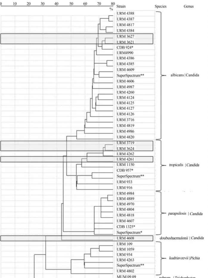
Figure 1- MALDI-TOF spectra based dendrogram of yeast isolates evaluated in this work. Discordances obtained by MALDI-TOF MS and classical phenotypic analyses are highlighted by grey boxes. CBD reference strains and Superspectra are marked by * and **, respectively.Trichophyton rubrum
(formerIssatchenkia orientalis) using the SARAMIS soft-ware package, which is the basionym teleomorphic name.
Isolates URM 4261, 3627, 3624 and 3621 were iden-tified as C. parapsilosis by phenotype. However, URM 4261 and 3624 wereC. tropicalisand URM 3627 and 3621 wereC. albicansby MALDI-TOF MS. Isolate URM 4261 was one of the six samples confirmed by VITEK-2 XL as
C. parapsilosis. This finding corresponds to 16.7% of dis-cordance in the overall isolates analysed by VITEK-2 XL in relation to the spectral phenotypic identification.
URM 4608 was identified asC. parapsilosis pheno-typically and was recorded as “unidentified” by MALDI-TOF MS, grouping separately on the dendrogram (Figu-re 1). This separation was confirmed by the molecular biol-ogy results presented below.
Finally, URM 3719 was identified as C. albicans
phenotypically and asC. tropicalisby MALDI-TOF MS.
ITS identification
All six isolates that presented discordant results from phenotypic and MALDI-TOF MS identifications were ana-lysed by ITS sequencing. URM 3621 and 3627 isolates were identified as C. parapsilosis by phenotype and C. albicans by MALDI-TOF MS (Table 1). ITS sequencing confirmed that these clinical yeast strains wereC. albicans (URM 3627, accession nº KF031307). URM 4261 (acces-sion nº KF031306) and 3624 (acces(acces-sion nº KF031304) were confirmed asC. tropicalisby ITS sequencing.
URM 3719 was identified asC. albicansby pheno-type but grouped withC. tropicalisby MALDI-TOF MS (Table 1 and Figure 1), and was confirmed asC. tropicalis
by ITS analysis (accession nº KF031305).
URM 4608 that was identified asC. parapsilosisby phenotype could not be identified by MALDI-TOF MS, grouping separately from theC. parapsilosiscluster in the MALDI-TOF MS dendrogram (Figure 1). However, it was identified as C. doubushaemulonii by ITS (accession nº KF031310) and when this new taxon information was added to the SARAMIS database, the isolate was recorded asC. doubushaemulonii(Figure 1).
Although the good concordance between pheno-typical and spectral identification ofPichia kudriavzevii, strains URM 1059 and URM 4802 were chosen for addi-tional ITS analysis. The sequencing results confirmed their identifications (accession nº KF031308 and KF031309, re-spectively).
Discussion
A comparison between the discriminative capability of phenotypic and MALDI-TOF MS characters of 40 clini-cal yeasts was reported herein. Moreover, discordant iso-lates were analysed by ITS sequencing which confirmed the MALDI-TOF MS identifications. Phenotype grouped the isolates into four distinct species:C. albicans,C. krusei
(P. kudriavzevii),C. parapsilosisandC. tropicalis. In
addi-tion, 6 out of 40 (15%) strains analysed by this method dis-agreed with the identification performed by MALDI-TOF MS. In line with described by Santos et al. (2011) the MALDI-TOF MS results were confirmed strongly by the ITS sequencing analyses. Through these combined ap-proaches five strains were renamed and one additional spe-cies was added to the collection:C. doubushaemulonii. The current data, when combined with data previously de-scribed (Marinach-Patriceet al., 2010; Jensen and Aren-drup, 2011; Santoset al., 2011), indicate that MALDI-TOF MS analysis is a powerful technique to identify clinical
Candidaisolates.
Because ribosomal proteins can be easily used as biomarkers in the proteomic-based technique by MALDI-TOF MS the reliability of this methodology is high and can discriminate between closely related Candida species. However, due to the biological variability of these yeasts, even for a single species, the availability of an extensive da-tabase is required. For example, MALDI-TOF MS gener-ated a distinctive spectrum forC. doubushaemuloniiURM 4608, separating it from the other isolates in the den-drogram, which is otherwise difficult to do. In this case, the database was not sufficiently extensive to provide straight-forward the correct identification due to a complete lack of data related withC. doubushaemulonii. As a matter of con-sequence, the culture collections with high quality biologi-cal materials are key elements to feed the MALDI-TOF MS databases with reference spectra. In contrast, with this new approach collections gain the possibility to requalify their holdings renaming misidentified strains.
MALDI-TOF MS is straightforward, rapid and em-ploys partially automated procedures. Results are obtained in approximately 30 sec per sample. Since the time for pathogen identification is an important determinant of in-fection-related mortality rates of hospitalised patients, this technique is an important tool in the fight to reduce mortali-ties when the time taken for classical methods is consid-ered.
In summary, the classical phenotypic approaches, in-cluding VITEK techniques are very important in the daily routine analyses in health centres and hospitals. However, it is associated with a high degree of misidentifications (Lockhartet al., 2008; Markleinet al., 2009; Putignaniet al., 2011) and it would be beneficial for these to be sup-ported by reference strains provided by culture collections and, if available, by MALDI-TOF MS with an appropriate database. MALDI-TOF MS is important to improve the ef-ficiency of the classical phenotypical methods for clinical
Candida polyphasic identifications even when isolates where preserved for decades in culture collections.
Acknowledgments
Action, under grant agreement No. FP7-228310 (EMbaRC project). Thanks are also due to Coordenação de Aper-feiçoamento de Pessoal de Nível Superior (CAPES, Brazil) for funding support.
References
Barnett JA, Paine RW, Yarrow D (2000) Yeasts: Characteristics and Identification. Cambridge University Press, Cambridge, U.K.
Boundy-Mills K (2012) Yeast culture collections of the world: meeting the needs of industrial researchers. J Ind Microbiol Biotechnol 39:673-680.
Cendejas-Bueno E, Gomez-Lopez A, Mellado E, Rodriguez-Tu-dela RL, Cuenca-Estrella M (2010) Identification of patho-genic rare yeast species in clinical samples: Comparison be-tween phenotypical and molecular methods. J Clin Microbiol 48:895-1899.
Correia A, Sampaio P, James S, Pais C (2006) Candida
bracarensissp. nov., a novel anamorphic yeast species
phe-notypically similar to Candida glabrata. Int J Syst Evol Microbiol 56:313-317.
De Hoog GS, Guarro J, Gené J, Figueras MJ (2000) Atlas of clini-cal fungi. Centraalbureau voor Schimmelculures, Uni-versitat Rovira i Virgili, Utrecht, Reus, The Netherlands. Essendoubi M, Toubas D, Bouzaggou M, Pinon JM, Manfait M,
Sockalingum GD (2005) Rapid identification of Candida
species by FT-IR microspectroscopy. Biochim Biophys Acta 1724:239-247.
Holland RD, Wilkes RD, Ralli F, Sutherland RD, Persons CC, Voorhees KJ, Lay JJO (1996) Rapid identification of intact whole bacteria based on spectral patterns using matrix-assisted laser desorption/ionization with time-of-flight mass spectrometry. Rapid Commun Mass Spectrom 10:1227-1232.
Jensen RH, Arendrup MC (2011)Candida palmioleophila: Char-acterization of a previously overlooked pathogen and its unique susceptibility profile in comparison with five related species. J Clin Microbiol 49:549-556.
Latouche GN, Daniel HM, Lee OC, Mitchell TG, Sorrell TC, Meyer WM (1997) Comparison of use of phenotypic and genotypic characteristics for identification of species of the anamorph genusCandidaand related teleomorph yeast spe-cies. J Clin Microbiol 35:3171-3180.
Leaw SN, Chang HC, Sun HF, Barton R, Bouchara JP, Chang TC (2006) Identification of medically important yeast species by sequence analysis of the internal transcribed spacer re-gions. J Clin Microbiol 44:693-699.
Lockhart SR, Messer SA, Pfaller MA, Diekema DJ (2008)
Lodderomyces elongisporus masquerading as Candida
parapsilosis as a cause of bloodstream infections. J Clin
Microbiol 46:374-376.
Macedo DPC, Farias AMA, Lima-Neto RG, Silva VKA, Leal AFG, Neves RP (2009) Opportunistic yeast infections and enzymatic profile of the etiological agents. Rev Soc Bras Med Trop 42:188-191.
Marinach-Patrice C, Fekkar A, Atanasova R, Gomes J, Djamdjian L, Brossas JY, Meyer I, Buffet P, Snounou G, Datry A, Hennequin C.; Golmard JL, Mazier D (2010) Rapid species diagnosis for invasive candidiasis using mass spectrometry. PLoS ONE 5:1-5.
Marklein G, Josten M, Klanke U, Müller E, Horré R, Maier T, Wenzel T Kostrzewa M, Bierbaum G, Hoerauf A, Sahl HG (2009) Matrix-assisted laser desorption ionization-time of flight mass spectrometry for fast and reliable identification of clinical yeast isolates. J Clin Microbiol 47:2912-2917. Medrano DJA, Brilhante RSN, Cordeiro RA, Rocha MFG,
Ra-benhorst SHB; Sidrim JJC (2006) Candidemia in a Brazilian hospital: The importance ofCandida parapsilosis. Rev Inst Med Trop S Paulo 48:17-20.
Mozina SS, Raspor P (1997) Molecular techniques for food iden-tification in food processing. Food Technol Biotechnol 35:55-61.
OECD (2007) Best Practices Guidelines for Biological Resource
Centres. Available at:
http://www.oecd.org/sti/biotechnologypolicies/38777417.p df. Accessed 14 December 2012.
Passarini MRZ, Santos C, Lima N, Berlinck RGS, Sette LD (2013) Filamentous fungi from the Atlantic marine sponge
Dragmacidon reticulatum. Arch Microbiol 195:99-111.
Putignani L, Del Chierico F, Onori M, Mancinelli L, Argentieri M, Bernaschi P, Coltella L, Lucignano B, Pansani L, Ranno S, Russo C, Urbani A, Federicibd G, Menichella D (2011) MALDI-TOF mass spectrometry proteomic phenotyping of clinically relevant fungi. Mol BioSyst 7:620-629.
Qian J, Cutler JE, Cole RB, Cai Y (2008) MALDI-TOF mass sig-natures for differentiation of yeast species, strain grouping and monitoring of morphogenesis markers. Anal Bioanal Chem 392:439-449.
Santos C, Fraga ME, Kozakiewicz Z, Lima N (2010b) Fourier transform infrared as a powerful technique for the identifica-tion and characterisaidentifica-tion of filamentous fungi and yeasts. Res Microbiol 161:168-175.
Santos C, Lima N, Sampaio P, Pais C (2011) Matrix-assisted laser desorption/ionization time-of-flight intact cell mass spec-trometry (MALDI-TOF-ICMS) to detect emerging patho-genicCandidaspecies. Diagn Microbial Infect Dis 71:304-308.
Santos C, Paterson RMR, Venâncio A, Lima N (2010a) Filamen-tous fungal characterisations by matrix-assisted laser desorption/ionisation time of flight mass spectrometry. J Appl Microbiol 108:375-385.
Schoch CL, Seifert KA, Huhndorf S, Robert V, Spouge JL, Le-vesque CA, Chen W, Fungal Barcoding Consortium Author List (2012) Nuclear ribosomal internal transcribed spacer (ITS) region as a universal DNA barcode marker for Fungi. Proc Natl Acad Sci USA, 109:6241-6246.
Simões MF, Pereira L, Santos C, Lima N (2013) Polyphasic iden-tification and preservation of fungal diversity: Concepts and applications. In: Malik, A., Grohmann, E., Alves, M.(eds). Management of Microbial Resources in the Environment. Springer, New York, USA, p. 91-117.
Smith D (2012) Culture collections. In: Sariaslani, S., Gaad, GM.(eds). Advances in Applied Microbiology. Academic Press, Burlington, USA, p. 73-118.
Sullivan DJ, Westerneng TJ, Haynes KA, Bennett DE, Coleman DC (1995)Candida dubliniensissp. nov.: phenotypic and molecular characterization of a novel species associated with oral candidosis in HIV-infected individuals. Microbiol-ogy 141:1507-1521.
Tavanti A, Davidson AD, Gow NAR, Maiden MCJ, Odds FC (2005) Candida orthopsilosis and Candida metapsilosis
spp. nov. to replaceCandida parapsilosisgroups II and III. J Clin Microbiol 43:284-292.
van Veen SQ, Claas ECJ, Kuijper EJ (2010) High-throughput identification of bacteria and yeast by matrix-assisted laser desorption ionization-time of flight mass spectrometry in conventional medical microbiology laboratories. J Clin Microbiol 48:900-907.
White TJ, Burns T, Lee S, Taylor J (1990) Amplification and di-rect sequencing of fungal ribosomal RNA genes for phylogenetics. In: Innis, M.A., Gelfand, D.H., Sninsky, J.J., Taylor, T.J.(eds.). PCR Protocols: a Guide for Methods and Applications. Academic Press, New York, USA, p. 315-322.