D iffe re ntial e xpre ssio n o f AMPA-type
glutamate re ce pto r subunits during
de ve lo pme nt o f the chick o ptic te ctum
1Departamento de Fisiologia e Biofísica, Instituto de Ciências Biomédicas,
Universidade de São Paulo, São Paulo, SP, Brasil
2Laboratório de Neurociências II, Universidade Cidade de São Paulo,
São Paulo, SP, Brasil S.S. Batista1,
R.S. Pires2 and
L.R.G. Britto1
Abstract
Glutamate receptors have been often associated with developmental processes. We used immunohistochemical techniques to evaluate the expression of the AMPA-type glutamate receptor (GluR) subunits in the chick optic tectum (TeO). Chick embryos from the 5th through the 20th embryonic day (E5-E20) and one-day-old (P1) chicks were used. The three types of immunoreactivity evaluated (GluR1, GluR2/3, and GluR4) had different temporal and spatial expression patterns in the several layers of the TeO. The GluR1 subunit first appeared as moderate staining on E7 and then increased on E9. The mature GluR1 pattern included intense staining only in layer 5 of the TeO. The GluR2/3 subunits presented low expression on E5, which became intense on E7. The staining for GluR2/3 changed to very intense on E14 in tectal layer 13. Staining of layer 13 neurons is the most prominent feature of GluR immunoreactivity in the adult TeO. The GluR4 subunit generally presented the lowest expression starting on E7, which was similar to the adult pattern. Some instances of transient expression of GluR subunits were observed in specific cell popula-tions from E9 through E20. These results demonstrate a differential expression of the GluR subunits in the embryonic TeO, adding infor-mation about their possible functions in the developmental processes of the visual system.
Co rre spo nde nce
S.S. Batista
Departamento de Fisiologia e Biofísica, ICB, USP Av. Prof. Lineu Prestes, 1524 05508-900 São Paulo, SP Brasil
Fax: + 55-11-3091-7426 E-mail: samuel@ fisio.icb.usp.br
Presented at the XVII Annual Meeting of the Federação de Sociedades de Biologia Experimental, Salvador, BA, Brazil, August 28-31, 2002.
Research supported by FAPESP, CAPES, CNPq and PRO NEX/MCT. S.S. Batista was the recipient of a fellowship from FAPESP.
Received April 5, 2002 Accepted June 20, 2002
Ke y words
•AMPA
•Development
•Glutamate receptors
•Neurotransmitters
•Receptor subunits
•Visual system
Glutamate receptors (GluRs) are present in most neurons of the vertebrate brain. They appear to be involved in diverse brain func-tions, such as learning, memory, synaptic plasticity, and developmental processes (1,2). The ionotropic branch of the GluRs includes three groups of receptors classified
accord-ing to their agonist selectivity: α
-amino-3-hydroxy-5-methylisoxazole-4-propionic acid (AMPA), kainate and N-methyl-D-aspartate (NMDA). The functional characteristics of
these GluRs result from the contribution of the four subunits that constitute the func-tional receptor (3). For instance, AMPA re-ceptors that present at least one GluR2
sub-unit show low permeability to Ca2+, while
the AMPA receptors formed by the other
subunits allow a significant Ca2+ influx (4).
This finding may have important
implica-tions during development, when Ca2+
cell death (5,6). A model that could be espe-cially useful to study these questions is the chick optic tectum (TeO). The avian TeO, in addition to expressing several types of gluta-mate receptors (7,8), exhibits a clear lamina-tion pattern and a well-characterized pattern of cell proliferation, migration, and differen-tiation (9). In the present study we deter-mined the immunoreactivity of the AMPA-type GluR subunits during development of the chick TeO.
Seventy-one chick embryos (Gallus
gal-lus) were used ranging in age from
embry-onic day 5 (E5) through the first post-eclo-sion day (P1). The embryos older than E12 were deeply anesthetized with ketamine and xylazine and perfused through the heart with phosphate-buffered saline and 4% parafor-maldehyde in 0.1 M phosphate buffer (PB, pH 7.4). Younger embryos were quickly de-capitated. All brains were removed and kept in fixative for 12 h and then transferred to a 30% sucrose solution in PB for cryoprotec-tion. Coronal sections (14-16 µm) of the frozen brains were cut on a cryostat and the sections including the TeO were selected for immunohistochemistry. Commercial anti-bodies (Chemicon, Temecula, CA, USA) against the GluR1, GluR2/3, and GluR4 AMPA receptor subunits were diluted 1:500 in PB containing 0.3% Triton X-100. Sec-tions were incubated with the primary anti-bodies for 14-18 h at room temperature (ca.
22oC), and then with a biotinylated goat
anti-rabbit antiserum (Vector Labs., Burlingame, CA, USA) for 60 min at room temperature. The sections were finally incubated for 1 h at room temperature with the avidin-biotin com-plex (ABC Elite, Vector Labs.) for 90 min. The sections were thoroughly washed with PB between steps. The immunoreaction was visualized using 0.05% diaminobenzidine and 0.01% hydrogen peroxide in PB. The intensity of staining for the GluR subunits was subjectively estimated. Nevertheless, a five-value scale was established to obtain a semiquantitative evaluation of
immunoreac-tivity for each antibody. No attempt was made to compare the intensity of staining between the subunits tested because of prob-able differences in the properties of the anti-bodies. For embryonic stages E5 through E18 we used a specific embryonic chick nomenclature to identify tectal layers (10). The identification of these layers after E18 was performed according to the stereotaxic atlas of the chick brain (11) using Cajal’s nomenclature (12). Controls for immuno-staining included the omission of the pri-mary antibodies or their replacement with normal rabbit serum. The staining was com-pletely eliminated in both situations. It should be added that these antibodies have been extensively characterized (7,8).
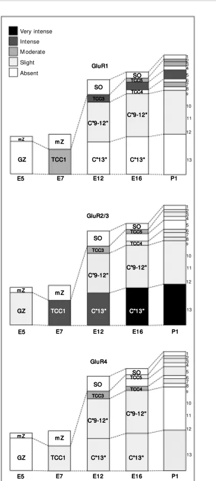
Differential patterns of expression were observed for the GluR subunits during de-velopment of the chick TeO. These data are described below and are summarized in Fig-ures 1 and 2.
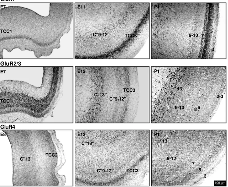
The GluR1 subunit started to be detected on E7. The immunoreactivity for this sub-unit first appeared in a single layer and was rated as moderate. This layer appeared to correspond to the outermost part of the first tectal transient cellular compartment (10). On E8, this stained layer apparently divided, with the outermost portion still presenting labeling for GluR1. The new innermost layer, which in the adult animal corresponds to Cajal’s layer 13, was not labeled on E8 or thereafter. On E9, there was an apparent increased labeling in the outer layer (second cellular transient compartment), just before this layer further divided in two, originating the presumptive superficial and intermedi-ate tectal layers. Staining for GluR1 was present in both of the new sublayers, with different levels of intensity. In general, stain-ing in the superficial layers was more intense than in the intermediate layers. The most distinctive feature of GluR1 staining in the chick tectum was clearly the labeling in layer 5.
appear during TeO embryonic development, starting on E5. On this day, the staining for GluR2/3 was weak and restricted to only one layer, i.e., the germinative zone. On E7, how-ever, immunoreactivity was already intense in most of that layer, which corresponds at this stage to the first transient cellular com-partment. Moderate staining for GluR2/3 also appeared in an external part of this transient compartment, which appears to be the same area that expresses GluR1 at this stage. After a subdivision on E8, both branches exhibited staining for GluR2/3. However, the inner-most branch presented in general more in-tense staining from E8 through P1. The out-ermost portion also presented staining for GluR2/3 which, despite some variations, only attained a slight level in our subjective scale. The neurons in layer 13 were by far those most intensely stained for any of the anti-bodies used in this study.
The GluR4 subunit began to be expressed on E7, with a weak intensity of labeling that was almost constant throughout develop-ment. Only one change was noted, which occurred between E11 and E16. During this period, staining for GluR4 appeared to in-crease transiently between E11 and E16 in a cellular compartment that generates some of the superficial tectal layers. Staining was only slight on P1, and the most distinctive feature of GluR4 was its presence in some large cells of layer 13, which did not label with any other antibody used here.
Figure 1. The temporal evolution of immunoreactivity for the glutamate receptor subunits GluR1, GluR2/3, and GluR4 during tectal development in the chick. The subjective scale for the semiquantitative analysis is show n at the top left. Dashed lines indicate the spatial changes of the different cellular compartments. GZ, germinative zone; mZ, marginal zone; TCC, transient cellular compartment; SO, stratum opticum; C” 9-12", compartment that generates the adult layers 9-12; C” 13", compartment that generates the adult tectal layer 13. The numbers on the right of each diagram indicate the tectal layers according to Cajal’s nomencla-ture (12). The nomenclanomencla-ture used here w as modified from Scicolone et al. (10).
Very intense Intense M oderate Slight Absent GluR1 GluR1 GluR1 GluR1 GluR1 C"9-12" C"9-12"C"9-12" C"9-12"C"9-12" C"13" C"13" C"13" C"13" C"13" m Z m Zm Z m Zm Z
SO SOSO SO SO TCC5 TCC5 TCC5 TCC5 TCC5 TCC4 TCC4 TCC4 TCC4 TCC4 TCC1 TCC1 TCC1 TCC1
TCC1 C"13"C"13"C"13"C"13"C"13"
GZ GZGZ GZGZ GluR2/3 GluR2/3 GluR2/3 GluR2/3 GluR2/3 SO SO SO SO SO TCC3 TCC3 TCC3 TCC3 TCC3 C"9-12" C"9-12"C"9-12" C"9-12" C"9-12" m Z m Z m Z m Z
m Z m Zm Zm Zm Zm Z
1 2 3 4 5 6 7 8 9 10 11 12 13 1 2 3 4 5 6 7 8 9 10 11 12 13 1 2 3 4 5 6 7 8 9 10 11 12 13 SO SO SO SO SO TCC5 TCC5 TCC5 TCC5 TCC5 TCC4 TCC4TCC4 TCC4 TCC4 SO SOSO SO SO TCC3 TCC3 TCC3 TCC3 TCC3 C"9-12" C"9-12" C"9-12" C"9-12" C"9-12" C"9-12" C"9-12" C"9-12" C"9-12" C"9-12" m Z m Z m Z m Z m Z C"13" C"13" C"13" C"13" C"13" TCC1 TCC1 TCC1 TCC1
TCC1 C"13"C"13"C"13"C"13"C"13" GZ
GZGZ GZGZ
m Z m Zm Z m Z
m Z m Zm Zm Zm Zm Z
C"9-12" C"9-12" C"9-12" C"9-12" C"9-12" C"9-12" C"9-12"C"9-12" C"9-12"C"9-12" SO SO SO SO SO TCC5 TCC5 TCC5 TCC5 TCC5 TCC4 TCC4 TCC4 TCC4 TCC4 SO SO SO SO SO TCC3 TCC3 TCC3 TCC3 TCC3 GluR4 GluR4 GluR4 GluR4 GluR4 E5 E5 E5 E5
E5 E7E7E7E7E7 E12E12E12E12E12 E16E16E16E16E16 P1P1P1P1P1
E5 E5 E5 E5
E5 E7E7E7E7E7 E12E12E12E12E12 E16E16E16E16E16 P1P1P1P1P1
E5 E5 E5 E5
E5 E7E7E7E7E7 E12E12E12E12E12 E16E16E16E16E16 P1P1P1P1P1 C"13" C"13"C"13" C"13" C"13" TCC1 TCC1TCC1 TCC1
TCC1 C"13"C"13"C"13"C"13"C"13" GZ
The results of this study indicate that there are different spatial and temporal patterns of expression of AMPA GluR1, GluR2/3, and GluR4 subunits in the chick TeO. Unfortunately, we have not been able to obtain independent, specific labeling for the GluR2 and GluR3 subunits. However, there is evidence that the mRNA coding for GluR3 is expressed at low levels in the ver-tebrate brain (7,8). Therefore, we consider that most of the staining that we have ob-tained in the TeO with the antibody against
the GluR2/3 subunits might have been due to the presence of large amounts of the GluR2 subunit. Accordingly, the discussion that follows mainly involves the GluR2 subunit when referring to the GluR2/3 data.
Possible associations between the differ-ential expression of various GluR subunits and developmental processes of the TeO may exist. For example, the early expression of all subunits tested here suggests some function in neurogenesis and migration since these processes occur at least until E8 in the
GluR1
GluR2/3
GluR4
TCC1E7 E11 P1
E7 E12 P1
E8 E12 P1
TCC1
TCC2 C"13"
C"13"
C"9-12"
TCC3
TCC3 C"9-12" C"13"
13
9-10 8
2-3
C"9-12" TCC3 9-10
5
3
6
13
9-12
7
5
3 7
100 µm
chick TeO. The especially precocious pearance of GluR2 subunits coincides ap-proximately with the peak of mitotic activity (13). Another important aspect of the pres-ent findings is related to the retinal innerva-tion of the TeO. Fibers from the contralateral retina start to innervate the chick TeO around E10 and these afferents, which represent the most prominent TeO innervation, continue to develop at least until E18 (9). Most of the changes in GluR expression were observed during this period, indicating a relationship between GluR expression and retinal affer-entation. This is also known as a period of intense synaptogenesis in the chick tectum (13). A typical example of the correlation of GluR temporal expression and synaptogene-sis appears to be the intense staining for GluR2 in the presumptive layer 13 of the TeO. A first increase of GluR2 expression in layer 13 neurons occurred on E10, followed by a marked peak on E14. Despite the fact that our data do not necessarily imply the occurrence of functional GluRs during de-velopment, it is noteworthy that calcium ac-cumulation mediated by glutamate also peaks around E10 in the retina, decreasing as syn-aptogenesis progresses (14). A relation be-tween the development of AMPA receptors and synaptogenesis has already been sug-gested for the development of the mouse cerebral cortex (15).
One of the most important aspects of the involvement of glutamate receptors in
de-velopmental functions is related to Ca2+
in-flux. Indeed, there is direct evidence that
Ca2+ flow through AMPA-type GluRs
regu-lates neurite outgrowth in developing retinal (16) and hippocampal (17) neurons. Since glutamate appears to be available early dur-ing development, activation of GluRs may have a strong impact on intracellular cal-cium and could therefore regulate a number of functions even before synaptogenesis (16). It is noteworthy that all subunits studied here may form GluRs with high calcium perme-ability, except for the edited form of GluR2 (18,19). The present data do not permit evalu-ation of the presence of the edited and non-edited GluR2 isoforms, and there is the pos-sibility that the edited form of GluR2 may start to be expressed at some point during development, when the developmental
func-tions of Ca2+ may compete with the possible
deleterious effects of high intracellular Ca2+.
This possibility has yet to be analyzed by molecular biological/electrophysiological techniques.
The chick TeO appears to represent a suitable model to directly evaluate the role of glutamate in developmental processes. The AMPA receptors exhibit temporal ex-pression changes during tectal development that are compatible with such a role for glutamate, which appears to also involve the NMDA receptors (1,20).
Ackno wle dgm e nts
Thanks are due to Dr. Andréa S. Torrão (USP) for critically reading the manuscript.
Re fe re nce s
1. Nguyen L, Rigo JM , Rocher V, Belachew S, M algrange B, Rogister B, Leprince P & M oonen G (2001). Neurotransmitters as early signals for central nervous system development. Cell and Tissue Research, 305: 187-202.
2. Kullmann DM , Asztely F & Walker M C (2000). The role of mammalian ionotropic receptors in synaptic plasticity: LTP, LTD and epilepsy. Cellular and M olecular Life
Sciences, 57: 1551-1561.
3. M adden DR (2002). The structure and function of glutamate receptor ion chan-nels. Nature Review s Neuroscience, 3: 91-101.
4. Pellegrini-Giam piet ro DE, Gort er JA, Bennett M V & Zukin RS (1997). The GluR2 (GluR-B) hypothesis: Ca(2+ )-permeable AM PA receptors in neurological disorders.
Trends in Neurosciences, 20: 464-470.
5. M cDonald JW & Johnston M C (1990). Physiological and pathophysiological roles of excitatory amino acids during central nervous system development. Brain Re-search Review s, 15: 41-70.
6. Ozaw a S, Kamiya H & Tsuzuki K (1998). Glutamate receptors in the mammalian central nervous system. Progress in Neu-robiology, 54: 581-618.
of AM PA-type glutamate receptor sub-units in the chick visual system. Brazilian Journal of M edical and Biological Re-search, 30: 73-77.
8. Theiss C, Hellmann B & Güntürkün O (1998). The differential distribution of AM PA-receptor subunits in the tectofugal system of the pigeon. Brain Research, 785: 114-128.
9. LaVail JH & Cow an WM (1971). The de-velopment of the chick optic tectum. I. Norm al m orphology and cytoarchitec-tonic. Developmental Brain Research, 28: 391-419.
10. Scicolone G, Pereyra-Alfonso S, Brusco A, Pecci SJ & Flores V (1995). Develop-ment of the laminated pattern of the chick tectum opticum. International Journal of Developmental Neuroscience, 13: 845-858.
11. Kuenzel WJ & M asson M A (1988). Ster-eotaxic Atlas of the Brain of the Chick
(Gallus dom est icus). Johns Hopkins Press, Baltimore, M D, USA.
12. Cajal SR (1911). Histologie du Système Nerveux de l’Homme et des Vertébrés. M aloine, Paris, France.
13. M ey J & Thanos S (2000). Development of the visual system of the chick. I. Cell differentiation and histogenesis. Brain Re-search Review s, 32: 343-379.
14. Sugioka M , Fukuda Y & Yamashita M (1998). Developm ent of glut am at e-induced intracellular Ca2+ rise in the em-bryonic chick retina. Journal of Neurobiol-ogy, 34: 113-125.
15. Arai Y, M izuguchi M & Takashim a S (1997). Developmental changes of gluta-mate receptors in the rat cerebral cortex and hippocampus. Anatomy and Embryol-ogy, 195: 65-70.
16. Catsicas M , Allcorn S & M obbs P (2001). Early act ivat ion of Ca(2+ )-perm eable AM PA receptors reduces neurite
out-grow th in embryonic chick retinal neurons.
Journal of Neurobiology, 49: 200-211. 17. M attson M P, Lee RE, Adams M E, Guthrie
PB & Kater SB (1988). Interactions be-tw een entorhinal axons and target hippo-campal neurons: a role for glutamate in the development of hippocampal circuitry.
Neuron, 1: 865-876.
18. Seeburg PH (1993). The molecular biol-ogy of mammalian glutamate receptor channels. Trends in Neurosciences, 16: 359-365.
19. Jonas P & Burnashev N (1995). M olecular mechanisms controlling calcium entry through AM PA-type glutamate receptor channels. Neuron, 15: 987-990. 20. Rashid NA & Cambray-Deakin M A (1992).