COMMUNICATION
AND
CHOICE
IN
YEAST
MATING
Maria
Joana
Patrício
Gonçalves
de
Sá
Dissertação
apresentada
para
obtenção
do
grau
de
doutor
em
Biologia
de
Sistemas
pelo
Instituto
de
Tecnologia
Química
e
Biológica
da
Universidade
Nova
de
Lisboa
Candidate:
Maria
Joana
Patrício
Gonçalves
de
Sá
Supervisor:
Andrew
W.
Murray,
Harvard
University
Em
memória
de
Maria
Videira,
que
me
ensinou
que
o
Saber
não
ocupa
lugar
The single biggest problem in communication is the illusion that it has taken place
TABLE OF CONTENTS
ABSTRACT ... 8
ACKNOWLEDGMENTS ... 10
CHAPTER 1: Introduction ... 13
CHAPTER 2: Regulated Cell-Cell Communication Makes Yeast Mating Economic
and Robust ... 23
CHAPTER 3: Sexual Identity in Yeast Mating ... 40
CHAPTER 4: Evolution and Specificity of the Ste2 Receptor ... 58
CHAPTER 5: Discussion ... 72
APPENDIX: Local Pheromone Degradation ... 81
MATERIALS AND METHODS ... 93
REFERENCES ... 104
TABLE OF FIGURES
Figure 1.1 – Schematic of the S. cerevisiae mating process... 15
Figure 1.2 - Components of yeast signaling cascade ... 17
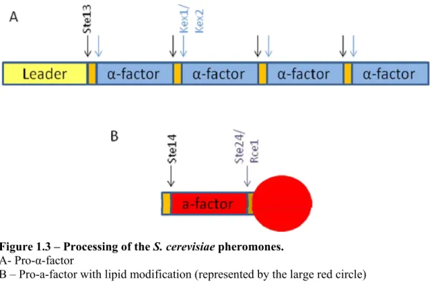
Figure 1.3 – Processing of the S. cerevisiae pheromones. ... 20
Figure 2.1 - Response of a-cells to pheromone ... 28
Figure 2.2 - The response of bar1∆ cells to homogenous stimulation by α-factor ... 30
Figure 2.3 - bar1∆ cells respond faster to α-factor gradients. ... 32
Figure 2.4 - Bar1 controls local pheromone concentrations to ensure efficient mating ... 35
Figure 2.5 – The role of Bar1 in mating ... 37
Figure 3.1.A – The Ascomycota and Basidiomycota species communicate via pheromones and GPCRs... 43
Figure 3.1.B – Artificial mating types ... 44
Figure 3.2 – Control crosses can mate ... 47
Figure 3.3 – Cells that communicate using only a-factor pheromones can mate ... 49
Figure 3.4 - Cells that communicate using only α-factor pheromones mate very poorly ... 51
Figure 3.5 – Mating efficiency of all the pairs tested ... 54
Figure 4.1 – Phylogenetic tree and synthesized pheromones ... 61
Figure 4.2 – Response of MATabar1 cells to different α-factor peptides ... 62
Figure 4.3 - Response of the cloned receptors to the corresponding pheromone ... 63
Figure 4.4 – Response of the cloned receptors to all pheromones ... 65
Figure 4.5 - Response of the cloned receptors to a subset of the pheromones ... 66
Figure 4.6 - dN/dS across the Ste2 receptor alignment ... 68
Figure 4.7 - The predicted transmembrane topology of the Ste2p protein from S.cerevisiae. ... 69
Figure A1.1 - -factor gradient in germinating spores. ... 83
Figure A1.2 – Ste2-GFP localization ... 84
Table A1.1 - Receptor mutants expressed ... 85
Figure A1.3 - BAR1-GFP localization ... 86
Figure A1.4 - SDS-PAGE and Western-blot with anti-TAP antibody for BAR1-TAP ... 87
Figure A1.5 – Biotin binding ... 88
Figure A1.6 – Cell wall bound chymotrypsin ... 89
Figure A1.7 - bar1MATa cells show alignment problems even in the presence of soluble protease. ... 90
ABSTRACT
Cell-cell communication is essential for all organisms and a hallmark of multicellularity. In the budding yeast, Saccharomyces cerevisiae, mating occurs when two haploid cells of opposite mating types (a and α), communicate through secreted pheromones and the corresponding transmembrane receptors, to find each other and fuse. I focused on the mating system of S. cerevisiae and used a quantitative approach to ask how yeast cells communicate with each other. I show that α cells advertise their presence strongly and devote about 1% of their protein synthesis to making just enough α-factor pheromone to initiate this communication. The a
SUMÁRIO
ACKNOWLEDGMENTS
This was a long and bumpy ride…
I must start by thanking Andrew Murray, my supervisor. Working with him has been an absolute privilege and I will always be grateful for the opportunity Andrew gave me. We have not always agreed, but I have always felt that I could express myself (and the scientific discussions just make it all worthwhile). Thank you, Andrew!
I also want to thank my “fake” thesis committee: Tim Mitchinson, Erin O’Shea and Michael Brenner, for agreeing to waste some time helping a stray Portuguese student. I first met Michael while still at the Medical School and he would sometimes take time off his sabbatical to have discussion over lunch with me. Michael’s enthusiasm is probably what kept me in Boston when I needed to change labs. Thank you, Michael! Erin was always available and put up with my indecisiveness. Erin tells it like she sees it and that was always immensely useful. Thank you, Erin! I will always be grateful for the opportunity to meet and discuss science with Tim. It is just so mind blowing and so much fun! Thank you, Tim!
not only to get a lot more done but, especially, not to despair. The more I teach you, the more I learn from you. Thank you, Perrine! And last but not least, the “girls”: Natalie, Mary, Lori, (Derek), Quincey, Melanie, Liedewij, Beverly, Lauren, and Linda. Thank you for putting up with my grumpiness!
Of course none of this would have been possible had I not been a Gulbenkian Fellow. I’m grateful to Miguel and Sukalyan for choosing me, Manuela for giving me a second chance and, more recently, Ana Maria Portocarrero. As the physicist trying to learn Biology I got a lot of help from my PGDB colleagues. A special hug to Tiago Carvalho, Marta Vitorino, Rita Tavares, Ulla Fiuza e Jovem Nómada. Vocês são os maiores! A big thank you to Mónica Dias, for always being available and for all the support, to Jorge Carneiro for so many discussions and to the IGC for providing such spectacular courses.
Outside of the lab, I relied on a number of people to keep my sanity. First, the Portuguese Máfia por tantas festas, copos, cangas, trivial, sardinhas, política, bola, marante… A minha vida em Boston divide-se entre o antes de vos conhecer e o depois. Ritinha, Armandinho e Franscisco, Kiiiiiiiika! Vizinha e Vizinho Godinho-Calado, Luís, Pedro e Cristina e Inês, Rui, Inês Grande, André, Eduardo e… Joaninha! Obrigada por TUDO! Depois, membros honorários da Máfia: Inbali, toda! Colo, Eyleen, Elenita, muchas gracias!
No entanto, há sempre alguém que nos faz falta, e ficam aqui os meus agradecimentos à Internet, por me deixarem mais perto dos “meus meninos”. MAV, Bruno, Daniel, Xavi, Rui, Rodrigo, Miguel, João: uns mais vezes, outros menos, mas sempre, sempre lá.
E porque não há tempo nem distância, bem-hajas, Tiago.
CHAPTER
1
Introduction
Cell-cell communication is essential for all organisms. In multicellular organisms this communication allows cells to differentiate into multiple cell types and tissues, grow and maintain their appropriate sizes, respond to internal and external signals and preserve homeostasis. Cell-cell signaling is also fundamental for single celled organisms to find food and mating partners. In unicellular as in multicellular organisms, one cell is constantly subjected to a very large number of signals, some of which they can sense and respond to. Appropriate integration and response to these stimuli is fundamental for the organism’s survival. For instance, a skin cell at the surface of the human body senses a number of environmental stimuli, like temperature and pressure, while integrating a number of stimuli from their fellow cells, such as signals to divide, stop dividing or even to commit suicide. If the outside temperature rises this information has to reach different parts of the body and, via cell-cell communication, elicit the correct responses, namely increased blood flow and fluid secretion.
There are at least three steps involved in cell-cell communication: 1) detection of an external signal; 2) signal interpretation and 3) response to the signal. Disruption in any of these steps can lead to disease: the loss of cell-cell communication is one of the hallmarks of cancer, while improper communication is one of the bases of auto-immune diseases. As a result, the manner in which cells integrate and respond to different signals is a fundamental and extensively studied problem in biology.
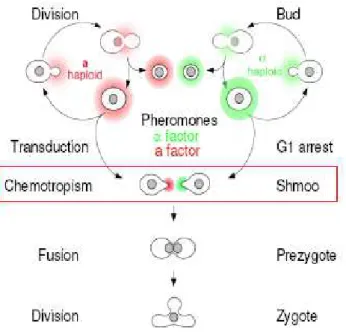
Approximately 15 minutes later, their nuclei fuse, and they re-bud as diploids. This full process can happen in as little as two hours (reviewed in [1]).
Figure 1.1 – Schematic of the S. cerevisiae mating process
Two haploid cells of the opposite mating type can polarize and grow towards each other (chemotropism). These cells form sexual protrusions called shmoos and can fuse their cytoplasms and nuclei to form diploid cells.
For these reasons, mating in budding yeast has been widely studied as an example of inter-cellular communication between eukaryotic cells. The work described in this thesis focuses on the signal sensing step and its implications for gradient detection, self vs. non-self discrimination and partner choice. In Chapter 3 we generate artificial and alternative mating types and ask what defines a possible mating partner by looking at the determinants of sexual identity. In Chapter 2 and Appendix A we look at what happens when one yeast cell is in the presence of several possible partners. We ask how they disentangle the signal gradients formed by large numbers of cells and how they distinguish between close, far away and equally distant partners. In Chapter 4 we extend these studies by looking at the specificity of different fungal pheromone receptors to self and non-self pheromones. Our goal is to look not only at the determinants of specificity but also at the role they might play in speciation.
the mating type loci, present some distinguishing features between the a-cells and the α-cells and discuss the role some of these differences might play in gradient shaping and signal detection. Finally, I compare the mating systems of different fungi and give a short overview of how these different species use the mating pathway to communicate
The yeast mating pathway and mating types
As described before, haploid yeast cells exist in two mating types: MATa and MATα. Both cells express at their surface seven-transmembrane G-protein coupled receptors (GPCRs) that detect the presence of small peptides secreted by the opposite mating type. MATa cells secrete a-factor and express at their surface the -factor receptor, Ste2, and MATα cells secrete -factor and express at their surface the a-factor receptor, Ste3. Pheromone binding triggers the dissociation of a trimeric G protein, whose subunits lead to the activation of a Mitogen-Activated Protein (MAP) kinase cascade. Fus3, the MAP kinase at the bottom of the cascade, phosphorylates and activates proteins that induce both cell cycle arrest (via Far1, a cyclin-dependent kinase inhibitor) and the transcription of genes involved in the process of mating triggered by the activation of the transcription factor Ste12. Free G also recruits Far1 that, in addition to arresting the cell cycle, recruits Cdc24, the guanine nucleotide exchange factor for Cdc42, the small G protein that controls actin polymerization and cell polarization [2] (Figure 1.2 adapted from [3]). This signaling cascade is conserved between the two mating types.
Figure 1.2 - Components of yeast signaling cascade (adapted from [3])
The mating response pathway for a MATa cell is shown. The α-factor pheromone binds the GPCR, leading to cell cycle arrest and actin polymerization (via Far1). MATα cells express the Ste3 receptor and respond to a-factor, but downstream of the receptor the signaling cascade is the same in both cell types.
Mcm1 to repress the a-specific genes and Matα1 binds both Mcm1 and Ste12 to activate the transcription of α-specific genes. The Mata1 regulator only plays a role in diploid cells, and at this time there is no function assigned to the Mata2 regulator (reviewed in [6]).
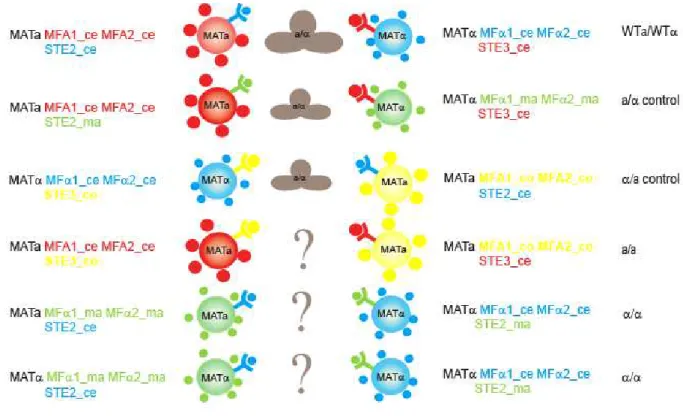
Some classic work in sexual specificity [7] has shown that mutant MATα cells can mate as either a or α cells depending on which set of receptors and pheromones are expressed, but this work did not rule out a contribution of the MATα and MATa loci to the mating process. We asked if the receptors/pheromones are also the major determinants of sexual identity by inducing same sex mating (MATa/MATa and MATα/MATα) when the mating pairs express complementary receptors and pheromones.
Yeast cells detect gradients and can mate over a wide range of conditions.
During mating, budding yeast cells need to detect the source of the signal coming from a potential partner. This process has been extensively studied using a cells that detect the small peptide pheromone α-factor. The pheromone’s presence induces the a cells to polarize towards the source of the attractant and grow mating protrusions, or shmoos. a cells also secrete a specific aspartyl protease (Bar1) that degrades and inactivates α-factor. a cells that lack Bar1 have been described as being supersensitive to α-factor-induced G1 arrest and exhibit mating difficulties [8], [9], but even in the complete absence of the protease, the cells can polarize and grow shmoos. Detection of chemical gradients is a necessary mechanism for living organisms, and it is often aimed at exploring the environment for nutrient sources, following prey or escaping poisonous environments or predators. Chemotaxis (polarized migration) and chemotropism
(polarized growth) are crucial for various biological events, such as developmental patterning, axon guidance, wound healing.
to a homogenous stimulation and return to a basal un-polarized state whereas yeast cells are able to detect gradients at concentrations at which they can spontaneously polarize [11], suggesting that suggesting that this adaptation mechanism might be missing.
Yeast cells can mate both as isolated pairs and in very dense mixtures, meaning that they have to be able to adapt to very different pheromone concentrations and be able to discriminate between many potential partners. A multitude of events in plant, fungal and animal development depends on a similar ability of cells to interact with only one of many possible partners. Examples include the interaction of neuronal growth cones with target cells (reviewed in [12]), myotube or vascular guidance (reviewed in [13-14]), the growth of pollen tubes to reach ovules (reviewed in [15]), and the mating of many fungi, including budding yeast (reviewed in [16-17]).
Bar1 homologues have been identified in different fungi, from Candida albicans [18] to
Schizosaccharomyces pombe [19], always in MATa cells, but these are the only known organisms that have a protease to degrade their own attractant, raising the question: why have yeasts evolved such an unusual and extracellular adaptation mechanism?
We looked at how Bar1 helps yeast cells detect α-factor gradients in controlled micro-fluidic devices, in the presence of single partners and in dense mating mixes.
The two pheromones are very different and this asymmetry is conserved.
There is no known equivalent of Bar1 being expressed by α-cells but this is not the only difference between the communication systems of the two haploid cells. The two pheromones, a-factor and α-factor, are secreted peptides that activate the corresponding receptors at the surface of the opposite cell. The α-factor, secreted by the α-cells, is encoded by two genes, MFα1 and
sequence tags the peptide for a covalent lipid modification. First, two other proteases, Rce1 and Ste24 cleave after the cysteine residue at the CAAX consensus sequence. Then, a complex of two proteins, Ram1 and Ram2, transfer a farnesyl group to the protein. Finally, Ste14 methylates the cysteine residue at the C-terminus. These highly hydrophobic peptides are then transported to the membrane and secreted via an a cell-specific transporter Ste6 (Figure 1.3.B, reviewed in [6]).
Figure 1.3 – Processing of the S. cerevisiae pheromones.
A- Pro-α-factor
B – Pro-a-factor with lipid modification (represented by the large red circle)
non-farnesylated mate 1000 fold worse [20]. This observation is also true for members of the Basidiomycota, such as the corn smut, Ustilago maydis [21].
Different yeasts share the same pheromones
The pheromone receptors belong to a ubiquitous family of GPCRs that play fundamental roles in cell signaling. S. cerevisiae has only three members of this family, but several hundreds have already been identified in humans where GPCRs represent the largest class of cell surface receptors (reviewed in [22]). These receptors are usually involved in transducing extracellular stimuli into intracellular signals and can be activated by light, neurotransmitters, odorants, lipids, hormones, chemokines, sugars, etc (recently reviewed in [23]).
As mentioned before, these receptors are characterized by an extracellular N-terminal domain, seven hydrophobic transmembrane domains and an intracellular C-terminus. Upon agonist binding, the receptor undergoes a conformational change that allows the receptor to associate (or release) its heterotrimeric G proteins, eliciting an intracellular response. These receptors are thought to heterodimerize and be very specific (recently reviewed in [24])
In S. cerevisiae, GPCRs are involved in detecting glucose, via Gpr1, and responding to mating stimulus, via the pheromone receptors Ste2 and Ste3. The Ste2 and Ste3 receptors have very little homology, although they are predicted to have the same topology, and signal by binding to the same intracellular machinery. These receptors are conserved and we can find homologs for both Ste2 and Ste3 in very distant fungi (reviewed in [22]).
Their conservation is even more striking in the Saccharomyces sensu stricto group. This is a collection of fermenting yeast species that includes S. cerevisiae, S. paradoxus, S. cariocanus, S. mikatae, S. kudriavzevii, S. bayanus, and the hybrid S. pastorianus [25], [26]. Genomic analysis of these species has revealed very high synteny and over 80% of their genes show over 80% sequence identity [27]. Additional to the high receptor similarity, S. cerevisiae, S. paradoxus and S. mikatae share the same a and α-factors; S. cerevisiae and S. castelii, an off-group Saccharomycete, share all but one amino acid in their a-factor pheromone and 10 out of 13 aminoacids in their α-factor-like peptides (this study). This is also known to be true in the Neurospora clade, another family of Ascomycetes, which are believed to share the same α-factor pheromone, although there is some variability between their receptors [28]. Co-existence between
species can be isolated and lab crosses have shown that there is no significant pre-zygotic isolation (reviewed in [31]) . The fact that the hybrids are sterile (unable to sporulate or giving rise to dead spores) raises the interesting question of why they have not diverged to prevent cross-talk between the pheromones and the receptors. Furthermore, it is not known how specific these receptors are to their own pheromones or how much different species, that may or may not share pheromones, use these GPCRs to communicate.
CHAPTER
2
Regulated
Cell
‐
Cell
Communication
Makes
Yeast
Mating
Economic
and
Robust
Budding yeast cells exist in two mating types, a and , which use peptide pheromones to communicate with each other and induce mating. We used quantitative measurements to understand how yeast cells mate efficiently under a wide range of conditions, ranging from isolated pairs of cells to dense mating mixtures. We show that cells devote 1% of their protein synthesis to making just enough -factor to initiate an inter-cellular positive feedback loop that induces cell cycle arrest, polarization, and fusion. Cells make a sharp transition between polarization and budding, despite substantial noise in the strength of signaling. They only respond to pheromone gradients over a narrow range, implying that cells should mate efficiently as isolated pairs but not in dense mating mixtures. We resolve this paradox by showing that a cells maintain the pheromone concentration at their surface within this range by regulating the expression of Bar1, the protease that degrades -factor. We conclude that yeast mate robustly by using the secreted protease to regulate ligand concentration and we discuss this as a general strategy for finding partner cells.
INTRODUCTION
Sex takes time and energy. Mating cells must signal to each other and arrest their cell cycles before they fuse. In nature, the budding yeast, Saccharomyces cerevisiae, is mostly found as diploid cells. Haploids exist in two mating types, a and which can proliferate asexually or mate with each other to form diploids. The a cells secrete a-factor and bear an -factor receptor, whereas cells secrete -factor and detect a-factor. Both express a common signal transduction pathway that is triggered by the binding of mating pheromones to their sex-specific receptors. The signal passes through a MAP kinase cascade and induces gene expression and polarized growth; polarization transforms ovoid cells into pear-shaped shmoos that grow towards each other (chemotropism) and fuse with each other at their tips (see Chapter 1 and Figure 1.1.A for more details, reviewed in [32]).
mating and has been proposed to help cells return to the cell cycle if they fail to mate [37], discriminate in favor of pheromone secreted by nearby cells [37], and modulate the direction of the pheromone gradient when an a cell is surrounded by multiple cells [38]. Despite these studies, we lack an integrated understanding of mating that answers the following questions: do decisions about mating show the same level of noise as signaling? How wide a range of pheromone gradients can cells detect? What is the physiological role of Bar1? How do cells regulate their pheromone response to mate efficiently under a wide range of conditions, varying from a pair of isolated a and cells to very dense mating mixtures, where each cell is surrounded by multiple partners of the opposite mating type?
accounts for about 1% of overall protein synthesis, 2) that this level can initiate courtship but not cell polarization, 3) that cells decide precisely, rather than noisily, whether to proliferate or polarize, and 4) that they only detect gradients over a narrow range of pheromone concentrations. In the second half of the paper, we explore the role of Bar1 in allowing yeast cells to mate efficiently over a wide range of conditions and we find that 5) the regulated secretion of Bar1 makes mating robust by keeping the pheromone concentration at the surface of cells within the narrow range needed for accurate gradient detection.
RESULTS
Measuring pheromone production and response
Unlike many unicellular eukaryotes, which only make pheromones when they are starved, haploid budding yeast are always ready to mate. This willingness implies a conflict between the benefit of making enough pheromone to signal to potential partners and the energetic cost of pheromone production.
For cells, this cost is set by the minimum pheromone concentration that a cells can detect and the physics of diffusion. At a minimum, cells must produce enough -factor to induce a response in a cells that lack Bar1, and thus cannot degrade -factor. This value has been reported to be as little as 0.1 [35, 39] and as much as 15 nM [36]. For simplicity, we assume that a single cell lies on the surface of an agar plate, very far from any other cells, that it secretes pheromone uniformly from its surface, and that the pheromone that it secretes diffuses into the agar, which acts as an infinite sink. Under these conditions, continuous pheromone secretion rapidly produces a spatial gradient in pheromone concentration that does not change over time. A spherical cell producing I molecules per second reaches a steady state concentration of
C0=I/(4aD) at its surface [40], where a is the cell radius (~2.5 m) and D is the diffusion
average protein of 450 amino acids [41]), but the pheromone concentration at the surface of an cell must still be far below 15 nM.
We started by inducing a bar1∆ cells with different concentrations of synthetic α-factor and scoring their morphology, by light microscopy, after 2h induction. With this information, we generated a calibration curve (Figure 2.1.A) where we make each pheromone concentration correspond to an observed percentage of shmoos. To measure the pheromone secretion rate, we grew cells exponentially in rich medium, transferred them to fresh medium for up to 2 hours, removed the cells, incubated the conditioned medium with abar1∆ cells, and scored the fraction
of the cells that had formed shmoos. We then compared the percentage of shmoos observed when the cells had been induced with the conditioned media, to the percentage of shmoos observed when the cells were responding to known concentrations of α-factor. This comparison allowed us to estimate that cells make -factor at 740 ± 220 molecules/cell/sec (Figure 2.1.B), corresponding to a steady state concentration at the cell surface of ~0.1nM -factor, far below the pheromone concentrations used in many previous studies, and only enough to occupy 1% of the -factor receptors (which have a Kd of about 6 nM [42]). Even at this low concentration, we estimate that -factor synthesis accounts for about 1% of total protein synthesis.
Can a cells detect such low levels of -factor and, if they can, what responses are induced? We began by studying the interactions of one a cell with one cell. Because we wanted to study the behavior of the a cell as the number of cells and the level of -factor increased, we kept the cells from mating by using the far1-H7 mutation; far1-H7 cells respond normally to pheromone but have polarization defects that reduce the efficiency of mating [43]. When an a
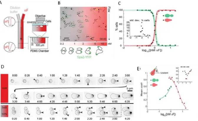
Figure 2.1 Response of a-cells to pheromone
A- Calibrating pheromone response and pheromone secretion. The response of a cells to -factor. Cells were treated with the indicated concentrations of -factor for 2 hours and the fraction of cells that had formed shmoos was determined by light microscopy. The calibration was performed on two strains, a bar1∆ (MP384) and a bar1∆ mfa1∆ (YTK277), to check that deletion of the major gene encoding a-factor had no effect on the response of cells to -factor. The assay was performed at least 3 times, at least 200 cells were counted for each time point. B - Bar graphs for the secretion of rates of -factor. Conditioned medium was collected from cells (MP634) for the indicated periods and assayed for its ability to induce shmooing in a bar1∆ cells
(MP384). The assay was performed 4 times, at least 200 cells were counted for each time point. C - The response of individual a cells to cells. Pairs of cells were micromanipulated to touch each other and observed. Bars show standard deviations. See Supplementary Table 2 for statistical tests.
We distinguished these possibilities by comparing the behavior of a bar1∆ cells that make
failed to arrest or arrested transiently (Figure 2.1.C), even though both types of a cells responded identically to synthetic pheromone (Fig 2-1-A). Thus unstimulated cells secrete just enough pheromone to make nearby a cells secrete more a factor and this reciprocation induces the cell to secrete enough pheromone to induce polarization of the a cell.
Do low concentrations of pheromone induce changes in gene expression? We monitored the transcriptional response of the FUS1 and BAR1 genes by fusing their promoters to YFP and measuring promoter activity as the rate at which fluorescence changed over time; FUS1 encodes a protein that plays an essential role in cell fusion and is the most commonly used reporter of pheromone signaling, whereas BAR1 encodes the -factor protease. Because we wanted to follow individual cells for long periods and because cells can remove -factor from the medium by endocytosis [45], we used microfluidics [46] (Figure 2.2.A) to expose a bar1∆ cells to a wide range of temporally stable concentrations of -factor. abar1∆cells expressing PBAR1-YFPand a
bar1∆ cells expressing PFUS1-YFP were distinguished from each other by fluorescent cell wall labeling, mixed together, placedin the same channel and exposed to a pheromone gradient. The
BAR1 promoter responded more strongly and more sensitively than the FUS1 promoter (Figure 2.2.B): BAR1 was induced 10 fold by 0.1 nM -factor, whereas FUS1 failed to respond at this concentration (Figure 2.2.B, inset). Our results are consistent with the observation that cell fusion needs higher levels of pheromone-induced signaling than cell polarization [47].
Cells respond precisely to pheromone
Figure 2.2 - The response of bar1∆ cells to homogenous stimulation by α-factor
A) Schematic of a device to produce a range of pheromone concentrations by using chaotic mixers in dilution chambers. The diagram shows a plan view (left) and cross sections at two magnifications (right). B) Induction of the FUS1 and BAR1 promoters by -factor. The BAR1
This precise morphological transition contrasts with the noisy expression from the BAR1
and FUS1 promoters, measured as the rate of YFP production. At 0.9 and 1.2 nM α-factor, there were enough cells of each type to perform t tests, which revealed that at each concentration, the expression levels in the three different morphologies were statistically indistinguishable from each other. However, for both cells that shmooed and budded, the response levels were statistically different between 0.9 and 1.2 nM α-factor (p < 0.025 for both comparisons) (Figure 2.2.D).
Cells polarize accurately over a narrow range of gradients
We also measured the response of a bar1∆ cells by exposing them to pheromone gradients in laminar flow chambers (Figure 2.3.A and 2.3.B). These experiments led to four conclusions: i) The transition between budding and shmooing occurs at the same concentration (1 nM) as it does for cells in homogenous pheromone concentrations (compare Figure 2.3.C with Figure 2.2.C), ii) cells in gradients respond faster than those treated with uniform pheromone concentrations (Figure 2.3.D), iii) cells can only detect gradients accurately over a narrow range (mean pheromone concentrations from 0.7 to 2.5 nM) (Figure 2.3.E), and iv) cells detect gradients most precisely at the mean concentration (1 nM), which equals the lowest concentration that induces shmooing in an isotropic field of pheromone (compare Figure 2.2.C and 2.3.E).
As long as we consider the mating of isolated pairs of cells, our results make sense. Single cells signal weakly, are induced to signal more strongly when they encounter a partner of the opposite sex, and cannot overstimulate their partners. But budding yeast also mate efficiently as dense mixtures of a and cells. In this situation, the same positive feedback would be disastrous because the mean pheromone concentration an a cell experiences depends on how many cells are nearby. We start by considering a single a cell on an agar surface surrounded by concentric rings of cells (Figure 2.4.A). The cells secrete -factor, which spreads by three-dimensional diffusion through the volume of the agar. If the radius of a ring is r, the contribution an individual cell makes to the pheromone concentration at the a cell falls as 1/r, but because the number of cells in a ring is proportional to r, each ring of cells contributes the same amount of pheromone to the location of the a cell and thus the pheromone concentration it experiences increases linearly with the number of rings of cells surrounding it (Figure 2.4.A). If we assume that
film of a and cells would exceed 100 nM, far above the concentration range at which a bar1∆
cells can detect gradients.
Figure 2.3 - bar1∆ cells respond faster to α-factor gradients.
Bar1 promotes mating by regulating global pheromone concentrations
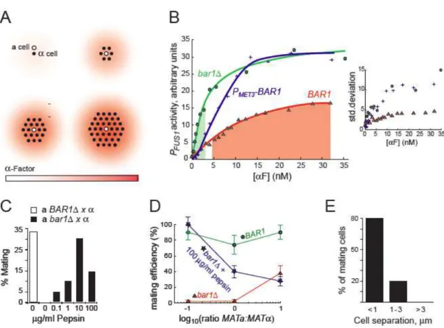
Regulated secretion of Bar1 is an attractive mechanism for controlling -factor concentrations. Bar1 limits the lifetime of diffusing -factor, preventing distant cells from contributing to the pheromone concentration at the surface of an a cell. We argue that the induction of Bar1 has been tuned to keep the factor concentration at the surface of a cells within the narrow range required for accurate gradient detection; the basal expression of Bar1 is low enough to allow a cells to detect the low levels of -factor made by a single cell, but maximal induction of Bar1 allows cells in dense mating mixtures to dramatically reduce what would otherwise be saturating pheromone levels.
To test the role of Bar1 in regulating the -factor response, we used the FUS1 promoter to report on the pheromone concentration cells are experiencing. In -factor-treated cells carrying
PFUS1-YFP, fluorescence accumulates at an average rate that is constant for at least 12 hours, suggesting that there is little or no adaptation in the pheromone response. Thus we can interpret the transcription of the FUS1promoter as a proxy for the pheromone concentration that the a cells experience. We compared the induction of PFUS1-YFP in three types of a cell: bar1∆ cells, cells
expressing BAR1 from its own promoter (BAR1), and cells expressing Bar1 from the MET3
promoter (PMET-BAR1) in medium lacking methionine (which represses PMET3). At every -factor concentration, the bar1∆ cells gave the strongest induction of PFUS1-YFP, but the relationship between BAR1 and PMET-BAR1 cells switched as the pheromone concentration rose (Figure 2.4.B). Below 3.5 nM -factor, cells expressing Bar1 from its own promoter responded more strongly than PMET-BAR1, demonstrating that the basal level of Bar1 expression is low. At higher pheromone concentrations, the BAR1 cells responded more weakly than the PMET-BAR1 cells. Thus the PMET-BAR1 cells express too much Bar1 in the absence of pheromone and too little at high pheromone levels, showing that tight regulation of Bar1 production is needed to allow a
cells to respond appropriately to a wide range of pheromone concentrations.
pheromone concentration required for accurate polarization of bar1∆ cells is 0.7 nM and BAR1
cells require just under 5 nM pheromone before their response reached the same level; but even at 30 nM, BAR1 cells are still responding more weakly than bar1∆ cells respond at 2.5 nM
pheromone, their upper limit for accurate gradient detection. These comparisons show that Bar1 induction reduces the pheromone concentration at the cell surface 7- to 12-fold relative to the concentration in bulk solution and that this reduction increases at higher pheromone levels, where BAR1 is more strongly induced.
The absence of Bar1 dramatically impairs the mating of a cells [8]. If the function of secreted Bar1 is to reduce the half life and diffusional range of -factor in mating mixtures, adding an exogenous protease should rescue the mating of abar1∆ cells. We mixed abar1∆and cells at high density and measured how efficiently the a cells mated by following individual cells by videomicroscopy. Because the cells are non-uniformly distributed, the mating efficiency of control BAR1 cells is only 30%. In the absence of exogenous protease, < 1% of the bar1∆a
cells mated. As we added increasing amounts of pepsin (the vertebrate protease most homologous to Bar1), the efficiency of mating rose to 30% at 10 g/ml pepsin and then fell, demonstrating that there is an optimum level of protease activity (Figure 2.4.C) and suggesting that the regulated production of Bar1 is essential for efficient mating. We tested this idea by varying the ratio of cells to three types of a cells: a BAR1, a bar1∆, and a bar1∆ cells with added pepsin (Figure
2.4.D). The results show that regulated protease production allows cells to mate efficiently under a wide range of conditions. The BAR1 cells mated efficiently over a 100 fold range of a: ratios, but the a bar1∆ cells only mated efficiently when a cells outnumbered cells. Adding an exogenous protease reverses this trend: the a bar1∆ cells with pepsin mated worse as cells became less frequent. This experiment demonstrates that the absence of protease, or unregulated protease activity make mating fragile for the same reason: only a small range of ratios of a: cells produce pheromone levels in the narrow range needed for gradient detection. This idea is supported by the observation that mixing BAR1 and bar1∆ cells allows cells lacking Bar1 to mate
Figure 2.4 - Bar1 controls local pheromone concentrations to ensure efficient mating
A) Each cell (black dot) produces the same amount of pheromone (red haze), but the contribution from different cells add to each other, increasing the pheromone concentration that an a cell (open dot) experiences as the number of cells rises at a constant cell density. B) FUS1
promoter activity (estimated from the rate at which the fluorescence of individual cells increased, see Materials and Methods for details) in bar1 (green), BAR1 (red), and PMET3-BAR1 (blue, Bar1
expressed from the MET3 promoter in the absence of methionine) cells as a function of pheromone concentration. The green shaded area shows the range of mean pheromone concentrations that allowed a bar1∆ cells to detect pheromone gradients, and the red area shows
If Bar1 is insulating a cells from the pheromone secreted by distant cells, the range of pheromone-based communication should be short. We studied the response of cells as a function of the distance between them. Cells that touch each other arrest and mate, cells that are separated from each other by a cell diameter (5 m) continue to proliferate, and at intermediate distances, a fraction of the cells arrest and mate (Figure 2.4.E). Because cells bud up pheromone gradients, they can approach a distant partner by budding rather than shmooing towards it, thus postponing the decision between arrest and proliferation.
DISCUSSION
We used quantitative measurements to ask how budding yeast cells can mate efficiently with each other under conditions that range from the equivalent of a desert island (two cells in isolation) to a crowded discotheque (dense mating mixtures). Yeast cells pay to advertise their sexuality by making and secreting pheromones. We show that to minimize this cost, cells produce the minimum quantity of -factor needed to induce an a cell to increase a factor secretion. This induction starts a positive feedback loop that eventually produces enough pheromone to arrest the two cells in G1 and induce them to polarize. Despite noise in their signaling pathway, pheromone-stimulated cells make a precise decision to shmoo or resume budding: a cells exposed to less than 1 nM -factor bud, and those exposed to more shmoo. Mating partners must polarize so that they grow towards each other, touch, and fuse. Since pheromone gradients direct polarization, cells must be able to detect the gradient produced by the nearest partner despite the presence of other potential partners that are more distant. For a
cells, this discrimination depends on Bar1, the protease that degrades -factor. In its absence, we find that cells only respond accurately to a narrow range of gradients, and bar1∆ cells only mate
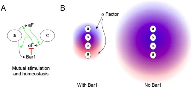
efficiently in conditions that keep the overall -factor concentration low. Figure 2.5.A illustrates the combination of positive feedback and homeostasis involved in mating: a- and -factor induce each other’s synthesis allowing the cells to produce the high pheromone levels needed for mating, and -factor induces Bar1 to prevent a cells from being over stimulated.
cells continue to proliferate rather than being arrested when they encounter cells that are too far away to mate with. Figure 2.5.B shows how Bar1’s ability to reduce the range of -factor diffusion helps to separate the contribution of pheromone from different cells.
Figure 2.5 – The role of Bar1 in mating
A) Shows the interaction between a and cells during mating. The secretion of -factor is stimulated by a factor and vice versa and -factor stimulates the secretion of Bar1 from a cells thus preventing the concentration of -factor at the surface of a cells from exceeding the levels needed for accurate gradient detection. B) The effect of Bar1 on insulating a cells from -factor secreted from distant cells. The left panel shows two cells secreting -factor, one marked red and the other blue, with the diffusional range of the -factor restricted by Bar1 secreted from the two a cells. In the right panel, the absence of Bar1 increases the diffusional range of the pheromone exposing the a cells to higher levels of -factor and preventing them from distinguishing the pheromone contribution from near and distant cells.
Morphological and transcriptional responses are poorly correlated
According to our transcriptional reporters, some cells that shmoo are signaling less strongly than those that bud, raising the question of how strongly the morphological responses of cells depends on the activity of the MAP kinase cascade that pheromones activate. Hartwell and his colleagues showed that cells that over express the pheromone-activated transcription factor (Ste12) can polarize towards mating partners in the absence of the MAP kinase cascade that normally transmits information about pheromones to the nucleus [47]. These two observations suggest that other aspects of the pheromone response must determine whether cells bud or shmoo and argue that behaviors under strong natural selection can evolve to be well insulated from the effects of transcriptional noise.
In the absence of Bar1, -factor-treated cells show two fates: they arrest in G1, fail to polarize, and eventually rebud, or they polarize and shmoo. The cells that shmoo do not adapt to the presence of pheromone. In nature, Bar1 is likely to play a critical role in adaptation by allowing a cells to recover quickly from situations that expose them to pheromone without providing them with a mating partner. For example, in an ascus that contains only three viable spores, two a and one , both a cells receive -factor, but only one can mate, and the disappointed suitor should return to the cell cycle as soon as possible.
Ecological and evolutionary aspects of mating
In nature, budding yeast is almost exclusively diploid [48], suggesting that the haploid phase of the life cycle is normally short. Germinating spores and asexually reproducing haploid cells secrete pheromones to advertise themselves to potential mating partners. In the absence of partners, making pheromone slows proliferation [49] and cells should minimize this cost, but in the competition for mates, the cell which makes the most pheromone wins [33-34]. These conflicting needs are resolved by making a factor induce cells to increase the production of -factor and vice versa. There are two advantages to making the threshold for pheromone induction lower than the one for cell cycle arrest: cells can begin courtship without sacrificing their proliferative potential and they can require that a potential partner demonstrate its fitness by producing the higher amounts of pheromone needed to cause arrest and ultimately mating.
least 10 fold as a and cells stimulate each other. Hartwell and his colleagues showed that a
cells efficiently chose partners who made more -factor than their competitors [33-34]. This behavior will select for increased -factor production, and because these cells threaten to overcome the ability of Bar1 to keep -factor concentrations within the range needed for accurate partner location, the spread of “sexier” cells will select for increased Bar1 expression in a cells, selecting for higher -factor production and so on.
ACKNOWLEDGMENTS
Matthieu Piel started with the Bar1 project, in the lab and, with help from Xin Jiang, set up the micro-fluidic system. Matthieu Piel did all of the flow chamber experiments described and looked at the mating efficiency of cells in dense mixes. Naama Barkai and Noa Rappaport are responsible for the theoretical argument of the linear increase in pheromone concentration. I would like to thank Chilin Guo and members of the Murray lab for discussions, Thomas Kramps and Kurt Thorn for strains and plasmids. This work was supported by grants from NIH (GM 68763 and GM 43987 to A. M), as well as fellowships to M. P. from HFSP (LT00271/2002-C), and to .J.G-S. from the Fundação para Ciência e Tecnologia (SFRH/BD/15220/2004).
CHAPTER
3
Sexual
Identity
in
Yeast
Mating
The higher fungi can be divided into two phyla: the Ascomycota and the Basidiomycota. Basidiomycetes, have complex mating systems, with one locus encoding regulatory transcription factors and the other encoding a variable number of pheromone and receptor genes. Most ascomycetes have two mating types, each of which expresses one receptor and a single pheromone. In the Ascomycota, both pheromones are small peptides and one of them (a-factor) is modified with a farnesyl lipid group that significantly alters its physical-chemical properties. In the Basidiomycota, all pheromones are lipid-modified and this difference is a distinguishing feature between the phyla. We asked whether the conservation in pheromone asymmetry, i.e. the fact that some are lipid modified and some are not, is required in ascomycetes and if the lipid modification of the pheromone plays a role in cell-cell fusion. We cloned receptor and pheromone genes from a homothallic Ascomycete and a heterothallic Basidiomycete and expressed these combinatorially into the yeast S. cerevisiae to generate novel, alternative mating pairs. We find that yeast cells can mate even when both mating pairs secrete a-like or α-like peptides. Importantly, this is true regardless of whether the cells express the a- or -mating type loci. Thus, we show that, in Saccharomyces cerevisiae, the only determinants of mating are the specificity of the receptors and their corresponding pheromones.
INTRODUCTION
Sex costs time and resources and represents a critical moment in an organism’s life cycle. Most eukaryotes are obligately sexual and, although some fungi do not have known sexual cycles, most are either homothallic (self-fertile) or heterothallic (self-sterile). The budding yeast,
expressed in the absence of Matα1 and Matα2. In diploid a/α cells, the cell contributes with Mat2, which still represses the MATa genes, and works with the a cell’s Mata1 regulator to block the expression of haploid specific genes. There is still no known role for the Mata2 regulator (reviewed in [50]). Haploid specific genes include those involved in all stages of the sexual development. As described in Chapter 1, the two haploid mating types sense each other’s presence by reciprocal sets of pheromones and pheromone receptors, with a cells secreting a -factor and responding to α-factor, and cells secreting α-factor and responding to a-factor.
Beyond the receptors, the signaling pathways are identical in both mating types, although the pheromones for the two mating types are asymmetric with respect to size and physic-chemical properties. While both pheromone are small hydrophobic peptides, a-factor is farnesylated and carboxymethylated at a C-terminal CAAX box and requiries a specific transporter for secretion, Ste6, a homolog of multidrug transporters. This asymmetry is conserved across the ascomycetes, but basidiomycetes only express the lipid-modified a pheromones (figure 3.1A). Mutations of the CAAX box result in non-farnesylated (or non-carboxymethilated) peptides and lead to significant reductions in mating efficiency, suggesting that the lipid tail is required for recognition and activation of the corresponding a-factor receptors [20]. The high hydrophobicity of the a-factor pheromone makes it very difficult to work with in a quantitative way and most studies looking at the yeast mating pathway are done with a cells being stimulated with -factor. Therefore, very little is known about the physical-chemical properties of the a -factor pheromone and how they might influence mating efficiency.
Figure 3.1.A – The Ascomycota and Basidiomycota species communicate via pheromones and GPCRs
The Ascomycota phylum, which includes species like S. cerevisiae and S. macrospora , communicate via asymmetric pheromones, α-factor like peptides and a-factor-like farnesyl modified peptides. The species from the Basidiomycota phylum, like S. commune, only use lipid-modified a-factor-like pheromones. The large circles represent modified pheromones and the small circles represent the α-factor-like peptides. The sequences for the α-factor like peptides from S. cerevisiae and S. macrospora are shown in blue and green, respectively. The sequences for the a-factor like pheromones for S. cerevisiae and S. commune are shown in red and yellow, respectively. This color code will be used in all the figures.
We created several artificial mating types and mating pairs and asked how important these haploid specific asymmetries are in sexual identity.
Figure 3.1.B – Artificial mating types
RESULTS
S. cerevisiae can mate using heterologous receptor and pheromone pairs.
We wanted to study how disrupting the asymmetry in either pheromone expression or MAT loci would reflect on mating efficiency. To do this we generated multiple artificial mating types by cloning the receptor and pheromone pairs from different fungal species. Since this is a synthetic system, we expected strains carrying the more distant protein homologues to mate worse than wild type S. cerevisiae strains. To measure the mating efficiency of these artificial mating types we generated strains that express the heterologous receptor proteins and mated them to strains carrying the matching heterologous pheromone genes. These sets of pairs served as positive controls and were expected to mate as all asymmetries were maintained: an a cell always mated with an α cell and they communicated via a- and α-like pheromones (Figure 3.2.B and 3.2.C)
To generate this system we chose two fungal species whose receptors had been successfully heterologously expressed in S. cerevisiae [52],[53]. Schizophyllum commune is a heterothallic Basidiomycete that is predicted to encode at least18 different receptors and more than 75 pheromones, all of which display the farnesylation CAAX motif. Expression of different combinations of the pheromones and receptors define more than 15000 possible mating types (for a review on S. commune mating see [4], especially chapter 18). To generate artificial a-mating types we cloned one of S. commune’s Ste3-like receptor (Bbr1) and one of the receptor’s matching a-like pheromones (Bbp2(4)), and expressed them in S. cerevisiae (Figures 3.1.A and 3.1.B). We will refer to this receptor as Ste3_co, to the pheromone as a_co and the pair has been color-coded in yellow in all of the figures and strain table.
Sordaria macrospora is a homothallic filamentous fungus closely related to Neurospora crassa (for a review on S. macrospora mating see [4], especially chapter 10). To generate artificial α mating types we cloned its Ste2-like receptor (Pre2) and its corresponding α-like pheromone (Ppg1) (Figure 3.1.A and 3.1.B). We will refer to this receptor as Ste2_ma, to the pheromone as α_ma and the pair has been color coded in green in all of the figures and strain table.
MFα2 for α-factor and MFA1 and MFA2 for a-factor. Ste2_ce (the α-factor receptor) and the corresponding pheromones, α1_ce and α2_ce have been color coded in blue and Ste3_ce (the a -factor receptor), a1_ce and a2_ce have been colored in red.
We started by making heterologous receptor green fluorescent protein (GFP) fusions and comparing their level of expression to that of the endogenous receptors in both a and α cells (Figure 3.2.A). STE2_ma was cloned at the STE2_ce locus in an a cell and STE3_co was cloned at the STE3_ce locus in an α-cell. Because these receptors are quite distant from S. cerevisiae and showed difficulties communicating with the downstream MAP kinase signaling components, a negative regulator of signaling gene, SST2, was deleted). Intrinsically, sst2 cells show decreased mating efficiency - around 5%, or 10 times worse than wild type cells (Figure 3.2.D and [54]), and this is taken into account when quantifying the alternative matings.
The MFα_ma gene was cloned into both α-factor loci and a-factor loci and a_co into both
a-factor loci. Both a and α-cells were found to express and secrete mature α_ma pheromone, albeit with significantly lower efficiency than cells secrete their own endogenous α-factor (Figure 3.2.B). We could not make the same type of quantitative measurements for a_co because the farnesyl group this pheromone makes it too hydrophobic and unwieldy to work with as it binds unspecifically to most labware surfaces.
We then assayed these strains for mating. The mating pairs constructed and their corresponding genotypes are shown in Figures 3.1.B and 3.2.C. The first mating pair listed served as a positive control of mating between two wild type S. cerevisiae strains: MATa STE2_ce MFA1/MFA2_ce x MATα STE3_ce MFα1/MFα2_ce. As expected these two strains mate with relatively high frequency, (figure 3.2.C and 3.2.D).
Figure 3.2 – Control crosses can mate
A- STE2-GFP fusions were made in strains carrying the receptor from S. cerevisiae and S. macrospora . Cells were incubated with 10μg/ml of the respective peptide pheromone (α_ce and
α_ma) for 2h. Cells were imaged on a Concavilin A coated slide and pseudo-colored. Both receptors are expressed and secreted to the membrane. B – The number of α-factor molecules being secreted per cell per second. The first bar shows the secretion rate for α_se being secreted by an α cell. The two green bars represent the secretion rate for α_ma when expressed in an a cell and in an α cell. Error bars represent standard deviations from at least 3 independent trials. C – Non-quantitative mating assay of the control crosses. Cell streaks were replica plated on top of each other and allowed to mate over night in complete media. They were then replica plated on selective media so that only diploids could grow. The figure shows that there is only mating in the diagonals. D – Quantitative mating assay for the control crosses. The shown crosses were allowed to mate on filters for 4 hours (in the case of the wild type) or for 7h (in the case of the
We then looked at mating efficiency using S. commune proteins by mating MATa
STE2_ce MFA1/MFA2_co with MATα STE3_co MFα1/MFα2_ce. This pair showed almost no mating at all. We hypothesized that the low mating efficiency could be explained by low levels in receptor and/or pheromone expression. Expressing the pheromones and receptors from a multi copy plasmid with a strong promoter showed that pheromone expression was the limiting factor, as increasing the receptor number didn’t significantly increase the number of mating events (Figure 3.3.A and data not shown). To correct for this bias, all subsequent experiments were done with the a_co expressing strains at a 5:1 ratio to the Ste3_co expressing strains. While mating efficiency was improved, it remained almost two orders of magnitude lower when compared to what was obtained by using the S. macrospora proteins. This could be rationalized as reflecting the large phylogenetic distance between S. commune and S. cerevisiae and a) its genes might not be expressed at the same levels and/or b) the receptor is likely to communicate with the MAP kinase signaling components less effectively.
Although efficiencies varied, all three pairs (expressing either S. cerevisiae, S. macrospora or S. communes proteins) can mate. We also found that the receptors are quite specific for their pheromones, as no off-diagonal mating can be observed in Figure 3.2.C.
We can recapitulate Basidiomycota matings in an Ascomycete
We then generated a mating pair where both cells express a-like pheromones and STE3-like receptors. In one a cell we replaced the endogenous STE2 receptor with the S. commune STE3-like receptor, STE3_co (yJS359). In another a cell we replaced the endogenous a-factor genes with the S. commune pheromone a_co and the STE2_ce receptor with the STE3_ce
receptor, usually expressed in α cells to make strain yJS360 (this also required the deletion of
Figure 3.3 – Cells that communicate using only a-factor pheromones can mate
The strains expressing the S. commune heterologous receptor and pheromone were mated with either α_ce or a_ce producing strains. A and B – the shown strains were mixed at different ratios (10:1, 5:1, 1:1, 1:5 and 1:10), spotted on complete media and allowed to mate over night (left panels). They were then replica plated on selective media that only allows diploid growth (right panels). We saw that consistent mating only happens when the S. commune pheromone secreting strains (yellow) were present in excess. This is true whether the mating pairs maintain the a/α asymmetry (A) or express only a-factor pheromones and MATa loci. C –Quantitative mating assay. Cells were mixed as described previously and allowed to mate on filters for 7h. Mating efficiency was calculated and error bars represent standard deviations from at least three independent mating trials.
This result was surprising for two main reasons. First, there appears to be no requirement for having asymmetric (farnesylated vs. non-farnesylated pheromones) for Ascomycete mating. This raises the question as to what the functional or evolutionary significance of this asymmetry might be. The second surprising finding follows from the fact that these fused cells are now a/a
The lipid tail is not-required for partner recognition and fusion
The mating pathway in S. cerevisiae has been extensively studied but the signal(s) for cell-cell fusion remains to be identified. We have shown that we can make two a cells fuse even in the absence of an -like pheromone. Because this situation appears to mimic mating in the Basidiomycota, we hypothesized that a-factor, or some unknown a-specific protein, might play a fundamental role in cell-cell fusion. If the farnesyl group of the pheromone is required for membrane fusion, a lipid-modified peptide should be required for mating to occur and one could expect that that two cells of opposite mating types that secrete only α-factor-like peptides to be able to form pre-zygotes but be unable to fuse.
To test this we constructed a mating pair that communicated using only -like pheromones. Starting from an -cell we replaced the Ste3 receptor with the Ste2-like receptor from S. macrospora , Ste2_ma (yJS220). This strain is now MATα STE2_ma MFα1/2_ce, producing S. cerevisiae’s α-factor and responding to the S. macrospora pheromone α_ma. Starting from an a cell, we replaced both a-factor producing genes with MFα_ma to make a cell that is MATaSTE2 MFα_ma (yJS214). This pair can communicate using only -like pheromones although the two cell backgrounds remain different, a and (Figure 3.1.B and 3.4.A). These cells can mate with efficiencies comparable to those of the S. macrospora controls (the mating of a
and cells in which the α-factor and α-factor receptor come from S. macrospora and the a-factor and a-factor receptor come from S. cerevisiae) of around 1% (compare second row in Figure 3.4.A with third row in Figure 3.2.D).
This suggests that there is no formal requirement for the lipid-modified pheromone in mating but didn’t rule out the possible contribution of the a cell to the mating process, for example through a role of a-specific genes other than the pheromone in mating. To address a putative role for a-specific genes in mating, we started from an cell and replaced both endogenous pheromone genes with MFα_ma, and replaced the naturally expressed STE3_ce
receptor with STE2_ce (yJS317) (Figure 3.4.A).
We now had two mating pairs that could communicate via α-factor like peptides only, but in one pair both cells express only the MATα locus. When we compared the mating efficiencies
we found that the MAT/MAT pair mated two orders of magnitude worse than the
MATa/MAT pair (Figure 3.4.A). Several factors could explain this phenotype: a) the α cells
pheromone; c) there is some a-specific protein that is important for efficient mating; or d) the / diploids have difficulties re-budding after fusion, as they could still self-stimulate and be arrested in G1.
Figure 3.4 - Cells that communicate using only α-factor pheromones mate very poorly
The strains expressing the S. macrospora heterologous receptor and pheromone were mated with either _ce or a_ce producing strains, expressing either the MATa or MAT loci. A – Mating efficiency was quantified as described above. Errors represent standard deviations of at least 3 independent trials. Mating pairs where both strains express the MAT locus mate 600 times worse than mating pairs that express different mating loci. B – STE2-GFP fusions were constructed and cells were induced with the respective pheromones and imaged for receptor localization. There are no significant differences in receptor expression.
The data presented in Figure 3.2.B shows that the cells actually produce slightly more
α-factor. The localization and expression of the receptor were analyzed by fluorescent microscopy (Figure 3.4.B). We found no significant difference in the expression of the receptor in the two different cell backgrounds.
A protease is required in the case of -factor matings
Using the available fluorescent markers to distinguish between different cells (see Supplementary Table 1), we mixed the yJS220 or yJS221 (MAT MF_ce STE2_ma) with its mating pair yJS317 (MAT MF_ma STE2_ce) and observed these mating mixes, under the microscope (Supplementary Movie 1). STE2_ma expressing cells were found to arrest and induce the mating pathway, as indicated by the activation of the FUS1 promoter, but have difficulty polarizing (as indicated by the movement of the Spa2 dot) and finding a mating partner. The _ma producing strains also arrest and turn on the FUS1 promoter but are over stimulated and make shmoos. On rare occasions two cells expressing the matching receptor and pheromone pairs find each other, align their polarities, and the fusion process proceeds normally. Although these cells take slightly longer to rebud, their subsequent cell cycles are of normal length indicating that these cells are not being pheromone stimulated.
We asked if the polarity/over-stimulating problems could be responsible for the lowered mating efficiency. A natural a-specific gene candidate was the already mentioned protease BAR1. Bar1 is fundamental in shaping the -factor gradient and helping a cells discriminate between possible partners, as argued in chapters 1 and 2. We deleted this protease in yJS214 (the
MATa mating pair, MF_ma STE2_ce) to make yJS385. _ma secretion is indistinguishable between this strain and its parent, yJS214, (data not shown) but mating efficiencies are now reduced 30 fold, the same reduction observed in wild type bar1 crosses (Figure 3.4.A), although they still mate 15 times better that the equivalent / pairs. Because the secretion levels are the same for both BAR1 and bar1 strains, we expect this protease not to cleave _ma and to act only on S. cerevisiae’s -factor, to prevent the saturation and directionality problems displayed by MAT MF_ma STE2_ce (yJS317) cells, thus improving the mating efficiency.
made long and sometimes multiple shmoos (Supplementray Movie 1). Adding an unspecific soluble protease would presumably help shmooing cells by reducing the overall concentration of
α_ce but would have the negative effect of making the MATα STE2_ma MFα_ce cells even less responsive. In this case, where both mating cells secrete and communicate via small unmodified peptides, the results would become harder to interpret. We tested the effect of adding different concentrations of both chymotrypsin and pepsin to the two heterologous mating pairs yJS220 (the Ste2_ma α cell) with both the MATa (deleted for Bar1) and MATα MFα_ma STE2_ce strains. Preliminary results indicat that adding soluble protease does increase the number of both a/α and
α/α diploids formed. We have not been able to raise the number of α/α diploids to the same level, consistent with the idea that these diploids behave like α cells and may have a harder time forming colonies.
Although we saw a decrease in mating efficiency when the two mating cells express the MAT
loci (yJS220 × yJS317) this reduction can be mimicked in MATaMAT matings by deleting Bar1 in the a cell (yJS220 × yJS385). The mating efficiency of these pairs was comparable to the one observed when only a-factor-like pheromones and MATa loci were expressed.
DISCUSSION
There is no inherent bias for a-a or - mating
![Figure 1.2 - Components of yeast signaling cascade (adapted from [3])](https://thumb-eu.123doks.com/thumbv2/123dok_br/15765722.640449/17.892.262.669.173.584/figure-components-yeast-signaling-cascade-adapted.webp)