Article
J. Braz. Chem. Soc., Vol. 23, No. 4, 593-601, 2012. Printed in Brazil - ©2012 Sociedade Brasileira de Química 0103 - 5053 $6.00+0.00
A
*e-mail: [email protected]
Electrocatalytic Oxidation of Dopamine, Ascorbic Acid and Uric Acid at
Poly-2,6-Diaminopyridine on the Surface of Carbon Nanotubes/GC Electrodes
Mohammad Ali Kamyabi* and Mohammad Ali Shafiee
Department of Chemistry, Zanjan University, PO Box 45195-313, Zanjan, Islamic Republic of Iran
A preparação e aplicação de poli-(2,6-diaminopiridina) sobre a superfície de um eletrodo de carbono vítreo (GCE) modificado com nanotubos de carbono de paredes múltiplas (CNTs) são descritas. Voltametria cíclica foi usada na síntese eletroquímica e na caracterização dos polímeros depositados sobre GCE. O eletrodo modificado mostra um efeito sinérgico das propriedades eletrocatalíticas e a elevada superfície ativa de ambos, polímero condutor e nanotubos de carbono, dando origem a uma melhora notável da oxidação eletrocatalítica de ácido ascórbico (AA), dopamina (DA) e ácido úrico (UA) com respeito a eletrodos modificados com polímero e eletrodos modificados com CNTs. Este tipo de eletrodo modificado mostra uma excelente atividade eletrocatalítica para a oxidação de AA, DA e UA dentro de um intervalo linear de 2,70 × 10⁻⁵-1,83 × 10⁻³, 8,33 × 10⁻⁷-1,00 × 10⁻⁵ e 4,16 × 10⁻⁶-2,25 × 10⁻⁴ mol L-1, respectivamente,
em pH 8.0 (solução padrão de fosfato). A mistura ternária que contém AA, DA e UA pode ser muito bem separada uma da outra a uma velocidade de varredura de 100 mV s-1 com uma
diferença de potencial de 184, 147 e 321 mV em voltametria de pulso diferencial (DPV) entre AA e DA, DA e UA, e AA e UA, respectivamente. Esta abordagem é tão fácil que pode ser usada para detectar seletivamente DA, AA e UA na presença uma das outras e também em algumas amostras reais.
The preparation and application of poly-(2,6-diaminopyridine) on the surface of a glassy carbon electrode (GCE) modified with multi-walled carbon nanotubes (CNTs) are reported. Cyclic voltammetry was used for both the electrochemical synthesis and characterization of the polymers deposited on GCE. The modified electrode shows a synergic effect of the electrocatalytic properties and high active surface area of both the conducting polymer and carbon nanotubes, giving rise to a remarkable improvement of electrocatalytic oxidation of ascorbic acid (AA), dopamine (DA) and uric acid (UA) with respect to polymer-modified electrodes and CNTs-modified electrodes. This kind of modified electrode shows and excellent electrocatalytic activity towards the oxidation of AA, DA and UA within the linear range of 2.70 × 10⁻⁵-1.83 × 10⁻³,
8.33 × 10⁻⁷-1.00 × 10⁻⁵ and 4.16 × 10⁻⁶-2.25 × 10⁻⁴ mol L-1, respectively, in pH 8.0 (phosphate buffer
solution). The ternary mixture, which contains AA, DA and UA, can be well separated from each other at a scan rate of 100 mV s-1 with a potential difference of 184, 147 and 321 mV in differential
pulse voltammetry (DPV) between AA and DA, DA and UA, and AA and UA, respectively. This approach is so simple and easy that can be used to selectively detect DA, AA and UA in the presence of each other and also in some real samples.
Keywords: 2,6-diaminopyridine, electropolymerized film, ascorbic acid, dopamine, uric acid
Introduction
Today, one of the main challenges is the development of methods to perform clinical analyses that are rapid in situ analyses. These methods must be sensitive and accurate, and able to determine various substances with different properties in real-life samples. Electrochemical
sensors for the measurement of analytes of interest in clinical chemistry are ideally suited for these new applications due to their high sensitivity and selectivity, portable field-based size, rapid response and low-cost.1
can accelerate transmission of electrons on the surface of electrode, it has high selectivity and sensitivity due to the film homogeneity in electrochemical deposition, strong adherence to the electrode surface and large surface area.2,3 Unlike conventional immobilization strategies
for biosensors, electropolymerization has no limit in terms of the geometry and area of the electrode, and offers advantages with respect to thickness control, reproducibility and uniformity of the polymer film on the electrode surfaces with more complex geometries.4 In
addition, electropolymerization permits simple electrode regeneration and can be easily extended to the production of microbiosensors. Many studies have indicated that polymer film modified electrodes show an enhanced response for the determination of various important biological and clinical species.5
Electropolymerization of conducting polymers, such as polypyrrole (PPy), polyaniline, polyacetylene, polyindole, polythionine and polythiophene, has been studied extensively for the development of biosensors.6-8 These polymers offer
great advantages due to their very good conducting and mechanical properties and good adhesion to the electrode substrate. However, it is of interest to extend such studies to non-conducting polymers like polyphenol, poly(o -phenylenediamine), poly(dichlorophenolindophenol) and overoxidized polypyrrole which have specific advantages for biosensor construction and electrocatalytic reactions.9-18
The non-conducting polymers provide very thin films due to their self-limiting growth, and hence the biosensors based on them have fast response. In addition, the permselectivity of the non-conducting films confer them improved biosensor selectivity and anti-fouling properties. Non-conducting membranes from polyphenylenediamines (PPDs) have been of particular interest because of their thin, dense films leading to both fast response and high H2O2 or O2
selectivity. Jang et al.19 reported the electropolymerization
mechanism for poly(1,2-diaminobenzene), and the film of electropolymerized poly(1,2-DAB) has been analyzed by an impedimetric technique.20 Also, enzyme glucose oxidase
(GOx) has been entrapped in a poly(1,2-DAB) film through the polymerization of 1,2-DAB on platinum-coated carbon fibers.21-23
In this work, an electropolymerized film of 2,6-diaminopyridine (2,6-DAP) was prepared on the surface of a GC electrode in 0.10 mol L-1 HCl in methanol
by cyclic voltammetry (CV). The poly(2,6-DAP) on the surface of GCE showed excellent catalytic activity toward oxidation of some biological compounds and conspicuously enhanced the redox peak currents. The ternary mixture, which contains ascorbic acid (AA), dopamine (DA) and uric acid (UA), can be well separated from each other at
the surface of this modified electrode. The separations of the oxidation peak potentials of AA-DA and DA-UA were over more than 184 and 147 mV, respectively. Using differential pulse voltammetry (DPV) technique, the calibration curves for AA, DA and UA were obtained over a wide range (2.70 × 10⁻⁵-1.83 × 10⁻³ mol L-1
for AA, 8.33 × 10⁻⁷-1.00 × 10⁻⁵ mol L-1 for DA and
4.16 × 10⁻⁶- 2.25 × 10⁻⁴ mol L-1 for UA at the poly(2, 6-DAP)
modified GCE). The theoretical limits of detection defined as 3σ of the proposed method for AA, DA and UA were 5.00 × 10⁻⁶, 4.16 × 10⁻⁸ and 7.10 × 10⁻⁷ mol L-1, respectively.
These results are somewhat similar (or worse) performances (in some cases) or superior ones (in most cases) than the previously reported modified electrodes in literatures.24-35
For the first time, the electropolymerization of 2,6-diaminopyridine was studied by Morea et al.36 in
acetonitrile solvent at a platinum electrode that led to the formation of two different films depending on the deposition potential. Their results revealed an ECE mechanism (electron transfer, chemical reaction, electron transfer) operating for film I formation and provided evidence for nucleation and growth in film II deposition.36 In the present
work, the electropolymerization of 2,6-diaminopyridine is reported on the surface of a GC electrode in 0.10 mol L-1
HCl in methanol. This film formation is different from the previously reported,36 and thus, the present study provides
a novel method for selective and sensitive detection of DA, AA and UA in the presence of the other two species.
Experimental
Reagents and solutions
All chemicals were of analytical reagent grade (from Merck or Sigma) unless otherwise specified and were used as received without further purification. Triply distilled water was used to prepare buffer and reagent solutions. The supporting electrolyte used in all the experiments was 0.1 mol L-1 phosphate buffer solutions (PBS).
Apparatus
Voltammetric experiments were performed using Metrohm Computrace Voltammetric Analyzer model 757 VA. A conventional three-electrode system was used with a bare or chemically modified GCE as working electrode, reference electrode Ag/AgCl, KCl 3 mol L-1, and
Preparation of the modified electrode
The glassy carbon electrode (2 mm diameter) was carefully polished with alumina powders (1.0, 0.3 and 0.05 µm) on polishing cloth. The electrode was placed in ethanol container and it was used bath ultrasonic cleaner in order to remove adsorbed particles. Then, 15 cycle scans were carried out in the potential range of −2.0 to +2.0 V vs. reference electrode in a solution of 1.0 mol L-1 sulfuric acid
(H2SO4),
35,37 this process was used to remove any impurities
of the electrode surface. Finally, the electrode thoroughly washed with triply distilled water, and for drying, it was heated for 5 min at 50 oC in oven. Multi-walled carbon
nanotubes (MWNTs) (10 mg) were dispersed in 10 mL dimethyl sulfoxide (DMSO) by ultrasonic stirring for about 30 min to give a black suspension of 1 mg mL-1. The
MWNT modified electrode was prepared by casting 10 µL of the MWNT suspension on the surface of GC electrode, which was dried in air for 24 h at room temperature. When the DMSO was volatilized, a MWNT film was formed. The surface modification of the MWNTs/GCE was performed in two steps:
(i) The poly(2,6-DAP) films were formed on the electrode surfaces by continuous potential cycling between −0.3 and 1.2 V at 50 mV s-1 in 0.10 mol L-1 HCl containing
4.0 mmol L-1 2,6-DAP, (total volume of 10 mL). Typically,
20 cycles were employed (although other cycle numbers were also studied).
(ii) In the second step, the poly(2,6-DAP) film on the GC electrode was converted to a conducting polymer by cycling the potential scan in 0.01 mol L-1 NaOH solution
at the scan rate of 50 mV s-1 for 10 times.
The surface area of the modified electrode (poly(2,6-DAP)/MWNTs/GCE) was 1.053 ± 0.006 cm2 whereas for
MWNTs/GCE 0.821 ± 0.005 cm2, which were evaluated
from cyclic voltammetry experiments. After surface modification of the GCE, the modified electrode was rinsed with distilled water and stored in triply distilled water for use. Solutions were purged with high purity nitrogen gas for at least 10 min before electrochemical measurements.
Results and Discussion
Electropolymerization of 2,6-DAP and its electrochemical properties
Because of difficulties related to the low solubility of 2,6-DAP, several aqueous media with different pH values were used for the electropolymerization of 2,6-DAP at the MWNTs/GCE. The final choice of medium was 0.10 mol L-1 HCl in methanol (concentrations of HCl were
examined from 0.01 to 1.00 mol L-1, and 0.10 mol L-1 was
used as optimum to give good solubility of 2,6-DAP). The continuous cyclic voltammograms that were recorded used GCE dipped in a mixture containing 4.0 mmol L-1 of
2,6-DAP and 0.1 mol L-1 HCl under deaerated conditions.
The effects of various factors on the poly(2,6-DAP) film formation on the surface of the MWNTs/GCE (such as the amount of monomer scan rate, number of scan, potential window, pH and also the conditions of the alkaline treatment) were evaluated according to the variation of the peak current of the cyclic voltammograms obtained for the modified electrode.
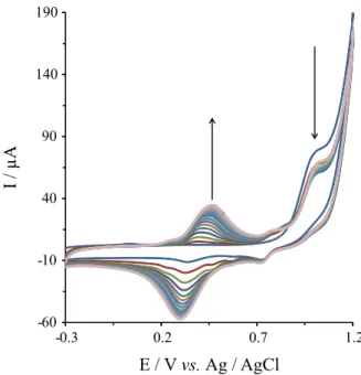
Figure 1 displays the CVs of 2,6-DAP electropoly-merization over the range of −0.3 to 1.2 V at 50 mV s-1 for
20 cycles. The forward scan of the first cycle revealed the presence of an irreversible oxidation and reduction peak of the monomer (Figure 1, first cycle, O1 and R1 peaks) with peak
potentials at about 1.01 and 0.68 V, respectively (vs. Ag/AgCl). In the subsequent cycles, the oxidation current decreased at 1.01 V with a simultaneous appearance of a pair of redox peaks at about 300/390 mV due to the formation of poly(2,6-DAP) film. This clearly shows the oxidation of the monomer 2,6-DAP and the formation of poly(2,6-DAP) film on the surface of the MWNTs/GCE. The peak-to-peak separation (∆E
p = Epa – Epc of the pair
redox peaks at about 300/390 mV) was bigger with increasing the number of cycles (90.0 mV in the second cycle and 200 mV in the last cycle). This fact suggests that the nonconductive polymer film was formed on the
surface of GC electrode with increasing scan cycles. A similar peak was also seen for the electropolymerization of o-phenylenediamine (oPD)38 and for 4-nitro-
1,2-phenylenediamine (4-NoPD).39
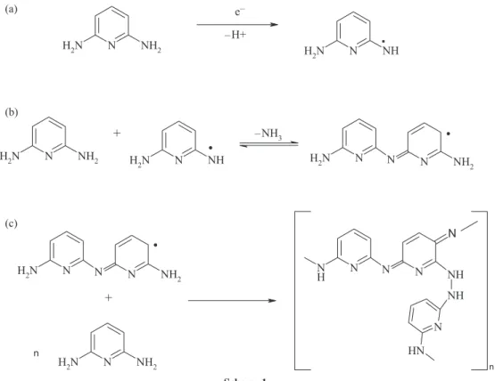
Scheme 1 represents the proposed mechanism for the formation of the active component during the electropolymerization.19,36,39 The polymerization
mechanism may be described as follows: 2,6-DAP was first oxidized to free radical (Scheme 1a). The processes occurring after this first step may follow various paths, the coupling of such radicals may occur or the elimination of ammonia group during electrochemical oxidation may occur (Scheme 1b).40 However, a possible structure for the
film is shown in Scheme 1c. The suggestion of a structure for the film is beyond the scope of this work. In fact, several possible polymeric backbones may be considered. Moreover, since 2,6-DAP has two virtual reaction sites (i.e., two amino groups), cross-linking reactions may also take place. This obviously enlarges the number of structures that may describe the film.
The poly(2,6-DAP) is a non-conducting membrane and this modified electrode does not show any electrocatalytic activity in aqueous solution. However, several scan cycles of the poly(2,6-DAP) film modified GCE in an alkaline solution (alkaline treatment) can improve the conductivity and also the electrocatalytic acitivity of the modified electrode (Figure S1). The cyclic voltammogram of MWNTs/ GCE modified with poly(2,6-DAP) film (after alkaline
treatment) shows a pair of reversible peaks in 0.1 PBS pH 8 at formal potential of about –80 mV with ∆Ep about
60 mV at scan rate of 100 mV s-1 in PBS pH 8.0 (Figure 2).
The good conductivity and electrocatalytic activity of the modified electrode after an alkaline treatment may be due to the reduction of some of the C−N bonds in the polymer and the rearrangement of the polymer. Similar behavior was also reported for poly(oPD).14,15,41 By this
alkaline treatment, the resulted conductive polymeric film shows a good electrocatalytic activity toward simultaneous detection of AA, DA and UA in the ternary mixture of them in 0.1 mol L-1 BPS, pH 8.
The effect of the scan rate on cyclic voltammogram of the poly(2,6-DAP) electrode (after alkaline treatment) in
Scheme 1.
the range of −0.3 to 0.5 V in the 0.1 mol L-1 PBS pH 8.0 was also investigated (Figure S2a). The anodic and cathodic peak currents are directly proportional to the scan rate in the range below 500 mV s-1 (Figure S2b) with the linear
equations: Ipa (µA) = 0.072 ν (mV s-1) – 4.488 (n = 10,
R2 = 0.995) and I
pc (µA) = −0.077 ν (mV s
-1) + 5.213
(n = 10, R2 = 0.994), respectively. The ratio of the anodic
to cathodic peak currents obtained at various scan rates was almost unity. The formal potential E° = (Epa + Epc)/2
is almost independent of the potential scan rate for scan rates below 500 mV s-1, suggesting facile charge transfer
kinetics over this range of scan rate.
The stability of the modified electrodes and the reproducibility of their electrochemical behavior were investigated by cyclic voltammetry after storing them in buffer solution (pH 8) for a long period of time and then recording the cyclic voltammograms. After immersing the modified electrodes for 24 h, the currents and the potential response remained almost unchanged. The recorded cyclic voltammograms after two week storing under ambient conditions were reproducible and unchanged. In addition, the stability and reproducibility of the modified electrodes were examined by repetitive recording of the cyclic voltammograms in buffer solution (pH 8). There was no change in the peak height and peak-to-peak separation after 50 cycles of repetitive cycling at scan rate of 100 mV s-1.
Electrocatalytic activity of the modified GCE
The catalytic oxidation of AA, DA and UA at the modified GCE was examined to evaluate the feasibility of using the modified electrode in electrocatalysis as well as in electroanalysis. In order to test the electrocatalytic activity of the modified electrodes, the cyclic voltammograms were obtained in the absence and presence of analytes in buffer solution.
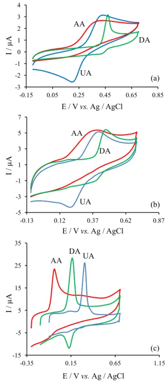
The results obtained under optimum conditions (pH 8.0) show that electrooxidation of ascorbic acid on the surface of the modified electrode occurs at a potential about 373 mV, less positive than that of at a bare GCE. The irreversible oxidation of ascorbic acid in pH 8.0 at a bare GCE occurs at 331 mV vs. Ag/AgCl (Figure 3a), while its oxidation peak at the modified electrode becomes about −0.042 mV vs. reference electrode (Figure 3c). The great increment of the current is mainly ascribed to the higher electroactive surface area of the poly(2,6-DAP)/MWNTs/ GCE. The poly(2,6-DAP) modified MWNTs/GCE before alkaline treatment had no electrocatalytic activity towards the electrooxidation of AA.
MWNTs could increase the surface area of the electrode, so the background current of the modified electrode is
higher than that of the bare surface (Figure 3b). In fact, the MWNTs/GC electrode can increase the current of the electrooxidation of AA but had no effect on decreasing the over-voltage of the electrooxidation of AA and also for DA and UA.
The oxidation of dopamine on the modified electrode is more reversible (∆E
p = 32 mV). As can be seen in
Figure 3a, cyclic volammogram of dopamine in pH 8.0 on the surface of a bare GC electrode showed a quasi-reversible behavior and the oxidation peak appeared at 411 mV vs. Ag/AgCl. Whereas, its oxidation peak potential at the surface of the modified electrode was shifted to 170 mV (Figure 3c). Also the oxidation of dopamine on the surface of poly(2,6-DAP) modified GCE before alkaline treatment is almost similar to a bare GCE.
The irreversible oxidation of uric acid in pH 8.0 at a bare GC electrode occurs at 465 mV vs. Ag/AgCl (Figure 3a), while its oxidation peak at the modified electrode becomes about 307 mV vs. reference electrode (Figure 3c). Again the poly(2,6-DAP) modified MWNTs/GC before alkaline treatment shows no catalytic effect on the electro-oxidation of uric acid.
The shift in potential values for oxidation of dopamine and uric acid at the surface of the modified GCE is smaller than that of ascorbic acid oxidation. Therefore, a separation of the oxidation peak potentials of dopamine, uric acid and ascorbic acid at the surface of the modified GCE occurred. Figure 3c shows that ascorbic acid, dopamine and uric acid are oxidized in pH 8.0 on the modified GCE at different potentials (Epa for oxidations
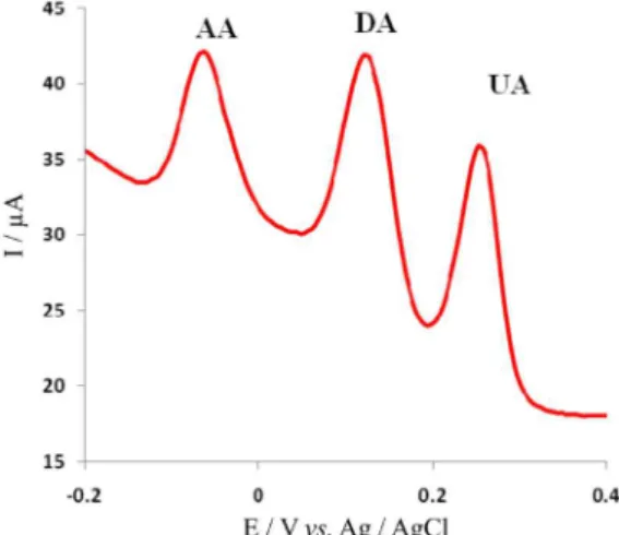
of ascorbic acid are 52 mV and for dopamine and uric acid 195 and 322 mV, respectively (vs. Ag/AgCl) with a suitable peak separation potentials which are about 143 and 127 mV between DA-AA and DA-UA, respectively). For simultaneous determination of DA, AA and UA, differential pulse voltammetry (DPV) was carried out in the potential range of −100 to 400 mV (Figure 4). Three well-defined peaks at about –63, 121 and 258 mV vs. Ag/AgCl were observed, corresponding to the differential pulse voltammograms of AA, DA, and UA, respectively. The
peak separations of 184, 147 and 321 mV between DA and AA, DA and UA, and UA and AA, respectively, allow us to detect DA, AA and UA simultaneously by using DPV. The value of this separation in peak potential depends on the pH value of the aqueous solution.
The influence of solution pH on the electrochemical responses of AA, DA and UA on the modified GCE was investigated by means of differential plus voltammetric method in 0.1 mol L-1 buffer solution at various pH values ranging
from 4.0 to 10.0. Further studies show that the oxidation peaks of dopamine and uric acid are separated in all testing pH (4.0 ≤ pH ≤ 10.0) and this separation for ascorbic acid and dopamine is maximum in lower pH values (Figure S3a), but maximum peak current for the oxidation of ascorbic acid, dopamine and uric acid occurred in the pH = 8 (Figure S3b). Therefore, pH 8.0 was selected as an optimum pH for determination of ascorbic acid, dopamine and uric acid.
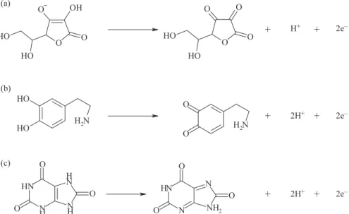
The electrocatalytic oxidation potential (Epa) of
DA and UA shifted to less positive potential by increasing pH (Figure S4) with a slope of –51.0 mV/pH and –57.0 mV/pH, respectively, being close to that expected for a monoelectronic/monoprotonic electrode reaction according to Nicholson equation42 (–59.2 mV/pH at 25 oC).
As DA and UA oxidation is a two-electron process, the number of protons involved is also predicted to be two. These accord with the mechanism of DA and UA oxidation (Scheme 2) as previously reported.43 The influence of the
solution pH in the electrochemical response of AA on the modified GCE showed that there is a linear relationship between electrocatalytic oxidation potential of AA and pH with the slop −33 mV/pH. This is expected for a dielectronic/monoprotonic electrode reaction and this accords with the mechanism of AA oxidation (Scheme 2), as the main form of AA (pKa1 = 4.7) is HA
–
in this pH range. Since differential pulse voltammetry has a much higher current sensitivity and better resolution than cyclic voltammetry, it was used in determination of AA, DA and UA concentration on the modified GCE and estimating the lower limit of detection. The oxidation peak currents of AA, DA and UA were measured in 0.10 mol L-1 pH 8.0 PBS, and
plotted against the bulk concentration of AA, DA and UA (Figure S5). The linear ranges for the determination of AA, DA, and UA using DPV were 2.70 × 10⁻⁵-1.83 × 10⁻³,
8.33 × 10⁻⁷-1.00 × 10⁻⁵ and 4.16 × 10⁻⁶-2.25 × 10⁻⁴ mol L-1,
respectively. The theoretical limits of detection defined as 3s of the proposed method for AA, DA and UA were 5.00 × 10⁻⁶, 4.16 × 10⁻⁸ and 7.00 × 10⁻⁷ mol L-1, respectively.
The relative standard deviations of 10 successive scans are 1.2, 1.4 and 1.3% for 1.0 mmol L-1 AA, 30 µmol L-1
DA and 90 µmol L-1 UA. The reproducibility of five different
electrodes was completed. The relative standard deviations
are 3.4, 3.9 and 4.5% for 1.0 mmol L-1 AA, 30 µmol L-1
DA and 90 µmol L-1 UA, respectively, which indicate that
the modified GCE had an excellent reproducibility. Table 1 shows a comparison between previously reported modified electrodes for determination of AA,
DA and UA and the poly(2,6-DAP)/MWNTs modified GC electrode. As can be seen, the proposed modified electrode shows somewhat similar (or worse) performances (in some cases) or superior ones (in most cases) than the previously reported modified electrodes.
Scheme 2. Mechanism of (a) AA, (b) DA and (c) UA oxidation at the modified GCE.
Table 1. Comparison of analytical parameters of several modified electrodes for AA, DA and UA determination
Electrode Method Analyte Linear range /
(µmol L-1)
Limit of detection / (µmol L-1)
Sensitivity /
(µA µmol-1 L) Reference
Modified carbon paste electrode by tetrabromo-p benzoquinone
DPV AA 10-600 0.62 0.005 44
DA 10-100 – 0.0074
UA 10-100 – 0.0022
Oxidation in mild acidic media CV AA 197-988 – – 46
DA 1.97-9.78 – –
UA 19.7-97.8 – –
Iron(II)-complex / MWNTs/GC DPV AA 11-1500 8 0.0118 35
DA 0.9-12000 0.2 0.059
UA 2-1500 1 0.0027
Pt/PF/Pd nano DPV AA in presence of ACOP 50-1000 7.1 5.92 50
DA in presence of ACOP 0.5-100 0.5 0.0213
CPE/CNF/Pd nano DPV AA 50-4000 15 – 49
DA 0.5-160 0.2 –
UA 2-200 0.7 –
Poly (3-(5-chloro-2-hydroxyphenylazo)-4,5dihydroxy naphthalene-2,7-disulfonic acid) film
DPV AA 5-240 1.43 0.013 45
DA 5-280 2.9 0.0157
UA 0.1-180 0.16 0.353
Novel choline and acetylcholine modified glassy carbon
DPV AA 7-90 0.9 – 48
DA 0.7-5 0.3 –
Dopamine solutions-phosphate buffer DPV AA 25-500 13 0.007 47
DA 1-20 0.11 0.006
UA 2.5-20 1.4 0.09
Poly(2, 6-DAP) filma /MWNTs/GC DPV AA 27-1830 5.0 – this work
DA 0.83-10.0 0.0416 –
UA 4.16-225 0.7 –
Interferences
For investigating the interferences, several compounds were selected. If the tolerance limit was taken as the maximum concentration of the foreign substances, which causes an approximately 5% relative error for 0.1 mmol L-1
AA, 25.0 µmol L-1 DA and 25.0 µmol L-1 UA, no interference
was observed for the following compounds (µmol L-1): K+,
Ca2+, Mg2+, Zn2+, starch, glutamic acid, tartaric acid and
glucose. The results are listed in Table 2.
Determination of AA in vitamin C injection, DA in dopamine hydrochloride injection solutions and UA in well water
In order to demonstrate the capability of this modified electrode for the catalytic oxidation of ascorbic acid, dopamine and uric acid in real samples, it was examined this ability in the voltammetric determination of AA and DA in some pharmaceutical preparation, such as vitamin C injection solution (standard content 100 mg per mL AA, 5 mL per injection) (Daro Pakhsh Co.) and dopamine hydrochloride injection (DHI) solution (standard content
of 40 mg per mL DA, 5 mL per injection) (Rasht Co.). The proposed modified electrode was also successfully applied to the determination of UA in spiked solution. Several spiked samples were prepared by adding aliquots of UA solution to Koshkan (a village near to Zanjan City in Iran) well water. All samples were diluted with phosphate buffer solution (pH 8.0) and then appropriate amounts of these diluted samples were transferred to the electrochemical cell to determine each species using DPV.
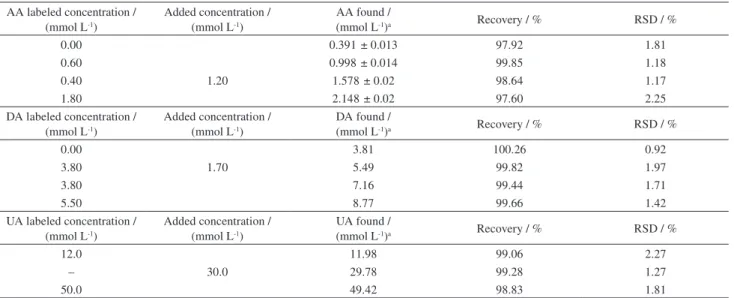
The standard addition technique was employed for AA, DA and UA determination. The results of AA, DA and UA determinations in the real samples and spiked samples with AA, DA or UA standard solutions are shown in Table 3. The recovery and precision were acceptable, revealing that the modified electrode could be efficiently applied for the determination of AA, DA and UA in pharmaceutical samples.
Conclusions
In this work, the advantageous features of polymerized film of 2,6-diaminopyridine (2,6-DAP) are demonstrated as electron transfer mediator onto a GC electrode surface. Due to the chemical stability, electrochemical reversibility and high electron transfer rate constant of the modified GCE, it can be used in electrocatalysis as electron transfer mediators to shuttle electrons between analytes and substrate electrodes. The modified electrodes showed excellent electrocatalytic ability for the oxidation of ascorbic acid, dopamine and uric acid. The ternary mixture of them can be well separated from each other at the surface of this modified GCE. Differential pulse voltammetry at the
Table 2. Interferences of some foreign substances for 0.1 mmol L-1 AA, 25.0 µmol L-1 DA and 25.0 µmol L-1 UA
Foreign substances Tolerance level / (µmol L-1)
Tartaric acid 300
K+, Mg2+, Ca2+, Zn2+ 250
Citric acid 10
Glutamic acid 250
Glucose 2500
Table 3. Recovery results obtained for determination of AA, DA and UA and the spiked of them in injection solutions (n = 5) AA labeled concentration /
(mmol L-1)
Added concentration / (mmol L-1)
AA found /
(mmol L-1)a Recovery / % RSD / %
0.00 0.391 ± 0.013 97.92 1.81
0.60 0.998 ± 0.014 99.85 1.18
0.40 1.20 1.578 ± 0.02 98.64 1.17
1.80 2.148 ± 0.02 97.60 2.25
DA labeled concentration / (mmol L-1)
Added concentration / (mmol L-1)
DA found /
(mmol L-1)a Recovery / % RSD / %
0.00 3.81 100.26 0.92
3.80 1.70 5.49 99.82 1.97
3.80 7.16 99.44 1.71
5.50 8.77 99.66 1.42
UA labeled concentration / (mmol L-1)
Added concentration / (mmol L-1)
UA found /
(mmol L-1)a Recovery / % RSD / %
12.0 11.98 99.06 2.27
– 30.0 29.78 99.28 1.27
50.0 49.42 98.83 1.81
modified GCE allows sensitive and selective determination of these biological compounds in the presence of common interferences in an aqueous solution.
Supplementary Information
Supplementary data are available free of charge at http://jbcs.sbq.org.br as PDF file.
Acknowledgement
The authors wish to express their gratitude to the Zanjan University Research Council for the support of this work.
References
1. Wang, J.; Electroanalysis 1991, 3, 255.
2. Ohnuki, Y.; Ohsaka, T.; Matsuda, H.; Oyama, N.; J. Electroanal. Chem.1983, 158, 55.
3. Volkov, A.; Tourillon, G.; Lacaze, P. C.; Dubois, J. E.;
J. Electroanal. Chem.1980, 115, 279.
4. Chung, T. D.; Biosens. Bioelectron.2001, 16, 1079. 5. Trojanowicz, M.; Microchim. Acta2003, 143, 75. 6. Vidal, J.-C.; Microchim. Acta2003, 143, 93.
7. Gerard, M.; Chaubey, A.; Malhotra, B.; Biosens. Bioelectron.
2002, 17, 345.
8. Saxena, V.; Malhotra, B. D.; Curr. Appl. Phys.2003, 3, 293. 9. Malitesta, C.; Palmisano, F.; Torsi, L.; Zambonin, P. G.; Anal.
Chem.1990, 62, 2735.
10. Centonze, D.; Guerrieri, A.; Malitesta, C.; Palmisano, F.; Zambonin, P. G.; Fresenius J. Anal. Chem.1992, 342, 729. 11. Bartlett, P. N.; Caruana, D. J.; Analyst1992, 117, 1287. 12. Groom, C. A.; Luong, J. H. T.; Anal. Lett.1993, 26, 1383. 13. Lowry, J. P.; McAteer, K.; El Atrash, S. S.; Duff, A.; O’Neill,
R. D.; Anal. Chem.1994, 66, 1754.
14. Palmisano, F.; Guerrieri, A.; Quinto, M.; Zambonin, P. G.; Anal. Chem.1995, 61, 1005.
15. Santhosh, P.; Gopalan, A.; Vasudevan, T.; Lee, K. P.; Appl. Surf. Sci.2006, 252, 7964.
16. Golabi, S. M.; Nozad, A.; Electroanalysis2003, 15, 278. 17. Nozad, A. G.; Golabi, S. M.; Maragheh, M. G.; Irannejad, L.;
J. Power Sources 2005, 145, 116.
18. Pournaghi-Azar, M. H.; Habibi, B.; J. Electroanal. Chem.2007,
601, 53.
19. Jang, D.-H.; Bull. Korean Chem. Soc.1995, 16, 392. 20. Martinusz, K.; Láng, G.; Inzelt, G.; J. Electroanal. Chem.1997,
433, 1.
21. Somasundrum, M.; Aoki, K.; J. Electroanal. Chem.2002, 530, 40. 22. Griffith, A.; Glidle, A.; Cooper, J. M.; Biosens. Bioelectron.
1996, 11, 625.
23. Yao, T.; Anal. Sci.2003, 19, 61.
24. Liu, A.; Honma, I.; Zhou, H.; Biosens. Bioelectron.2007, 23, 74.
25. Lin, X.; Zhuang, Q.; Chen, J.; Zhang, S.; Zheng, Y.; Sens. Actuators, B: Chemical 2007, 125, 240.
26. Ozcan, L.; Sahin, M.; Sahin, Y.; Sensors 2008, 8, 5792. 27. Yao, H.; Suna, Y.; Lin, X.; Tang, Y.; Huang, L.; Electrochim.
Acta2007, 52, 6165.
28. Gopalan, A. I.; Lee, K.-P.; Manesha, K. M.; Santhosh, P.; Kim, J. H.; Kang, J. S.; Talanta 2007, 71, 1774.
29. Zare, H. R.; Nasirizadeh, N.; Ardakani, M. M.; J. Electroanal. Chem.2005, 577, 25.
30. Lin, L.; Chen, J.; Yao, H.; Chen, Y.; Zheng, Y.; Lin, X.;
Bioelectrochemistry2008, 73, 11.
31. Sudhakara Prasad, K.; Muthuraman, G.; Zen, J.-M.;
Electrochem. Commun. 2008, 10, 559.
32. Lin, X.; Zhuang, Q.; Chen, J.; Zhang, Sh.; Zheng, Y.; Sens. Actuators, B: Chemical2007, 125, 240.
33. Yao, H.; Sun, Y.; Lin, X.; Tang, Y.; Huang, L.; Electrochim. Acta2007, 52, 6165.
34. Jiao, Sh.; Li, M.; Wang, C.; Chena, D.; Fang, B.; Electrochim. Acta2007, 52, 5939.
35. Kamyabi, M. A.; Narimani, O.; Monfared, H. H.; J. Braz. Chem. Soc.2011, 22, 468.
36. Morea, G.; Guerrieri, A.; Malitesta, C.; Torsi, L.; J. Chem. Soc., Faraday Trans. 1991, 87, 3515.
37. Kamyabi, M. A.; Narimani, O.; Monfared, H. H.; J. Electroanal. Chem.2010, 642, 67.
38. Dai, H. P.; Wu, Q. H.; Sun, S. G.; Shiu, K. K.; J. Electroanal. Chem. 1998, 456, 47.
39. Yu, B.; Khoo, S. B.; Electrochim. Acta2005, 50, 1917. 40. Bacon, J.; Adams, R. N.; J. Am. Chem. Soc.1968, 90, 6596. 41. Bilal, S.; Holze, R.; J. Electroanal. Chem. 2006, 592, 1. 42. Nicholson, R. S.; Anal. Chem. 1965, 37, 1351.
43. Zhao, Y.; Gao, Y.; Zhan, D.; Hui, H.; Zhao, Q.; Kou, Y.; Shao, Y.; Li, M.; Zhuang, Q.; Zhu, Z.; Talanta2005, 66, 51. 44. Kalimuthu, P.; John, S. A.; Bioelectrochemistry2009, 77, 13. 45. Poh, W. C.; Loh, K. P.; Zhang, W. D.; Triparthy, S.; Ye, J. S.;
Shen, F. S.; Langmuir2004, 20, 5484.
46. Antiochia, R.; Lvagnini, I.; Magno, F.; Valentini, F.; Palleschi, G.; Electroanalysis2004, 16, 1451.
47. Wang, J. X.; Li, M. X.; Shi, Z. J.; Li, N. Q.; Gu, Z. N.; Anal. Chem. 2002, 74, 1993.
48. Wang, L.; Wang, J. X.; Zhou, F. M.; Electroanalysis2004, 16, 627.
49. Wang, J.; Peng, T.; Vince, V.; J. Electroanal. Chem. 1987, 234, 119.
50. Fei, J.; Wu, K.; Yi, L.; Li, J.; Bull. Korean Chem. Soc. 2005,
26, 1403.
Submitted: April 10, 201