Histopathological features of post-mortem pituitaries:
A retrospective analysis
FRANCISCO JOSÉ TORTOSA VALLECILLOS1*, SANTIAGO ORTIZ FERNÁNDEZ2
1MD, specialist in Pathological Anatomy – Department of Pathology, Centro Hospitalar Lisboa Norte, Lisboa. Lecturer, Faculdade de Medicina, Universidade de Lisboa. PhD Student at the Department of Medicine, Universitat Autònoma de Barcelona (UAB), Barcelona, Spain
2MD, specialist in Pathological Anatomy – Department of Pathology, Centro Hospitalar Lisboa Norte, Lisboa. Lecturer, Faculdade de Medicina, Universidade de Lisboa, Lisbon, Portugal
SUMMARY
Study conducted at Centro Hospitalar Lisboa Norte, EPE, Hospital de Santa Maria, Lisbon, Portugal Article received: 3/5/2015 Accepted for publication: 3/25/2015 *Correspondence: Serviço de Anatomia Patológica CHLN, EPE, Hospital de Santa Maria Address: Av. Prof. Egas Moniz Postal code: 1649-035 Lisboa – Portugal [email protected] http://dx.doi.org/10.1590/1806-9282.62.05.399
Objective: As a result of the use of neuroimaging techniques, silent pituitary le-sions are diagnosed more and more frequently; however, there are few published post-mortem studies about this gland. Incidence data of pituitary lesions are rare and in Portugal they are outdated or even non-existent. The aim of this study is to determine the prevalence of normal patterns and incidental post-mortem pi-tuitary pathology at Centro Hospitalar Lisboa Norte, analyzing the associations with clinical data and assessing the clinical relevance of the indings.
Method: We reviewed retrospectively and histologically 167 pituitaries of a con-secutive series of autopsies from the Department of Pathology of this centre. They were done between 2012 and 2014, and in all cases medical records were reviewed. The morphological patterns observed, were classiied into three major groups: 1) Normal histological patterns and variants; 2) Infectious-inlamma-tory pathology, metabolic and vascular disorders; 3) Incidental primary prolif-eration and secondary to systemic diseases.
Results: The subjects included in this study were of all age groups (from 1 day to 91 years old), 71 were female and 96 male. Fifty-seven of these glands didn’t show any alteration; 51 showed colloid cysts arising from Rathke cleft; 44 pre-sented hyperplasia in adenohypophysis and we identiied 20 adenomas in 19 glands (immunohistochemically, eight PRL-producing and ive ACTH-produc-ing tumors), ten of which associated with obesity, 11 to hypertension and six to
diabetes mellitus. There were two cases with metastasis.
Conclusion: Subclinical pathology in our country is similar to that seen in oth-er parts of the world, but at oldoth-er ages.
Keywords: pathology, pituitary gland, autopsy.
I
NTRODUCTIONStudies on the incidence and prevalence of pituitary ad-enomas (PA) and related injuries have varied over time and depending on the population studied. These varia-tions are related to advances in health including increased access to modern imaging studies and the increased num-ber of specialists in endocrinology. As a result of the de-velopment and widespread use of neuroradiology imag-ing studies, namely computed tomography (CT) and magnetic resonance imaging (MRI), silent pituitary le-sions are diagnosed increasingly often.1-3 In a recent re-view of autopsy and MRI studies, the estimated global
prevalence of PA was 16.7%.4 Recently it has been recog-nized that the prevalence of clinically diagnosed PA is 3.5 to 5 times more common than previously thought, ac-cording to a Belgian study that showed a prevalence of approximately 1 per 1,000 persons.5
atrophy, etc.) that may go unnoticed for long periods of time or even fail to be diagnosed due to their limited clinical signiicance. The incidental discovery of PA has become a subject of growing interest. However, the fre-quency with which systemic diseases (granulomatous inlammation, metabolic processes, metastatic tumors, etc.) can affect secondarily the structure of this gland is not known.2,7,8
Data on incidence of PA in the population are scarce, and the series based on MRI and autopsies are discordant with the surgical series in tertiary centers. Since the prev-alence of values in different ethnic groups or populations is unclear, further studies are needed community-based, deining the real burden of pituitary lesions in the usual clinical practice, as well as geographical differences.
In Portugal, data on pituitary disease are scarce, ob-solete or non-existent, which means that the national health system lacks reliable and updated epidemiologi-cal data to ensure adequate resource allocation, propor-tional to the impact of such tumors in the community.
Most post-mortem studies were done in forensic au-topsies and many of them recommended, due to the high frequency of occult pituitary lesions found, anatomical and clinical autopsy studies in order to evaluate the as-sociated clinical features.9
The objective of this study is to determine the prev-alence of normal patterns and incidental post-mortem pituitary pathology in a consecutive series of patients of Centro Hospitalar Lisboa Norte (CHLN) – the larg-est referral center in Portugal –, which includes Hospi-tal Santa Maria and HospiHospi-tal Pulido Valente in Lisbon, analyzing possible associations with the available clin-ical data and highlighting the medclin-ical relevance of the indings.
M
ETHODFor the preparation of this work, 167 pituitary glands of a consecutive series of autopsies conducted by the Path-ological Anatomy Service of CHLN were studied retro-spectively. Autopsies were conducted between 1/1/12 and 12/31/14. Pediatric necropsies (newborns and children) were included.
In all cases examined, the corresponding medical re-cords were reviewed, with analysis of the following vari-ables: age, gender, reason for inclusion, personal medical history and causes of death, including in the case of neo-natal autopsies, the study of the placenta.
During the autopsy, the pituitary stalk was cut as high as possible to leave the gland intact. The procedure involved opening the sellar diaphragm and fracturing the
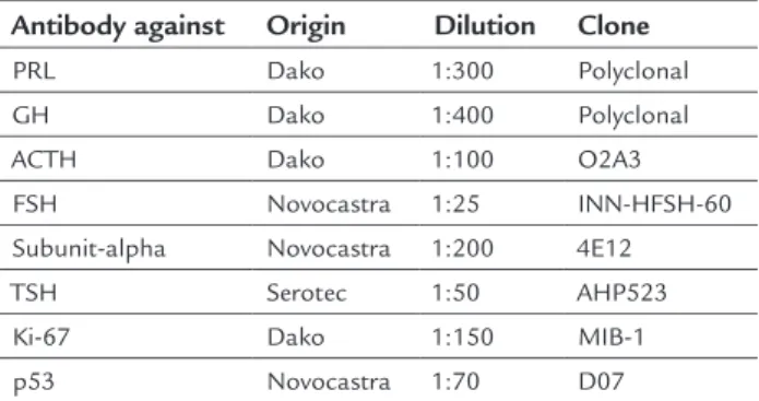
dorsum of the sella turcica, subsequently pushed to al-low the gland to be removed intact. Immediately after ex-traction, the pituitary glands were ixed in 10% buffered formaldehyde for at least 24 hours and up to 72 hours. Subsequently, the glands were assessed macroscopically, weighed, measured, cut sagittally, and processed accord-ing to the usual technical procedures for ixaccord-ing in paraf-in blocks for a complete histological evaluation. Sections were cut at 2 micra for hematoxylin-eosin (H&E) stain-ing, 4 micra for reticulin staining (Gomori’s method), and 2 micra for immunohistochemical study, if necessary. In the latter case, the glands were deparafinized, subjected to antigen retrieval and incubated with individual anti-bodies directed against speciic pituitary hormones or cellular proteins (Table 1).
TABLE 1 Antibodies used, origin, dilution and clone.
Antibody against Origin Dilution Clone
PRL Dako 1:300 Polyclonal
GH Dako 1:400 Polyclonal
ACTH Dako 1:100 O2A3
FSH Novocastra 1:25 INN-HFSH-60
Subunit-alpha Novocastra 1:200 4E12
TSH Serotec 1:50 AHP523
Ki-67 Dako 1:150 MIB-1
p53 Novocastra 1:70 D07
PRL: prolactin; GH: growth hormone; ACTH: adrenocorticotropic hormone; FSH: follicle-sti-mulating hormone; TSH: thyroid-stifollicle-sti-mulating hormone.
The preparations were stained with H&E and reticulin, as well as pituitary hormones (PRL, GH, ACTH, FSH, sub-unit-alpha, and TSH) or Ki-67 (indicative of cell prolifer-ation) and p53 (tumor suppressor gene) where necessary. PAs found were sorted according to the 2004 version of the World Health Organization (WHO) classiication of tumors of endocrine organs.10
Cases in which necropsy was partial and limited to the thoracic and/or abdominal cavity; cases in which the time elapsed from death to completion of the examina-tion was over 24 hours; and those cases in which the corpse was in poor conditions, and the glands showed evidence of autolysis were not included in this study.
We deined as pituitary incidentalomas all unexpect-ed pituitary sellar lesions found during the necropsy due to any unrelated reason. This deinition excludes subjects who experienced symptoms described as typical for PA, including visual changes and syndromes of defective or excessive pituitary hormone secretion.
the observed morphological patterns were classiied into three groups:
1. Histologically normal patterns and variants (develo-pmental anomalies and cystic lesions);
2. Infectious-inlammatory disease, metabolic disorders and vascular disorders;
3. Incidental primary proliferation and secondary to systemic diseases.
R
ESULTSGender distribution was 71 (42.5%) females and 96 (57.5%) males, with 153 corpses of white race and 14 colored. Age ranged between 1 day and 91 years; 22.8% of the autop-sies were conducted on subjects between 70 and 79 years old (Chart 1). All autopsies derived from the different Ser-vices offered at the CHLN.
Histologically normal patterns and variants
Fifty-seven (34.1%) of the glands examined showed no histologic abnormalities with possible pathological sig-niicance, 20 of which (out of 24) accounted for
autop-sies in individuals under 18 years. Considering only adult glands (aged between 18 and 65 years), the average weight after ixation was 0.74+/-0.25 g, and the average size of their longest axis was 13.94+/-1.72 mm.
Fifty-one of the 167 glands studied (30.5%) had col-loid cysts derived from Rathke’s cleft. In all cases, the cysts were located in the intermediate lobe; in two subjects, they were also located in the pars tuberalis. These were single cysts (in 11 of the glands) or, more frequently, multiple (40 cases; 78.4%) small cysts between less than 1 and 9 mm. Three percent of the glands studied (ive cases) had inframillimetric foci of squamous metaplasia with glan-dular remnants. They were observed in groups, mainly in the pituitary stalk and, less often, in the pars tuberalis.
In 23 of the 167 cases (13.8%), rows of basophil en-docrine cells immunohistochemically positive for ACTH were observed extending from the interface of the ante-rior and posteante-rior lobes to the depth of the neurohypoph-ysis. All cases were related to individuals older than 50 years (except for the gland of a 23 years old subject).
Herring bodies were also found in the neurohypoph-ysis of two males (73 and 82 years old).
CHART 1 Distribution of the number of autopsies by gender and age range. 0-9 year
s
10-19 year
s 20-29 year
s 30-39 year
s 40-49 year
s 50-59 year
s 60-69 year
s 70-79 year
s 80-89 year
s 90-99 year
s
Number of cases
Female 25
20
15
10
5
0
In one gland in this series, we observed a concentric arrangement of unmyelinated nerve ibers comprising the neurohypophysis, which gave it a nodular appearance that reminded schwannomas (“nodular” neurohypoph-ysis). It measured 1 mm and was located at the interface of the pars nervosa up to the infundibulum; there was no association with cellular pleomorphism, anisokaryosis, mitosis or other changes.
The cells in the neurohypophysis had granular trans-formation in three subjects, one female and two males, aged 62-78 years, one with amyloidosis and two with chronic renal failure (CRF).
None of the autopsies performed revealed atrophy or lattening of the pituitary gland similar to that described in empty sella syndrome (intrasellar arachnoidocele that occurs as a result of a change in the diaphragm),11,12 and no cases of pan-hypopituitarism was found in the review of clinical histories.
None of the indings correlated with clinical symp-toms that were mentioned in the medical history.
Infectious-inlammatory disease, metabolic disorders and vascular disorders
The presence of isolated lymphocytic foci within the ad-enohypophysis was a inding found in six of 167 autop-sies (3.6%). In these cases, the subjects were between 61 and 90 years old (except for a patient aged 39 years), and lymphocytes were mature and positioned as isolated cells, not causing destruction of the gland. Autoimmune or lymphoproliferative diseases were not constant.
There was nonspeciic chronic inlammation in two newborns whose placentas revealed infection and hypox-emia, and in a 70-year-old with a history of epilepsy and meningitis. There was also an active chronic periglandu-lar inlammatory iniltrate in a 75-year-old who died due to complications of a lu-like illness.
The presence of intracellular deposits of pigment in the neurohypophysis was a relatively common inding (six of 167 autopsies). They are dispersed in the paren-chyma and were associated in two cases with cerebrovas-cular accident (stroke) and hemolysis; in other cases, no relationship was found.
We must highlight the incidence of pituitary vascu-lar disease in this series of clinical autopsies (3%); ive of the examined glands had ischemic strokes. Age ranged between 41 and 82 years. In all cases, the review of the pa-tients’ medical history showed vascular risk factors such as high blood pressure (hypertension), diabetes mellitus
(DM) or stroke, and more.
The presence of benign vascular proliferations was not observed in any of the 167 cases.
Incidental primary proliferation and secondary to systemic diseases
Forty-four of the studied pituitary glands (26.3%) showed adenohypophysis hyperplasia, with expansion of the aci-ni demonstrated by reticulin histochemical staiaci-ning. In some cases there was a focal pattern, and in others a dif-fuse pattern, involving one or more cell types. Sixteen of the affected subjects were women and 28 men. Age ranged from 23 to 91 years. Considering only the glands of adults (aged 18 to 65 years), the mean size after ixation was 14.69+/-2.02 mm, which implies a slight increase with
re-spect to the average normal pituitary.
Since we did not ind in the clinical histories of pa-tients whose pituitary glands had hyperplasia any data that could be related to etiology, these cases were con-sidered as asymptomatic or incidental idiopathic hyper-plasia.
Twenty “typical” PAs were found in 19 subjects (one gland with two tumors), all smaller than 10 mm (mi-croadenomas). Nine subjects were female (12.7% of the total cases in females) and 11 male (11.5% of the total males). Age ranged from 10 to 88 years, with 14 cases (70%) between the 7th and 9th decades of life. Immuno-histochemically, four were PRL-producing tumors, two PRL/GH, one PRL/GH/FSH, one PRL/GH/TSH, one GH/TSH, ive ACTH (one of which with pituitary apo-plexy), one FSH, three plurihormonal tumors, and two
null-cell. Clinically, 10 cases were associated with obe-sity (one morbid), 11 HBP and six DM; there were no cases associated with hypo- or hyperthyroidism. Atyp-ical cases of PA or primary pituitary carcinomas were not identiied.
The PAs were not large enough to produce clinical symptoms. After the clinical histories were reviewed, none of the patients with lesions described had information pointing to compressive, visual or endocrine symptoms that could be related to the pathologic indings.
D
ISCUSSIONKeep in mind that the collection of data from the 167 subjects was retrospective, and clinicopathological cor-relation is based on data previously collected on clinical history. Thus, it is possible that this was not an accurate and comprehensive relection of the patient’s situation, or that the pituitary disease generated symptoms that were not so relevant within the context of morbidity, which implies a limitation to this study.
We must also consider that the sample size does not allow for statistical signiicance necessary to some of the indings, and that we can only assume the trends. Never-theless, interest is still great since these data derive from a series of anatomical and clinical autopsies.
Since the pediatric population is included, as well as a wide range of subjects of different age groups, we consider this a representative sample of the general population.
Within the patterns of histological normality and vari-ants, our study showed in normal pituitary glands: col-loid cysts, foci of squamous metaplasia, rows of endocrine cells in the neurohypophysis, Herring bodies, nodular transformation of the neurohypophysis and modiication of its cells, adopting aspect of granular cells similar to those observed in other organs.
Colloid cysts are relatively frequent, small and asymp-tomatic. These remnants of Rathke’s cleft are common incidental indings in autopsies. Studies in normal pitu-itary gland demonstrated that they appear in 32% of cas-es13 (30.5% in our series); of these, 80% (100% in our se-ries) were located in the intermediate lobe and the other in pars tuberalis.
Sometimes, the cysts are large enough to produce symptoms due to compression of the pituitary gland, op-tic chiasm and hypothalamus.14 Sometimes the infection of a cyst can cause the formation of abscesses.15 These cysts are benign and usually heal after they are excised.16 Subjects who undergo partial excision and drainage may have recurrences.
Squamous metaplasia of glandular remains is an adap-tive process that appears with some frequency (3% in our series). Sometimes it leads to the formation of cysts large enough to produce symptoms,17 which are primarily visu-al. Resection is often incomplete since it can affect vital structures, but relapse occurs very slowly over the years. Malignant transformation to squamous cell carcinoma is rare, but can occur.18
The presence of rows of ACTH-immunoreactive ba-sophil cells in the posterior pituitary (“baba-sophil invasion”) is an age-related inding not customarily associated with endocrinopathies.
Sometimes it is possible to discern using H&E in the neurohypophysis swollen axons that store oxytocin or va-sopressin, with eosinophilic ibrillar appearance, which are called Herring bodies or neurosecretory bodies and constitute a normal inding.
Aggregates of polygonal cells with granular cytoplasm in the neurohypophysis or infundibulum are incidental indings at autopsy.19 They are usually asymptomatic but, if they grow, they can compress the gland, the optic chi-asm or the hypothalamus.
Not often, infectious-inlammatory disease, meta-bolic disorders and vascular disorders can occur, which may affect the primary or secondary structure of this gland. In our study, we highlight the presence of isolat-ed lymphocytic foci, nonspeciic inlammation, ferric pigment deposits in the posterior pituitary, and some ischemic strokes.
Occasionally, a small number of lymphocytes can be seen on the interface between the anterior and posterior lobes of the pituitary.20 Histologically, the cells are easily distinguished from the extensive and destructive iniltra-tion observed in lymphocytic hypophysitis. Microfoci of lymphocytes have no clinical signiicance. Lymphocytic hypophysitis, by contrast, is a well-described inlamma-tory process,21 which can resemble PAs clinically and ra-diologically. Approximately 80% of cases occur in preg-nancy, mostly postpartum, and they are characterized by total or partial pituitary insuficiency due to an autoim-mune process,22 with men affected on rare occasions.
The presence of intracellular deposits of ferric pig-ment in macrophages of the neurohypophysis (conirmed in our series by histochemical study using Perls staining) was a relatively common inding (observed in six of 167 autopsies). These deposits seem to indicate minor bleed-ing similar to those that occur in other organs durbleed-ing small coagulation disorders, hypersideremia or mild lo-cal trauma.7 We did not ind in our study a possible asso-ciation between some sort of speciic pathology and the presence of such deposits. Given our sample size, it is dif-icult to know whether this is a signiicant inding or if it is caused by chance.
In our study, the ive subjects with ischemic infarcts also had risk factors (obesity, HBP, DM and/or stroke). Age range was wide, between 41 and 82 years, although
three of these ive subjects (60%) were relatively young (41, 43 and 56 years old). However, it is noted that for clinical
As for pituitary incidental primary proliferation and secondary to systemic diseases, we point out the occur-rence of hyperplasia, adenomas and metastases.
Although in the past it was believed that the focal hyperplasia did not occur in the pituitary gland, cur-rently there is no doubt that it exists and that it may even appear clinically and biochemically.24,25 In this series, ac-inar hyperplasia was observed in 44 of the 167 pituitary glands. The medical history of these subjects did not in-clude information (either clinical or biochemical) that could be connected to the etiology of hyperplasia. While hyperplasia is a well-described phenomenon, there are still questions that remain without clear answer: Are all forms of hyperplasia primary or they result from hypo-thalamic changes? Are these cellular adaptation process-es leading to adenomas? Are hyperplastic cells more prone to malignant transformation? What are the mo-lecular mechanisms related to this hyperplasia?26,27 The predominance of males (63.6% of cases) in our series stands out, contrary to what is classically described for adenomas.
PAs comprise 10-20% of all intracranial tumors.4 They affect mainly women between the 3rd and 6th decade, but can occur in any age group.28,29 Small incidental adeno-mas can occur with a probability of 20% in pituitary glands examined at autopsy.30,31
The incidence of adenomas in this study is consis-tent with that found in other series. Twenty adenomas (12.1%) were found; other studies4,32 show incidences at 10-20%. Coinciding with the classically described in the literature, women were slightly more affected than men. In our study series, ages ranged from 30 to 88 years (ex-cept for a 10 year old child), with 14 cases between the 7th and 9th decades of life, slightly higher than that found in other reviews.1,8,30 The most frequently found adeno-mas are prolactinoadeno-mas.30,32,33 In our series, of the 20 PAs founds, four were PRL-producing tumors, other four cases were PRL/GH, and ive corresponded to ACTH-producing adenomas. If we compare these data with the analysis of the surgical series held between 2004 and 2013 in the same hospital, in which 220 PAs were ana-lyzed, we observe a prevalence of adenomas positive for GH (60) and FSH (59), followed by ACTH-producing tu-mors (44). Only 24 adenomas were PRL-secreting (a good response to medical treatment lead to low prevalence in surgical series).
The pathogenesis of apoplexy, an uncommon ind-ing, is also little known. Pituitary apoplexy is deined as the sudden manifestation of symptoms such as severe headache, nausea, vomiting, loss of vision, paralysis of
the cranial nerves and altered consciousness with radio-logic evidence of hemorrhagic stroke in PA.34 Several neu-rosurgical series indicate that the incidence of pituitary apoplexy ranges from 2 to 7% when the clinical signs are associated with histopathological evidence of hemor-rhage or necrosis. All kinds of gland tumors have simi-lar risk of apoplexy. Men are more affected than women (2:1). Randeva et al. conducted a retrospective study of cases of pituitary apoplexy in order to establish the clin-ical presentation, predisposing factors, treatment and patient outcome.34 They concluded that the most com-mon symptom is headache, and that HBP may be an im-portant predisposing factor. In another similar study, also retrospective, carried out by Da Motta et al., the au-thors concluded that pituitary apoplexy is not an infre-quent complication of adenomas.35 Only one of the 20 observed adenomas (ACTH-secreting) presented this phenomenon in a 30-year-old man with HBP.
Comparatively, primary tumors of the neurohypoph-ysis are rare and, in general, similar to primary tumors of the central nervous system. This is conirmed in our study, which found no cancer in this part of the gland.
Two of the 25 cases of cancer in different organs and tissues (8%) had pituitary metastases (one LL/CLL and one lung carcinoma). Other series showed similar inci-dence, between 2 and 25%,36-38 although most metastat-ic tumors are clinmetastat-ically asymptomatmetastat-ic. It should be not-ed that breast and lung cancers are the most common primary neoplasms leading to metastases in the pitu-itary.39-43 In this study, there were only two cases of lung tumors (one of which metastasized) and no cases of breast cancer.
C
ONCLUSIONThe prevalence of pituitary pathology found in our midst in this series of anatomical and clinical autopsy is simi-lar to that described elsewhere in the world. However, con-trary to what has been described in the literature, we have observed an increase in age at diagnosis. As seen in oth-er studies, post-mortem pituitary adenomas predomi-nate slightly among women and strongly differ from those found in surgical series regarding types of adenoma.
To our knowledge, this is the irst comprehensive and updated study to estimate the prevalence of post-mortem incidental pituitary pathology conducted in Portugal.
A
CKNOWLEDGMENTSR
ESUMOCaracterísticas histopatológicas de hipóises post mortem: análise retrospectiva
Objetivo: como resultado da utilização de técnicas de neuroimagem, cada vez se diagnosticam mais lesões hi-poisárias silentes; porém, há poucos estudos post mortem
publicados sobre essa glândula. Os dados de incidência existentes sobre lesões hipoisárias são raros, sendo em Portugal desatualizados ou inexistentes. O objetivo é de-terminar a prevalência dos padrões normais e da patolo-gia hipoisária incidental post mortem no Centro Hospita-lar Lisboa Norte, analisando as associações com dados clínicos e avaliando a relevância clínica dos achados.
Método: revisaram-se histologicamente de forma retros-pectiva 167 hipóises de uma série consecutiva de autóp-sias do Serviço de Anatomia Patológica desse centro, rea-lizadas entre 2012 e 2014, sendo revisadas em todos os casos as histórias clínicas. Os padrões morfológicos ob-servados classiicaram-se em três grandes grupos: 1) pa-drões histológicos de normalidade e variantes; 2) patolo-gia infeccioso-inlamatória, distúrbios metabólicos e transtornos vasculares; 3) proliferação primária inciden-tal e secundária a doenças sistêmicas.
Resultados: os doentes incluíam todas as faixas etárias (de 1 dia a 91 anos), sendo 71 do sexo feminino e 96 do masculino. Cinquenta e sete das glândulas não apresen-taram qualquer alteração; 51 mostraram cistos coloides derivados da issura de Rathke; em 44, observou-se hiper-plasia da adeno-hipóise e identiicaram-se 20 adenomas em 19 glândulas (oito imuno-histoquimicamente produ-tores de PRL e cinco de ACTH), dos quais dez associados à obesidade, 11 à hipertensão arterial e seis a diabetes mel-litus. Houve dois casos com metástases.
Conclusão: a patologia subclínica em nosso meio é simi-lar à observada em outras partes do mundo, mas em ida-des mais avançadas.
Palavras-chave: patologia, hipóise, autopsia.
R
EFERENCES1. Teramoto A, Hirakawa K, Sanno N, Osamura Y. Incidental pituitary lesions in 1000 unselected autopsy specimens. Radiology. 1994; 193(1):161-4. 2. Sanno N, Oyama K, Tahara S, Teramoto A, Kato Y. A survey of pituitary
incidentaloma in Japan. Eur J Endocrinol. 2003; 149(2):123-7.
3. Vernooij MW, Ikram MA, Tanghe HL, Vincent AJ, Hofman A, Krestin GP, et al. Incidental indings on brain MRI in the general population. N Engl J Med. 2007; 357(18):1821-8.
4. Ezzat S, Asa SL, Couldwell WT, Barr CE, Dodge WE, Vance ML, et al. The prevalence of pituitary adenomas: a systematic review. Cancer. 2004; 101(3):613-9.
5. Daly AF, Rixhon M, Adam C, Dempegioti A, Tichomirowa MA, Beckers A. High prevalence of pituitary adenomas: a cross-sectional study in the province of Liege, Belgium. J Clin Endocrinol Metab. 2006; 91(12):4769-75. 6. Fainstein Day P, Guitelman M, Artese R, Fiszledjer L, Chervin A, Vitale NM,
et al. Retrospective multicentric study of pituitary incidentalomas. Pituitary. 2004; 7(3):145-8.
7. Sano T, Rayhan N, Yamada S. Pathology of pituitary incidentaloma. Nippon Rinsho. 2004; 62(5):940-5.
8. Hurley DM, Ho KK. MJA Practice Essentials-Endocrinology. 9: Pituitary disease in adults. Med J Aust. 2004; 180(8):419-25.
9. Kisungi S. The prevalence and classiication of occult pituitary lesions at autopsy in Kenyatta National Hospital, City Mortuary and Armed Forces Memorial Hospital in Nairobi. [Dissertation]. Nairobi: Department of Human Pathology, University of Nairobi; 2010.
10. Lloyd RV, Kovacs K, Young Jr WF, Farrel WE, Asa SL, Trouillas J, et al. Tumours of the pituitary gland. In: DeLellis RA, Lloyd RV, Heitz PU, Eng C (eds.). Pathology and genetics of tumours of endocrine organs. WHO/ IARC Classiication of Tumours. 3.ed. v.8. Lyon: IARC Press; 2004. p.9. 11. Jaffer KA, Obbens EA, El Gammal TA. “Empty” sella: review of 76 cases.
South Med J. 1979; 72(3):294-6.
12. Ammar A, Al-Sultan A, Al Muhim F, Al Hassan AY. Empty sella syndrome: does it exist in children? J Neurosurg. 1999; 91(6):960-3.
13. Noronha BE, Panda NK, Mann SB, Mehra YN, Banerjee CK. Incidence of pharyngeal hypophysis in neonates: a histologic study. Ann Otol Rhinol Laryngol. 2001; 110(4):364-8.
14. Tomlinson FH, Scheithauer BW, Young WF Jr. Rathke’s cleft cyst: a clinicopathologic study of 31 cases (abstract). Brain Pathol. 1994; 4:453. 15. Israel ZH, Yacoub M, Gomori JM, Dotan S, Felling Y, Shoshan Y, et al.
Rathke’s cleft cyst abscess. Paediatr Neurosurg. 2000; 33(3):159-61. 16. Falavigna A, Ferraz FA, Madalosso FA, Hohmann FB. Rathke’s pouch cyst:
case report. Arq Neuro-psiquiatr. 2003; 61(2A):281-4.
17. Rhodes RH, Davis RL, Beamer YB, Marantz C. A suprasellar epidermoid cyst with symptoms of hypothalamic involvement: case report and a review of pathogenetic mechanisms. Bull Los Angeles Neurol Soc 1981; 46:26-32.
18. Lewis AJ, Cooper PW, Kassel EE, Schwartz ML. Squamous cell carcinoma arising in a suprasellar epidermoid cyst: case report. J Neurosurg. 1983; 59(3):538-41.
19. Tomita T, Gates E. Pituitary adenomas and granular cell tumours. Incidence, cell type and location of tumour in 100 pituitary glands at autopsy. Am J Clin Pathol. 1999; 111(6):817-25.
20. Shanklin WM. Lymphocytes and lymphoid tissue in the human pituitary. Anat Rec. 1951; 111(2):177-91.
21. Leung GK, Lopesn MB, Thorner MO, Vance ML, Laws ER Jr. Primary hypophysitis: a single-center experience in 16 cases. J Neurosurg. 2004; 101(2):262-71.
22. Takao T, Nanamiya W, Matsumoto R, Asaba K, Okabayashi T, Hashimoto K. Antipituitary antibodies in patients with lymphocytic hypophysis. Horm Res. 2001; 55(6):288-92.
23. Mooney EE, Toner M, Farrell MA. Selective necrosis of the posterior pituitary gland – case report. Clin Neuropathol. 1995; 14(1):42-4.
24. Li YN, Tao W, Ren ZY, Su CB, Wang RZ. Magnetic resonance imaging of pituitary hyperplasia in a child with growth arrest and primary hypothyroidism. Zhongguo Yi Xue Ke Xue Yuan Xue Bao. 2001; 23(4):412-4.
25. Hoogenberg K, van Tol KM. Pituitary hyperplasia during primary hypothyroidism. Thyroid. 2003; 13(8):831-2.
26. Scheithauer BW, Kovacs K, Horvath E. The adenohypophysis. In: Lechago J, Gould VE (eds.). Bloodworth’s endocrine pathology. 3.ed. Baltimore: Williams and Wilkins; 1997. p.140.
27. Arrechea MA, Tuñón T, Díaz MJ, Córdoba A, Martínez-Peñuela JM. Patología hipoisaria silente. Estudio de una serie de autopsias clínicas In: IXº Congreso Virtual Hispanoamericano de Anatomía Patológica y II Congreso de Preparaciones Virtuales por Internet 2007, Mayo 1-31. Conganat; 2007. [conferencia Nº 812].
28. Ostrom QT, Gittleman H, Farah P, Ondracek A, Chen Y, Wolinsky Y, et al. CBTRUS statistical report: primary brain and central nervous system tumors diagnosed in the United States in 2006-2010. Neuro Oncol. 2013; 15(Suppl 2):ii1-ii56.
29. Laws ER Jr, Scheithauer BW, Groover RV. Pituitary adenomas in childhood and adolescence. Prog Exp Tumor Res. 1987; 30:359-61.
31. Kontogeorgos G, Kovacs K, Horvath E, Scheithauer BW. Multiple adenomas of the human pituitary. A retrospective autopsy with clinical implications. J Neurosurg. 1991; 74(2):243-7.
32. Coulon G, Fellmann D, Arbez-Gindre F, Pageaut G. [Latent pituitary adenomas. Autopsy study]. Sem Hosp. 1983; 59(40):2747-50.
33. Raappana A, Koivukangas J, Ebeling T, Pirilä T. Incidence of pituitary adenomas in Northern Finland in 1992-2007. J Clin Endocrinol Metab. 2010; 95(9):4268-75.
34. Randeva HS, Schoebel J, Byrne J, Esiri M, Adams CB, Wass JA. Classical pituitary apoplexy: clinical features, management and outcome. Clin Endocrinol (Oxf). 1999; 51(2):181-8.
35. da Motta LA, de Mello PA, de Lacerda CM, Neto AP, da Motta LD, Filho MP. Pituitary apoplexy. Clinical course, endocrine evaluations and treatment analysis. J Neurosurg Sci. 1999; 43(1):25-36.
36. Teears RJ, Silverman EM. Clinicopathologic review of 88 cases of carcinoma metastatic to the pituitary gland. Cancer. 1975; 36(1):216-20.
37. Megan Ogilvie C, Payne S, Evanson J, Lister TA, Grossman AB. Lymphoma metastasizing to the pituitary: an unusual presentation of a treatable disease. Pituitary. 2005; 8(2):139-46.
38. Heshmati HM, Scheithauer BW, Young WF Jr. Metastases to the pituitary gland. Endocrinologist. 2002; 12(1):45-9.
39. Komninos J, Vlassopoulou V, Protopapa D, Korias S, Kontogeorgos G, Sakas DE, et al. Tumors metastatic to the pituitary gland: case report and literature review. J Clin Endocrinol Metab. 2004; 89(2):574-80.
40. Coutinho L, Furian R. Metástase de carcinoma de mama na hipóise: relato de um caso. Arq Neuro-Psiquiatr. 1978; 36(4):365-70.
41. Marin F, Kovacs KT, Scheithauer BW, Young WF Jr. The pituitary gland in patients with breast carcinoma: a histologic and immunocytochemical study of 125 cases. Mayo Clin Proc. 1992; 67(10):949-56.
42. de la Monte SM, Hutchins GM, Moore GW. Endocrine organ metastases from breast carcinoma. Am J Pathol. 1984; 114(1):131-6.