304
Rev Dor. São Paulo, 2014 oct-dec;15(4):304-7
ABSTRACT
BACKGROUND AND OBJECTIVES: Brain magnetic reso-nance is a major exam to evaluate thunderclap headache, after excluding subarachnoid hemorrhage. his study aimed at report-ing a case of brainstem cavernous angioma (cavernoma) where clinical presentation and computerized tomography have sug-gested intraventricular hemorrhage.
CASE REPORT: Female patient, 55 years, was referred to the hospital with a history of new headache 10 days ago. Pain on-set was sudden, pressutype, severe, located in the occipital re-gion with irradiation throughout the head, followed by severe photophobia, nauseas, diplopia and blurred vision. Neurologi-cal evaluation has revealed anisocoria, complete ophthalmople-gia and right eyelid ptosis. Cranial CT has shown blood in the third ventricle. Conventional brain arteriography has not shown aneurysm, arteriovenous malformation or venous sinus throm-bosis. At lumbar puncture, an opening water pressure of 45cm was found and liquor analysis was normal. Brain resonance has shown oval lesion (1.0x1.0x0.6cm) of exophytic aspect in the interpeduncular cistern and third ventricle, compatible with brainstem cavernoma.
CONCLUSION: In this case, magnetic resonance was essential for the diagnosis, since routine exams (brain tomography, liquor puncture and arteriography) could not deine it. Further studies are needed to explain how magnetic resonance impacts investigation. Keywords: Central nervous system cavernous angioma, Disor-ders secondary to headache, Magnetic resonance.
Investigation of thunderclap headache in cavernous angioma: when
magnetic resonance makes the difference. Case report*
Investigação de cefaleia em trovoada em angioma cavernoso: quando a ressonância magnética
faz a diferença. Relato de caso *
Jose Bras de Souza Junior1, Karen dos Santos Ferreira1, Roberto Satler Cetlin1, Fabíola Dach1
*University of São Paulo, Clinicas Hospital, School of Medicine of Ribeirão Preto, Department of Neurosciences and Behavioral Sciences, Ribeirão Preto, SP, Brazil.
1. University of São Paulo, School of Medicine, Clinicas Hospital, Department of Neurosci-ences, Ribeirão Preto, SP, Brazil.
Submitted in October 07, 2014.
Accepted for publication in November 04, 2014. Conlict of interests: none – Sponsoring sources: none.
Correspondence to: Jose Bras de Souza Junior
Av. Bandeirantes nº 3900, 4º A. – Departamento de Neurociências e Ciências do Com-portamento
14048-900 Ribeirão Preto, SP, Brasil. E-mail? [email protected]
© Sociedade Brasileira para o Estudo da Dor
RESUMO
JUSTIFICATIVA E OBJETIVOS: A ressonância magnética cerebral é um exame importante na investigação da cefaleia em trovoada, após a exclusão de hemorragia subaracnoidea. O ob-jetivo deste estudo foi relatar um caso de angioma cavernoso (cavernoma) no tronco cerebral, em que a apresentação clínica e tomograia computadorizada sugeriram uma hemorragia in-traventricular.
RELATO DO CASO: Paciente do gênero feminino, 55 anos, foi encaminhada ao hospital com uma história de cefaleia nova há 10 dias. A dor teve início súbito, do tipo pressão, de forte intensidade, localizada na região occipital com irradiação para toda a cabeça, acompanhada de fotofobia intensa, náuseas, dip-lopia e visão turva. O exame neurológico revelou anisocoria, oftalmoplegia completa e ptose palpebral à direita. TC de crânio mostrou sangue no terceiro ventrículo. Arteriograia cerebral convencional não apresentou aneurisma, malformação arterio-venosa ou trombose de seios venosos. Na punção lombar, uma pressão de 45cm de água de abertura foi encontrada e a análise do líquido cefalorraquidiano foi normal. A ressonância de crânio revelou lesão oval (1,0x1,0x0,6cm) de aspecto exofítica na cis-terna interpeduncular e terceiro ventrículo compatível com cav-ernoma de tronco cerebral.
CONCLUSÃO: No caso descrito, a ressonância magnética foi essencial para o diagnóstico, uma vez que os exames de rotina (tomograia de crânio, punção de líquor e arteriograia) não conseguiram deini-lo. Maiores estudos são necessários para es-clarecer como a realização de ressonância magnética impacta a investigação.
Descritores: Hemangioma cavernoso do sistema nervoso central, Ressonância magnética, Transtornos secundários da cefaleia.
INTRODUCTION
hunderclap Headache (TC) is a type of headache with sud-den or hyper-acute onset. Most common phenotype of this headache, followed or not by neurological deicits, is asso-ciated to the rupture of intracranial aneurysm, or subarach-noid hemorrhage (SAH), but there might be other causes such as brain venous thrombosis, other intracranial
hemor-CASE REPORT
305
Investigation of thunderclap headache in cavernous angioma: when magnetic resonance makes the diference. Case report
Rev Dor. São Paulo, 2014 oct-dec;15(4):304-7
rhages, pituitary apoplexy or reversible brain vasoconstriction syndrome1. In general, intracranial hemorrhages have high morbidity and mortality rates. Among them, intraventricu-lar hemorrhage (IVH) is associated to poorer prognosis and higher need for care and assistance2.
With regard to investigation, the irst exam should be brain computerized tomography (CT) to detect possible intracranial bleeding. If this exam is normal, CSF puncture is indicated to rule out any bleeding which might have gone undetected by CT. If bleeding is detected, next step is arteriography to locate a possible brain aneurysm. If there is no brain aneurysm, brain magnetic resonance (MRI) becomes an important exam for the etiological investigation of thunderclap headache. It may detect, for example, some brain tumors such as cavernous an-giomas, which may not be visible by conventional angiography and have unique indings on resonance. So, starting from the evidence of primary IVH, MRI would be a critical exam3. his study aimed at reporting a case of thunderclap headache caused by minor third ventricle hemorrhage, secondary to midbrain cavernous angioma (dorsal tegument), not previ-ously diagnosed, as well as at reviewing major etiologies and exams for investigation.
CASE REPORT
Female patient, Caucasian, 55 years old, presented in April 2014 with a new explosive, occipital headache with whole brain irradiation, with severe photophobia, nauseas, followed by diplopia and blurred vision. Brain CT has shown small hy-perdense image in third ventricle, compatible with bleeding. Patient was admitted to the Emergency Unit 10 days after ictus, and was scheduled for brain angiography.
Patient was in good general status, conscious, oriented, com-plaining of severe headache with the same initial characteris-tics, causing irritability and discomfort. Associated morbidi-ties were systemic hypertension, smoking, chronic obstructive pulmonary disease (COPD) and major depressive disorder. Patient denied family history of neurologic diseases. Physi-cal evaluation has shown anisocoric (D>E) photoreactive and brady-reactant pupils, in addition to left eye inferomedial de-viation. Extrinsic eye movement and fundus evaluation was impaired by severe photophobia. Remaining neurological evaluation was normal.
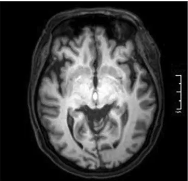
At this moment, brain CT had no evidence of hemorrhage. Pa-tient was submitted to arteriography, which has not shown evi-dence of aneurysm, vascular malformation or venous thrombo-sis. he day after admission, lumbar puncture was performed in L3-L4 with opening pressure of 45cm of water. CSF was clear, colorless, with 2 cells, 1.6 red cells and normal biochemistry. Brain MRI was requested and has shown oval exophytic le-sion (1.0x1.0x0.6cm) in interpenducular cistern and third ventricle, with hyperintense signal at T1-T2 sequence and hy-pointense signal halo at T2 sequence (Figures 1 to 4), com-patible with brainstem cavernous angioma. Patient evolved with partial headache remission with topiramate (treatment of intracranial hypertension, since acetazolamide was
con-traindicated – COPD hypercapnia). Patient maintains mild converging strabismus and is under Neurosurgery clinical-imaging follow-up.
Figure 1. Resonance magnetic in axial cut, lair sequence
Oval formation with hyperintense signal at T1 and T2 sequenc-es, located in central and anterior region of midbrain tegu-ment, with exophytic aspect for the interpeduncular cistern and third ventricle, measuring approximately 1.0x1.0x0.6 cm, with hypointense signal halo at T2 sequence.
here has been no contrast enhancement. here has been no opaciication of altered circulation at angio-MRI sequences.
306
Souza Junior JB, Ferreira KS, Cetlin RS and Dach F Rev Dor. São Paulo, 2014 oct-dec;15(4):304-7
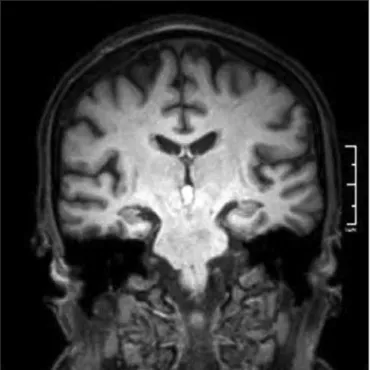
Figure 3. Weighted coronal resonance magnetic in T1
Figure 4. Sagital weighted magnetic resonance in T1
DISCUSSION
Approximately 30% of IVH are primary, that is, originated from the ventricular system itself. hey may come from in-traventricular structure of from lesion close to the ventricle. hey appear in situations of intraventricular trauma, aneu-rysm rupture or vascular malformation and tumor complica-tion, for example. he other 70% of IVH are secondary, cor-responding to the extension of intraparenchimal hemorrhage or SAH2.
Cavernomas (synonym to cavernous angiomas) are slow low capillary malformations, made up of roughly dilated vascular channels lined with a single layer of endothelial cells, without elastic tissue and smooth muscle. Blood breakdown and reac-tive gliosis products may be found in adjacent brain paren-chyma and are described as looking like raspberries3.
In our case, initial suspicion was SAH, of probable vascular origin by the evidence of primary IVH. However, brain ar-teriography was normal. In the meantime, patient remained symptomatic, with signs of intracranial hypertension (ICH) – diplopia and blurred vision – possibly associated to hemo-ventricle products breakdown and intervention on CSF low4,5. Hypertensive CSF has conirmed such hypothesis, and topira-mate was started. Considering previous COPD and associated hypercapnia, we decided to rule out acetazolamide to manage ICH. Brain MRI was essential to clarify the diagnosis.
Cavernomas are reported in 0.1-0.5% of general population, with bimodal presentation from 3 to 11 years of age (30%) and from 30 to 40 years of age (60%). Above 40 years of age, only 10% of diagnoses are made5,10. here is a well-known predominance of females (1.8:1), in addition to higher preva-lence in Caucasian patients6.
Solitary cavernous angioma occurs in 80% of cases and multi-ple cavernous angiomas in 20%. With regard to genetic predis-position, 80% are sporadic and 20% are familial7. Our patient was a 55-year old female with isolated and sporadic lesion. With regard to location, 70% are supratentorial (predominance of frontal lobe), 25% are infratentorial and 5% are located in spinal cord. In brainstem, 57% of cavernomas are located in the pons, 29% in midbrain and 14% in the bulbus3,10. Bleeding location, size and presence lead to a broad variation of neuro-logical repercussions, from asymptomatic to sudden death by acute hemorrhage, identiied in 20% of cases7.
Clinical manifestations may be nausea, vomiting, dizziness and epileptic crises8. Focal motor deicits are seen in approxi-mately 46% at presentation. A range of 10% to 90% with reports of headache is due to diferent situations, from in-cidental diagnoses, where there is association with common migraine, to headache induced by ICH9. In our case, it was thunderclap headache with focal deicits, caused by expansive midbrain lesion.
Cavernomas are part of a group of vascular malformations an-giographically occult or cryptic, like venous angiomas, capil-lary telangiectasis and some arteriovenous malformation. So, brain angiography is an exclusion exam.
Brain CT may evidence isodense lesion or with nonspeciic focal hyperdensity due to recent hemorrhages or to microcal-ciications and with poor contrast enhancement. Such ind-ings are nonspeciic and the diagnosis of cavernoma is often ignored3,10.
307
Investigation of thunderclap headache in cavernous angioma: when magnetic resonance makes the diference. Case report
Rev Dor. São Paulo, 2014 oct-dec;15(4):304-7
Risk factors predisposing to bleeding are gestation, cavernomas above 1.0cm, age below 35 years and, especially, previous bleed-ing. In the general mean, based on natural history evidences, bleeding rate per year in cases of supratentorial cavernomas is 3%, increasing to 5% for rebleeding. For infratentorial caverno-mas, the same rates are 5% increasing to 15%, respectively3,10. he option for expectant follow-up is well established in cases of asymptomatic cavernomas, incidental indings or for pa-tients without surgical conditions (due to inaccessibility of the lesion or to patient’s clinical conditions). In this thera-peutic modality, control is regularly performed with MRI to evaluate growth or new hemorrhages. In addition, gestation, intensive physical exercises and anticoagulants should be dis-couraged3,7.
CONCLUSION
his case shows the importance of brain MRI in patient with thunderclap headache, however further studies are needed in this area to explain how this impacts the investigation.
REFERENCES
1. Kapoor S. Headache attributed to cranial or cervical vascular disorders. CurrPainHe-adache Rep 2013;17(5):334.
2. Hinson HE, Hanley DF, Ziai WC. Management of Intraventricular Hemorrhage. Curr Neurol Neurosci Rep. 2010;10(2):73–82.
3. Maranha LA, Araújo JC. Central Nervous System Cavernomas. J BrasNeuroci-rurg2012;23(4):316-322.
4. Welch KM, Nagesh V, Aurora S, et al. Periaqueductal grey matter dysfunction in migraine: cause or the burden of illness? Headache2001;41:629–37.
5. Afridi S, Goadsby PJ. New onset migraine with a brain stem cavernous angioma. J NeurolNeurosurgPsychiatry2003;74:680-681.
6. Stacey RJ, Findlay GFG, Foy PM, Jefreys RV. Cavernomas in the central nervous system and the relevance of multiple intracranial lesions in the familial form of this disease. J NeurolNeurosurgPsychiatry1999;66:117.
7. Schwarz N, Nohl F, Dang L, et al. Acute headache in a case of cerebral cavernomas. Praxis (Bern 1994) 2007;96(19):775-8.
8. Seltmann S, Wellnitz EM. Defective Signaling Pathway Leads to Vascular Malfor-mations in the Brain. Joint Press Release of the German Cancer Research Center (DeutschesKrebsforschungszentrum) and the University Medical Center Mannheim 2010;Nr.39.
9. Gohary M, Tomita T, Gutierrez FA, McLone DG. Angiographically occult vascular malformations in childhood. Neurosurgery1987;20:759.