r e v b r a s o r t o p .2 0 1 4;4 9(3):313–316
w w w . r b o . o r g . b r
Case report
Solitary ischial osteochondroma: an unusual cause of sciatic
pain: case report
夽
,
夽夽
Frederico Barra de Moraes
a,∗, Paulo Silva
b, Rogério Andrade do Amaral
b,
Frederico Faleiro Ramos
a, Rômulo Orlando Silva
a, Diogo Azevedo de Freitas
aaDepartamento de Ortopedia e Traumatologia do Hospital das Clínicas da Faculdade de Medicina da Universidade Federal de Goiás,
Goiânia, GO, Brazil
bHospital Geral de Goiânia, Goiânia, GO, Brazil
a r t i c l e
i n f o
Article history: Received 23 April 2013 Accepted 7 June 2013 Available online 25 April 2014
Keywords: Oncology Orthopedics Sciatic nerve Pelvis Sciatica
a b s t r a c t
The aim was to report on a rare case of osteochondroma of the left ischium, which evolved with compression of the sciatic nerve, thus causing sciatic pain in the homolateral lower limb. The patient was female and presented sciatic pain that was treated clinically for one year. However, the pain evolved with increasing intensity and worsened with hip movement. This was associated with diminished motor force and paresthesia of the homolateral lower limb. Radiological investigation of the region showed a bone lesion in the external portion of the left ischium, in the path of the sciatic nerve. Tomographic reconstruction showed cortical continuity with the bone of origin, i.e., a pattern characteristic of osteochondroma. En-bloc resection of the lesion was performed using the Kocher-Langerbeck route, and the anato-mopathological analysis proved that it was an osteochondroma. The patient’s neurological symptoms improved and, after two months of follow-up, she remained asymptomatic and without any signs of recurrence. Since osteochondroma is the commonest benign bone tumor, it should be taken into consideration in the diagnostic investigation of compressive tumor lesions that could affect the sciatic nerve.
© 2014 Sociedade Brasileira de Ortopedia e Traumatologia. Published by Elsevier Editora Ltda. All rights reserved.
Osteocondroma solitário de ísqueo: uma causa não usual de ciatalgia:
relato de caso
Palavras-chave: Oncologia Ortopedia Nervo ciático Pelve Ciática
r e s u m o
Relatar um caso raro de osteocondroma do ísqueo esquerdo, que evoluiu com compressão no nervo ciático e provocou ciatalgia no membro inferior homolateral. Paciente do sexo feminino apresentou ciatalgia e foi feito tratamento clínico por um ano. Porém a dor evoluiu, aumentou de intensidade e piorou com a movimentac¸ão do quadril, associada a diminuic¸ão da forc¸a motora e a parestesia do membro inferior homolateral. A investigac¸ão radiológica da região mostrou uma lesão óssea na porc¸ão externa do ísqueo esquerdo e no trajeto do
夽Please cite this article as: de Moraes FB, Silva P, do Amaral RA, Ramos FF, Silva RO, de Freitas DA. Osteocondroma solitário de ísqueo:
uma causa não usual de ciatalgia: relato de caso. Rev Bras Ortop. 2014;49:313–316. 夽夽
Work performed at the Orthopedics and Traumatology Clinic, Goiânia, Goiás, Brazil.
∗ Corresponding author.
E-mail: frederico [email protected] (F.B. de Moraes).
314
r e v b r a s o r t o p .2 0 1 4;4 9(3):313–316nervo ciático. A reconstruc¸ão tomográfica evidenciou continuidade cortical com o osso de origem, padrão característico de osteocondroma. Fez-se a ressecc¸ão em bloco da lesão pela via de Kocher-Langerbeck e o estudo anatomopatológico provou ser um osteocondroma. Os sintomas neurológicos da paciente melhoraram e, após dois anos de acompanhamento, ela permanece assintomática e sem sinais de recorrência. Por ser o tumor ósseo benigno mais comum, o osteocondroma deve ser considerado na investigac¸ão diagnóstica de lesões tumorais compressivas, que podem acometer o nervo ciático.
© 2014 Sociedade Brasileira de Ortopedia e Traumatologia. Publicado por Elsevier Editora Ltda. Todos os direitos reservados.
Introduction
Osteochondromas (exostoses or endochondromatous exos-toses) are common bone tumors. They account for 8.5% of bone tumors and 36% of benign bone tumors.1They can occur
as solitary tumors or as multiple exostoses.
The predominant location for osteochondromas is the region proximal to the knee (distal femur and proxi-mal tibia), followed by the humerus and proxiproxi-mal femur.2
Osteochondromas are generally diagnosed during childhood and adolescence. The clinical manifestations depend on occurrences of fractures at the base of the exostosis and inflammation and compression in the structures surrounding the tumor mass. Pelvic locations are uncommon and account for 5.6% of the cases, while cases affecting the ischium are even less common and account for only 0.4% of the cases.3
This location has complex anatomy, allows compression of the sciatic nerve and evolves with sciatic pain that is difficult to investigate clinically.
The objective of this study was to report on a case of ischial osteochondroma in which the unusual location allowed com-pression of the sciatic nerve and caused chronic sciatic pain.
Case report
The patient was a 42-year-old female patient who presented sciatic pain in her left leg, from the hip to the foot, with pares-thesia on the anterolateral face of the left leg and foot, without alteration of the patellar reflex (L4) or Achilles reflex (S1). She had a negative Lasègue test. The patient underwent radiogra-phy and computed tomograradiogra-phy (CT) of the lumbar spine and the results obtained were normal. She was started on clini-cal treatment with anti-inflammatory drugs and opioids, but without improvement. Over a one-year period, the pain pro-gressed and started to worsen with movement of the left hip, along with diminished motor strength (grade 4) on dorsiflex-ion of the foot (L4), extensdorsiflex-ion of the hallux (L5) and plantar flexion (S1). Imaging examinations of this region were then requested.
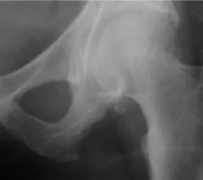
Radiography of the left hip (AP view) showed slight and poorly defined alteration of the external portion of the ischium (Fig. 1). CT of the pelvis was then requested, on which tumor development with bony characteristics was observed in the left ischium. It measured approximately 4 cm, was peduncu-late and well-delimited, lay on the path of the sciatic nerve and was compatible with an osteochondroma (Fig. 2).
En-bloc resection surgery was performed on the tumor development in the left ischium, by means of the posterior
Fig. 1 – Preoperative radiograph on the left hip, in AP view, showing slight bone alteration in the ischium.
Kocher-Langerbeck route, and the sciatic nerve was decom-pressed (Fig. 3). Anatomopathological examination of the surgical specimen confirmed the hypothesis of osteochon-droma. After the surgery, the patient’s neurological symptoms improved and she remains asymptomatic after two years of follow-up.
Discussion
The initial approach in cases of lumbar sciatic pain is difficult because the differential diagnosis is extensive. There are dif-ferent forms of vertebral involvement and clinical conditions without direct involvement that may mimic radiculalgia. The investigation therefore needs to integrate signs, symptoms, physical examinations, imaging examinations and laboratory tests, in order to guide logical management. The imaging investigation should be done carefully because the findings are often nonspecific and thus should be interpreted within a broader clinical context.
This patient arrived with complaints of sciatic pain and the radiological evaluation complemented with CT on the lumbar spine did not show alterations and ruled out the hypothesis of radicular compression. CT is the best method for viewing the bone architecture, but it is inferior to magnetic resonance imaging for evaluating soft tissues.4At that time, the
r e v b r a s o r t o p .2 0 1 4;4 9(3):313–316
315
Fig. 2 – Three-dimensional tomographic reconstruction of the pelvis showing pedunculate tumor development in bone of the left ischium, along the path of the sciatic nerve, in front view (A) and rear view (B).
suspicion of systemic illness. Moreover, clinical examination showed grade 4 motor impairment of the nerve roots of L4 (dorsiflexion of the foot), L5 (extension of the hallux) and S1 (plantar flexion). The investigation was facilitated by the spe-cific complaint of worsening of the pain with hip movement. AP radiographs of the region were requested and a lesion was found in the left ischium (Fig. 1). Tomographic reconstruction of the pelvis made it clear that this was a tumor development on the path of the left sciatic nerve that was compatible with osteochondroma, which explained the condition of progres-sive sciatic pain.
Osteochondromas are bone exostoses formed by cortical and medullary bone that is continuous with the originating bone and is covered with a coating of hyaline cartilage. Osteo-chondromas have their own growth plate, which produces bone that forms the exostosis. It is believed that osteochondro-mas arise from a change in growth direction of the epiphyseal disc, which grows persistently and later on undergoes endo-chondral calcification. The lesion continues to grow from the cartilaginous coating, just like a normal epiphyseal disc, and therefore growth after skeletal maturity is reached during puberty is not expected. This explains why the peak inci-dence of osteochondromas is in the second decade of life.4
The patient reported here was in her fifth decade of life (42 years of age), an age group in which only 5% of the diag-noses are made.5
Fig. 3 – After the operation, with en-bloc resection of the tumor development.
Osteochondromas can affect any bone within endochon-dral ossification. The greatest incidence of osteochondromas occurs in the knee region (distal metaphysis of the femur and proximal metaphysis of the tibia), followed by the prox-imal regions of the humerus and femur. Occurrences in the ischium are uncommon and account for 0.4% of the cases.5
They can be solitary or multiple. The latter are associated with multiple hereditary exostosis, which is an autosomal dom-inant syndrome. Complications occur more frequently with this syndrome and include deformity (cosmetic or bone), frac-tures, vascular impairment, formation of pouches, malignant transformation and neurological sequelae. Despite this vast range of clinical manifestations, osteochondromas are gener-ally asymptomatic and they are diagnosed by chance.
Osteochondromas are diagnosed radiologically. The lesions are characterized by continuation of the cortical and spongy bone with the underlying bone (Fig. 1). The hyaline cartilage is not seen on radiographic examination, unless it has become calcified, when it acquires an appearance of cotton wool-like stains, which suggests that osteochondromas have a benign nature and are long lasting. Thick cartilage that is invisible on radiographs is predictive of malignity.3CT is essential for
evaluating cases in complex anatomical sites (for example, in the pelvis, scapular belt, limb roots and spine).4Tomographic
reconstruction defined the present case and precisely showed a pedunculate lesion measuring 4 cm, located along the path of the left sciatic nerve (Fig. 2).
The presence of an osteochondroma is not an absolute indication for surgical resection. An expectant approach is taken in cases in which there are no clinical manifestations. Surgery is indicated in cases of pedunculate tumors (which are generally associated with complications) or when there is compression of nerves, arteries or tendons, or functional and anatomical alterations. Therefore, in the present case, the progressive neurological deficit in the leg comprised a formal indication for surgery. En-bloc resection was performed (Fig. 3). The Kocher-Langerbeck route was used and no intercurrences were observed either during the immediate postoperative period or later on. The importance of ample resection is that the presence of remainders of the perichondrium and the cartilaginous coating may enable local recurrence of the osteo-chondroma.
316
r e v b r a s o r t o p .2 0 1 4;4 9(3):313–316warning signs are rapid growth of the lesion, appearance of pain, thickening of the cartilage coating or discontinuity of the exostosis with the underlying cortical bone, when the demarcation of the lesion surface is radiologically lost. The transformation generally occurs to grade 1 chondrosarcoma. The anatomopathological examination on the surgical speci-men confirmed the radiological suspicion of osteochondroma, and the two-year postoperative radiographic and clinical follow-up on the patient ruled out the possibility of malignant transformation or local recurrence of the lesion.
The neurological impairment that was presented regressed completely consequent to the surgical decompression and strength grade 5 was achieved for the myotomes correspond-ing to L4, L5 and S1. Complete remission of the compressive symptoms relating to osteochondromas in other topograph-ical areas has generally been observed,7–12 for example
Horner’s syndrome in cases of cervical osteochondroma and radicular compression syndrome in cases of spinal osteochon-droma. Because of the scarcity of cases, these is a lack of data for making specific prognostic evaluations on sciatic pain secondary to compression of the sciatic nerve due to ischial osteochondromas.
Conflicts of interest
The authors declare no conflicts of interest.
r e f e r e n c e s
1. Defino HL, Pereira CU, Barbosa CVP. Tumores benignos e lesões pseudotumorais da coluna vertebral. Rio de Janeiro: Revinter; 2002.
2. Radu AS. Síndromes lombares. In: Lopes AC, editor. Tratado de clínica médica. São Paulo: Roca; 2006. p. 1732–42.
3. Garcia RJ. Diagnóstico e tratamento de tumores ósseos. Rio de Janeiro: Elsevier; 2005.
4. Murphey MD, Choi JJ, Kransdorf MJ, Flemming DJ, Gannon FH. Imaging of osteochondroma: variants and complications with radiologic–pathologic correlation. Radiographics.
2000;20(5):1407–34.
5. Zhao CQ, Jiang SD, Jiang LS, Dai LY. Horner syndrome due to a solitary osteochondroma of C7: a case report and review of the literature. Spine (Philadelphia, PA, 1976). 2007;32(16): E471–4.
6. Han IH, Kuh SU. Cervical osteochondroma presenting as brown-sequard syndrome in a child with hereditary multiple exostosis. J Korean Neurosurg Soc. 2009;45(5): 309–11.
7. Gürkanlar D, Aciduman A, Günaydin A, Koc¸ak H, Celik N. Solitary intraspinal lumbar vertebral osteochondroma: a case report. J Clin Neurosci. 2004;11(8):911–3.
8. Byung-June J, Seung-Eun C, Sang-Ho L, Hyeop JS, Suk PS. Solitary lumbar osteochondroma causing sciatic pain. Joint Bone Spine. 2007;74(4):400–1.
9. Xu J, Xu CR, Wu H, Pan HL, Tian J. Osteochondroma in the lumbar intraspinal canal causing nerve root compression. Orthopedics. 2009;32(2):133.
10. Ohtori S, Yamagata M, Hanaoka E, Suzuki H, Takahashi K, Sameda H, et al. Osteochondroma in the lumbar spinal canal causing sciatic pain: report of two cases. J Orthop Sci. 2003;8(1):112–5.
11. Bess RS, Robbin MR, Bohlman HH, Thompson GH. Spinal exostoses: analysis of twelve cases and review of the literature. Spine (Philadelphia, PA, 1976). 2005;30(7): 774–80.