Lipid Profile and Severity of Atherosclerotic Disease in Acute
Coronary Syndrome
Rafaela Andrade Penalva, Marçal de Oliveira Huoya, Luis Cláudio Lemos Correia, Gilson Soares Feitosa, Ana Marice
Teixeira Ladeia
Escola Bahiana de Medicina e Saúde Pública, Hospital Santa Izabel, Salvador, BA - Brazil
Summary
Background: The relationship between coronary artery disease (CAD) and dyslipidemia in acute coronary syndromes
has been rarely demonstrated in clinical and epidemiological studies.
Objective: To evaluate the association between lipid profile and severity of CAD in patients with acute coronary syndrome without ST-segment elevation.
Methods: In this retrospective study, the authors reviewed medical records of 107 consecutive patients diagnosed with acute coronary syndrome (ACS) without ST-segment elevation admitted within a one-year period and who had undergone coronary angiography during hospitalization. Laboratory evaluation included serum levels of lipid fractions. Severity of CAD was determined by evaluating the number, degree, and score of coronary artery obstructions. For statistical analysis, the Student’s t test, chi-square test and ANOVA with statistical significance set at p<0.05, as well as multivariate analysis were performed.
Results: A total of 107 patients were included; 94(88%) had CAD, of which 50 (53.2%) were males with predominance
of multivessel disease. As regards the lipid profile, 4(59.8%) patients were observed to have TC<200mg/dl, 33(30.8%) had HDL<40mg/dl, and 38(35.5%) had LDL<100mg/dl. The analysis of coronary angiographies showed that 94(88%) patients had CAD, and 84% had > 70% stenosis. In the association between lipid profile and CAD, we observed a higher TC/HDL ratio in the multivessel and two-vessel groups in comparison with the one-vessel group (4.3±2, 4.0±1.7, 2.9±1., respectively) with ANOVA p=0.049. In the multivariate analysis, the TC/HDL ratio remained a significant predictor (p = 0.01).
Conclusion: The TC/HDL ratio was a marker of severity of CAD in relation to the number of vessels affected, thus demonstrating that the lipid profile can be a determinant of severity in patients with ACS without ST-segment elevation. (Arq Bras Cardiol 2008;90(1):24-29)
Key words: Coronary atherosclerosis; atherosclerosis; dyslipidemias.
Mailing address: Rafaela Andrade Penalva •
Rua Altino Seberto de Barros, 295/1004 - Itaigara - 41825- 010, Salvador, BA - Brazil
E-mail: [email protected]
Manuscript received December 15, 2006; revised manuscript received June 29, 2007; accepted October 08, 2007.
Introduction
Dyslipidemia is a well-established risk factor for the development of coronary artery disease (CAD), and this has been demonstrated in several clinical and epidemiological studies1-7. High plasma low-density lipoprotein (LDL-C)
concentrations are directly correlated with the development of coronary artery disease8, and low high-density lipoprotein
(HDL-C) concentrations have been pointed out as one of the strongest independent risk factors for coronary atherosclerotic disease9. New evidences show that mild increases in
triglycerides lead to increased risk of coronary events and progression of coronary artery disease, as well as to the formation of new lesions10-12.
The relationship between lipid profile and obstructive coronary artery disease is well known13-14. However, studies on the role
of lipoprotein levels as markers of severity of Acute Coronary Syndrome (ACS) are still scarce in the literature. Although clinical manifestations of ACS do not depend exclusively on the extent of CAD15, the obstructive impairment and number of vessels
affected can interfere with the therapeutic strategy. Therefore, we can presume that if one of the lipid fractions is predictive of the degree of anatomical impairment on coronary angiography, it will potentially be able to influence the decision of a strategy on invasive investigation in patients with ACS.
Thus, the objective of this study was to evaluate the existence of an association between lipid profile and severity of coronary atherosclerotic disease in patients with acute coronary syndrome without ST-segment elevation.
Methods
In the statistical analysis, the clinical characteristic age was assessed using the mean and respective standard deviation; the categorical clinical variables (gender, cardiovascular risk factors and use of medication) were expressed as proportions. To evaluate the association between lipid profile and number of vessels affected, the means were compared using the ANOVA test. The Student’s t test was used to compare the means between the groups with or without the presence of obstructions. Statistical significance was set at p < 0.05. Multivariate analysis was also performed. In the logistic regression, the dependent variable was the number of vessels affected; this variable was dichotomized in one or more than one vessel affected. The independent variables were the significant variables in the univariate analysis. Data were entered in and analyzed by the SPSS version 10.0 software program.
This study was submitted to and approved by the Research and Ethics Committee of Hospital Santa Izabel.
Results
Of the 107 patients included, 94 (88%) had CAD, of whom 50 (53.2%) were males with a predominance of multivessel disease. Mean ages were higher in the two-vessel and multivessel groups in comparison with the one-vessel group (68±9 vs. 61±13 years, p=0.035). DM and hypertension were more frequent in the multivessel group (p=0.004 and p=0.042, respectively), and no differences between the groups were observed as for the other risk factors. Unstable angina was the most frequent clinical manifestation in this population, affecting 73% of the patients in the one-vessel group and 69% in the multivessel group (Table 1).
Analysis of the lipid profile showed that 64 (59.8%) of the patients had TC <200md/dl, 18 (16.8%) had TC > 240md/dl, 33(30.8%) had HDL <40md/dl, 23 (21.5%) had HDL >60mg/ dl, and 52 (48.6%) had TG<150mg/dl. The mean values of TC, LDL, triglycerides and NHDLC were 190.71±55.12 md/dl, 109.03±49.42 md/dl, 180.81±95.99 md/dl and 141.43±56.55 md/dl, respectively, with no significant difference in lipid and September, 2004 with a diagnosis of acute coronary
syndrome (ACS) without ST-segment elevation, and who had undergone coronary angiography during hospitalization. These medical records belonged to the database of the Chest Pain Unit (CPU) of Hospital Santa Izabel (HSI), city of Salvador, State of Bahia, Brazil. The diagnosis of ACS without ST-segment elevation included non-ST-segment elevation myocardial infarction (NSTEMI) and unstable angina (UA). The diagnosis of NSTEMI was made in patients who presented with retrosternal chest pain radiating or not to the neck, jaw, epigastrium, shoulder and left arm, with a sudden onset and duration of 30 minutes or more and varying degrees and symptoms; these patients had no ST-segment elevation on ECG, but elevated levels of biochemical markers of myocardial necrosis – total CK and CK-MB. Unstable angina was characterized by a constrictive chest pain with duration between 5 and 30 minutes, radiating to the left arm, jaw, or right shoulder, associated with cold sweat, nausea, vomiting, or relieved by nitrate; by laboratory tests based on troponin T levels > 0.01 and < 0.1 with normal CPK-mass and the following electrocardiographic (ECG) changes: T wave inversion
≥1.0mm, segment depression ≥1 mm and transient ST-segment elevation ≥ 1 mm (spontaneous reversal or with
nitrate)16. The evaluation of clinical and epidemiological
variables included the identification of risk factors for CAD and data regarding blood pressure, radial pulse, respiratory rate, outpatient therapy, as well as ECG results.
Laboratory evaluation included data regarding the levels of total cholesterol (TC), HDL cholesterol (HDL), and triglycerides (TG), as determined using the enzymatic method; LDL cholesterol (LDL), using the Friedewald formula17;
and non-HDL cholesterol (NHDLC), as calculated by the difference between TC and HDL. According to the CPU protocol, all tests were performed in blood samples collected at admission in the HSI clinical laboratory. Failure to fast was not an exclusion criterion. The lipid profile was classified according to the III Brazilian Guidelines on Dyslipidemias and Guideline on the Prevention of Atherosclerosis of the Brazilian Cardiology Society18.
All coronary angiographies (ANGI) were performed using Judkins technique and were subjectively analyzed by professionals from the Catheterization Service of HSI. Based on the angiography results, data regarding the percentage of obstructions were noted down separately into three categories
(< 50 %, ≥ 50 and < 70%, ≥ 70%); the number of vessels
affected, with specific identification of main and secondary vessels were also noted down. CAD was defined as the presence of any obstruction quantified angiographically. For the analysis of severity of CAD, two criteria were considered: percentage of obstruction and number of vessels affected. Also, the Nikita et al score system19,20 was used for quantitative
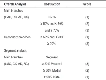
analysis of CAD. The main arteries (anterior descending – AD, circumflex – CX, right coronary – RC, and left main coronary – LMC) were analyzed and scored as for the presence of lesions in the proximal, medial or distal segments. The secondary arteries were scored only as for the degree of obstruction, regardless of its location. Severity of CAD was defined by the sum of scores of all lesions, and the score for location in main arteries was attributed only for lesions ≥ 50% (Table 1).
Table 1 – Quantitative analysis of CAD (Score System)
Overall Analysis Obstruction Score
Main branches
(LMC, RC, AD, CX) < 50% (1)
≥ 50% and < 70% (2)
and ≥ 70% (3) Secondary branches ≥ 50% and < 70% (1)
≥ 70%. (2) Segment analysis
Main branches Segment
(LMC, CX, AD, RC) ≥ 50% Proximal (3)
≥ 50% Medial (2)
variables between patients with and without obstruction (Table 2).
The analysis of the association between lipid profile and number of vessels affected showed that the total cholesterol/ HDL ratio was higher in the multivessel group with a statistically significant difference, and p ANOVA equal to 0.049 (Table 3).
Although LDL-cholesterol had been higher in the multivessel CAD group in comparison with the one-vessel group with means of 115.98±53.88 and 85.54±32.31, respectively, no statistically significant difference was found (Table 4).
In the multivariate analysis, using the number of vessels affected as the dependent variable and TC/HDL ratio, hypertension, DM and age as independent variables, we observed that the TC/HDL ratio remained as a significant predictor (p=0.01). In addition, hypertension and age were also predictive of the number of vessels affected; DM lost significance after adjustment (p=0.75).
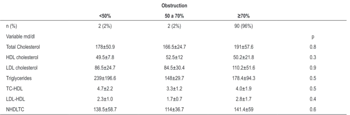
The analysis of the association between lipid profile and percentage of obstruction did not show any difference
between the three degrees of obstruction (< 50 %, ≥ 50 and < 70%, ≥ 70%) for any of the lipid variables analyzed
(Table 5).
The analysis of severity of CAD using the score system showed that the mean and median scores among patients with obstruction were 10.8±4.6 and 11, respectively. Based on the mean and median, the ANGI were classified into
three score ranges: <6; ≥ 6 and < 15; ≥15. The frequency
of distribution by score range showed that 12.8% (12) of the patients had a score lower than six; 66% (62) between six and 15; and 21.3% (20) higher than 15. No difference between the groups was observed as regards the assessment of severity of CAD using the score system and lipid profile for any of the variables studied. However, the lipid fractions total cholesterol, LDL-C and NHDLC tended to be lower in the group with a
score ≤ 6 (Table 6).
Table 2 – Clinical characteristics of the population
One-vessel Two-vessel Multi-vessel p
Sample number 15 24 55
Age (years) 61±13 68±9 68±9 0.035
Male gender 8 (53%) 10 (42%) 32 (58%) 0.400
Hypertension 10(67%) 24(100%) 50(91%) 0.004
Dyslipidemia 10 (67%) 19(79%) 42(76%) 0.660
Sedentary life style 8(53%) 13(54%) 29(53%) 0.993
Diabetes Mellitus 4(27%) 15(63%) 20(36%) 0.042
FH CAD 5(33%) 4(17%) 12(22%) 0.473
Smoking 3(20%) 2(8%) 6(11%) 0.523
NSTEMI 4(27%) 12(50%) 17(31%) 0.341
Unstable angina 11(73%) 12(50%) 38(69%) 0.341
Lipid-lowering drug 5(33%) 9(38%) 20(36%) 0.965
ASA 13(87%) 12(50%) 34(62%) 0.069
ASA - acetylsalicylic acid; CAD - coronary artery disease; FH CAD - family history for coronary artery disease; NSTEMI - non-ST-segment elevation myocardial infarction; p - probability of signiicance.
Table 3 – Lipid proile vs CAD
Variable md/dl General Population Without obstruction With obstruction p
Sample Size 107(100%) 13 (12%) 94 (88%)
Total Cholesterol 190.7±55.1 194.2±43.2 190±56.8 0.8
HDL cholesterol 49.7±20.5 47.5±11.4 50±21.5 0.6
LDL cholesterol 109±49.4 109.1±38 109±50.8 0.9
Triglycerides 180.8±95.9 185.9±104.4 180±95.3 0.8
TC/HDL 4.0±1.9 4.3±1.3 4.0±1.3 0.6
LDL/HDL 2.8±1.5 2.0± 1.9 2.7±1.9 0.5
NHDLTC 141.4±56.6 146.7±43.5 140.7±58.2 0.7
Table 4 – Laboratorial lipid proile in the study population
Variable md/dl One-vessel Two-vessel Multivessel p ANOVA
Total Cholesterol 162.9±39.7 193.3±52.8 196.4±60.9 0.1
HDL cholesterol 48.9±21.2 51.5±16.4 49.7±23.6 0.9
LDL cholesterol 85.5±32.3 104.6±48.6 115.9±53.9 0.1
Triglycerides 147.9±81.2 186.4±80.9 184.5±104.2 0.4
TC-HDL 2.9±1.6 4.1±1.7 4.3±2.0 0.049
LDL-HDL 2.0±1.5 3.7±1.6 2.5±1.8 0.2
NHDLTC 117.3±39.1 141.8±52.5 146.7±63.9 0.2
HDL cholesterol - high-density lipoprotein cholesterol; LDL cholesterol - low-density lipoprotein cholesterol; LDL-HDL- low and high-density lipoprotein cholesterol ratio; NHDLTC - non high-density lipoprotein cholesterol; p - probability of signiicance; TC-HDL - total cholesterol and high-density lipoprotein cholesterol ratio.
Table 5 – Association between lipid proile and degree of obstruction
Obstruction
<50% 50 a 70% ≥70%
n (%) 2 (2%) 2 (2%) 90 (96%)
Variable md/dl p
Total Cholesterol 178±50.9 166.5±24.7 191±57.6 0.8
HDL cholesterol 49.5±7.8 52.5±12 50.2±21.8 0.3
LDL cholesterol 86.5±24.7 84.5±30.4 110.2±51.6 0.9
Triglycerides 239±196.6 148±29.7 178.4±94.3 0.5
TC-HDL 4.7±2.2 3.3±1.2 4.0±1.9 0.5
LDL-HDL 2.3±1.0 1.7±0.7 2.8±1.7 0.4
NHDLTC 138.5±58.7 114±36.7 141.4±59 0.6
HDL - cholesterol - high-density lipoprotein cholesterol; LDL - cholesterol - low-density; lipoprotein cholesterol; LDL-HDL - low and high-density lipoprotein cholesterol ratio; NHDLTC - non high-density lipoprotein cholesterol; p - probability of signiicance; TC-HDL - total cholesterol and high-density lipoprotein cholesterol ratio.
Table 6 – Association between lipid proile and Score
Variable md/dl <6 ≥ 6 and < 15 ≥ 15 p ANOVA
Total Cholesterol 164±38 198±61 178 ±45 0.09
HDL cholesterol 49±12 51±24 46±13 0.6
LDL cholesterol 84±31 116±55 101±41 0.1
Triglycerides 161±65 187±106 162±63 0.5
TC-HDL 3.2±1.5 4.2±2.1 3.9±1.5 0.2
LDL-HDL 1.9±1.2 2.8±1.3 2.9±1.8 0.7
NHDLTC 115±37 148 ±64 132±44 0.1
HDL - cholesterol - high-density lipoprotein cholesterol; LDL - cholesterol - low-density; lipoprotein cholesterol; LDL-HDL - low and high-density lipoprotein cholesterol ratio; NHDLTC - non high-density lipoprotein cholesterol; p - probability of signiicance; TC-HDL - total cholesterol and high-density lipoprotein cholesterol ratio.
Discussion
The analysis of ANGIO showed that most patients in this study population had obstructions ≥70% in main vessels, with multivessel lesions. This predominance is consistent with that described in the literature, demonstrating that CAD preferably affects the main vessels14; also, with the progression of the
disease, multivessel lesions are more frequently observed4.
In addition, laboratory evaluation showed that the mean
values obtained for LDL and TG were higher than those considered of low risk for CAD, according to the III Brazilian Guidelines on Dyslipidemias and Guideline on Prevention of Atherosclerosis of the Department of Atherosclerosis of the Brazilian Society of Cardiology18. These findings validate the
of the coronary arteries.
In this study, the mean levels of lipid variables did not allow the discrimination between the presence and absence of CAD; however, we demonstrated that the variable TC/ HDL was associated with the number of vessels affected, and was higher in the two-vessel and multivessel groups when compared to the one-vessel group. It is noteworthy that although hypertension and age proved to be predictive of the number of vessels affected, the TC/HDL ratio was maintained in the multivariate analysis, in an independent form. As regards the variable TC/HDL and its relationship with the presence and severity of CAD, clinical and angiographic studies have correlated it with the progression or regression of CAD3,21,22. Therefore, this finding reinforces the importance
of the measurement of the TC/HDL ratio as an individual risk factor for CAD, as well as an indicator of extent and severity of the disease, even in the presence of cholesterol levels considered normal, thus suggesting that the imbalance between TC and HDL levels plays a more important role in the pathophysiology of atherogenesis. It is important to consider that the atheroprotective function of HDL is not restricted to reverse cholesterol transport, but can also transport antioxidant enzymes, break down oxidized lipid fractions, and neutralize their proinflammatory effects23. It is noteworthy that 38.8%
of our population had HDL < 40md/dl, and only 21.5% had levels considered atheroprotective.
The analysis of LDL showed that this variable increased with the number of vessels affected. Patients with one-vessel disease had LDL levels of 85.54±332.31md/dl, whereas those with multivessel disease had levels of 115.98±53.88 md/dl (p=0.077). Although no statistical significance had been observed, a datum which may have resulted from the small number of patients in each subgroup, the difference observed in serum LDL levels between the two groups suggests an important clinical value, since patients with multivessel disease had LDL levels >100md/dl, which is considered the cut-off point for the prevention of CAD in high risk patients10.
Several studies agree that high levels of LDL are correlated with the presence and severity of CAD2,15,21. Korhonen et al4 study
adds that high levels of LDL are associated with an increased risk of CAD, and the more vessels obstructed, the higher the serum LDL level in the group, thus corroborating the findings of the present study.
In our study, it is noteworthy that although the TC/HDL ratio had proven to be associated with a higher number of vessels affected, it was not associated with the degree of stenosis in patients with non-ST-segment elevation ACS, unlike the other lipid variables. This can be explained by the fact that the lesions that are more potentially unstable and prone to rupture are frequently non-occlusive and not diagnosed by angiography24. On the other hand, these lesions
have a large lipid nucleus with signs of active inflammation and macrophage accumulation at the site of plaque rupture24.
However, the absence of a relationship between serum LDL-cholesterol and oxidized LDL-LDL-cholesterol in the plaque has been demonstrated in ACS15.
The analysis of severity of CAD using the score system did not show differences for any of the variables, although the lipid
fractions total cholesterol, LDL-c and non-HDL cholesterol had shown a slight trend to be lower when associated with a score lower than six. The score system included the degree of obstruction in its analysis, and this may have contributed to a restricted discriminative power, since, according to the literature, the degree of obstruction is probably not the most important of the factors which precipitate the manifestations of ACS24.
This study may have limitations resulting from its retrospective design, and from the fact that the ANGIO were analyzed by several different professionals. However, it is noteworthy that the possible limitation resulting from the variability of angiography results does not affect their validity, since the study was conducted in a specialized referral hospital for cardiology, where the catheterization team members are highly skilled in the analysis of coronary obstruction. Another aspect that could be considered a limitation was the determination of the lipid profile from blood drawn at admission to comply with the CPU protocol, thus failing to observe a 12-hour fast. Nonetheless, it is important to point out that triglycerides are basically the lipid fraction that has a significant postprandial variation25,26. In addition, although the
criteria used to define unstable angina had been those valid at the moment the database was beginning to be created, they may deserve a reassessment in light of the more recent definitions suggesting that TnT elevation above the 99th percentile (0.01) and below the 10% coefficient of variation (0.03) as an indicator of necrosis should be evaluated within the clinical setting27. However, this limitation does not affect
the objective of the study, since the patients were closely evaluated according to the current CPU protocols, and by professionals able to identify the presence of myocardial necrosis even in unclear clinical situations.
Even though atherosclerosis does not result simply from lipid accumulation, in this study, which included a population with severe CAD characterized by multivessel lesions and predominance of obstructions > 70% and in main vessels, TC and the TC/HDL ratio were markers of severity of CAD in relation to the number of vessels affected, thus demonstrating that the lipid profile may be a determinant of a more significant anatomical impairment also in patients with non-ST-segment elevation ACS. Therefore, considering that a reduction in lipid variables usually occurs a few hours after the manifestations of acute coronary syndrome28, the findings of this study
emphasize the need for assessment of the lipid profile of these patients to be made at admission, so as to identify patients at a higher potential risk.
Potential Conflict of Interest
No potential conflict of interest relevant to this article was reported.
Sources of Funding
There were no external funding sources for this study.
Study Association
References
1. Smith SC, Jackson R, Pearson TA, Fuster V, Yusuf S, Faergeman O, et al. Principles for national and regional guidelines on cardiovascular disease prevention: a scientific statement from the world heart and stroke forum. Circulation. 2004; 109: 3112-21.
2. Watts GF, Mandalia S, Brunt JNH, Slavin BM, Coltart DJ, Lewis B. Independent associations between plasma lipoprotein subfraction levels and the course of coronary artery disease in the St. Thomas`Atherosclerosis Regression Study (STARS). Metabolism. 1993; 42: 1461-7.
3. Ladeia AM, Guimarães AC, Lima JC. Perfil lipídico e doença arterial coronariana. Arq Bras Cardiol. 1994; 63: 101-6.
4. Korhonen T, Savolainen MJ, Koistinen MJ, Ikäheimo M, Linnaluoto MK, Kervinen K, et al. Association of lipoprotein cholesterol and triglycerides with the severity of coronary artery disease in men and women. Atherosclerosis. 1996; 127: 213-20.
5. Saku K, Zhang B, Ohta T, Arakawa K. Quantity and function of high density lipoprotein as an indicator of coronary atherosclerosis. J Am Coll Cardiol. 1999; 33: 436-43.
6. Larosa JC. Prevention and treatment of coronary heart disease. Circulation. 2001; 104: 1688-92.
7. Rocha ASC, Matiello ME, Cunha AB, Paris DS, Pittella FJM, Melhado JC, et al. Perfil clínico e angiográfico da doença arterial coronária em pacientes com idade inferior a 50 anos. Editorial Laranjeiras. 2002; 1 (1): 1.
8. Rudel LL, Kesainiemi A. Low-density lipoprotein particle composition: what is the contribution to atherogenicity? Curr Opin Lipidol. 2000; 11: 227-8. 9. Miller GL, Miller NE. Plasma high-density lipoprotein concentration and the
development of ischaemic heart disease. Lancet. 1975; 1: 16-9.
10. Assmann G, Schulte H. Role of triglycerides in coronary artery disease: lessons from the prospective cardiovascular munster study. Am J Cardiol. 1992; 70: 10H-13H.
11. Austin MA. Plasma triglyceride and coronary heart disease. Arterioscler Thromb. 1991;11: 2-14.
12. Hokanson JE, Austin MA. Plasma triglyceride level is a risk factor for cardiovascular disease independent of high-density lipoprotein cholesterol level: a meta-analysis of population based prospective studies. J Cardiovasc Risk. 1996; 3: 213-9.
13. Libby, P. Current concepts of the pathogenesis of the acute coronary syndromes. Circulation. 2001; 104: 365-72.
14. Braunwald E, Antman EM, Beasley JW, Califf RM, Cheitlin MD, Hochman JS, et al. ACC/AHA guideline update for the management of patients with unstable angina and non-ST-segment elevation myocardial infarction. 2002. Summary article: a report of the American College of Cardiology / American Heart Association Task Force on Practice Guidelines (Committee on the Management of Patients with unstabel angina. Circulation. 2002; 106: 183-90.
15. Ehara S, Ueda M, Naruko T, Haze K, Itoh A, Otsuka M, et al. Elevated levels of oxidized low density lipoprotein show a positive relationship with the severity of acute coronary syndromes. Circulation. 2001; 103: 1955-60. 16. Sociedade Brasileira de Cardiologia. Diretrizes sobre angina instável e infarto
agudo do miocárdio sem supradesnível do segmento ST. Arq Bras Cardiol. 2001; 77 (supl 2): 1-22.
17. Friedwald WT, Levy RI, Fredrickson DS. Estimation of concentrations of low density cholesterol in plasma without the use of preparative ultracentrifuge. Clin Chem Baltimore. 1972; 499-502.
18. Sociedade Brasileira de Cardiologia. III Diretrizes Brasileiras Sobre Dislipidemias e Diretriz de Prevenção da Aterosclerose. Arq Bras Cardiol. 2001; 77 (supl 3): 1-48.
19. Nikkila E, Viikinkoski P, Valle M. Prevention of progression of coronary atherosclerosis by treatment of hiperlipidaemia: a seven yar prospective angiographic study. Br Med J. 1984; 289: 220-3.
20. Ladeia AM. Correlação angiográfica entre o perfil lipídico e a intensidade da doença aterosclerótica coronariana. [dissertação]. Salvador: Universidade Federal da Bahia; 1992.
21. Schwertner HÁ, Fischer JR. Comparison of various lipid, lipoprotein and bilirubin combinations as risk factors for predicting coronary artery disease. Atherosclerosis. 2000; 150: 381-7.
22. Cobbaert C, Jukema JW, Zwinderman AH, Withagen AJAM, Lindemans J, Bruschke AVG. Modulation of lipoprotein(a) atherogenicity by high density lipoprotein cholesterol levels in middle-age men with symptomatic coronary artery disease and normal to moderately elevated serum cholesterol. J Am Coll Cardiol. 1997; 30: 1491-9.
23. Libby P, Ridker P, Maseri A. Inflammation and atherosclerosis. Circulation. 2002; 105: 1135-43.
24. Ross R. Atherosclerosis: an inflammatory disease. N Engl J Med. 2003; 340: 115-23.
25. Karpe F, Steiner G, Uffelman K, Olivecrona T, Hamstan A. Postprandial lipoprotein and progression of coronary atherosclerosis. Atherosclerosis. 1994; 106: 83-97.
26. Lima JG, Nóbrega LHC, Nóbrega MLC, Bandeira F, Sousa AGP. Dislipidemia pós-prandial como achado precoce em indivíduos com baixo risco cardiovascular. Arq Bras Endocrinol Metabol. 2002; 46: 249-54.
27. Apple F, Quist H, Doyke P, Otto A, Murakami M. Plasma99th percentile reference limits for cardiac troponin and creatine kinase MB mass for use with European Society of Cardiology/ American College of Cardiology Consensus Recommendations. Clin Chem. 2003; 49 (8): 1331-6.