An Assessment of Neutrophils/Lymphocytes Ratio in Patients
Suspected of Acute Coronary Syndrome
Ana Denise Zazula, Daniel Précoma-Neto, Aline Maria Gomes, Heidi Kruklis, Giovano Franco Barbieri, Rafael Yared
Forte, André Ribeiro Langowiski, Giuseppe Facin, Luiz Cesar Guarita de Souza, José Rocha Faria Neto
Catholic University of Paraná, Santa Casa de Misericórdia Hospital, Curitiba, PR - Brazil
Summary
Background: Leukocytes total count is an independent risk marker for cardiovascular events. The ratio between neutrophils and lymphocytes (N/L) count has been investigated as a new predictor for cardiovascular risk, although its diagnostic role when assessing patients suspected of an acute coronary syndrome (ACS) condition is not yet known.
Objective: To evaluate the diagnostic power of N/L ratio in patients who have been admitted at a Chest Pain Unit (CPU) with the suspicion of ACS.
Methods: Evaluation was conducted in 178 patients admitted with chest pain. Diagnostic flowchart including clinical, electrocardiographic, and laboratory data. Diagnosis obtained was: acute myocardial infarction (AMI) with (AMI-STE) and with no segment T elevation (AMI-NSTE), unstable angina (UA ) and non-cardiac pain (NC). Total and differential leukocyte count was conducted in peripheral blood sample collected at admission.
Results: Patients diagnosed with non-cardiac pain reported the lowest N/L ratio (n=45; 3.0 ± 1.), followed by UA (n=5; 3. ± 2.9), AMI-NSTE (n=33; 4.8 ± 3.7) and AMI-STE (n=35; .9 ± 5.7); p < 0.0001. N/L ratio above 5.7 (highest quartile) reported 91.1% specificity, 4.51 odds ratio (CI 95% 1.51 to 13.45) for the final diagnosis of ACS when compared to the groups at lower quartiles.
Conclusion: The N/L ratio presents correlation with final diagnosis of patients with suspicion of ACS at admission. Considering this is a low cost, good reproductibility test, new studies should ellucidate whether the ratio may be of relevance for diagnosis flowcharts currently in use. (Arq Bras Cardiol 2008;90(1):30-35)
Key words: Coronary arteriosclerosis; chest pain; neutrophiles; lymphocytes.
Mailing address: José Rocha Faria Neto •
Rua Desembargador Otávio do Amaral, 741/802 - Bigorrilho - 80730-400, Curitiba, PR - Brazil
E-mail: [email protected]; [email protected]
Manuscript received March 19, 2007; revised manuscript received July 04, 2007; accepted July 23, 2007.
Introduction
Atherosclerosis is a multifactor condition involving dyslipedemia, hyperglycemia, smoking habits, hypertension and other endothelial lesion causes in its pathogenesis1,2.
Atherogenesis is an active inflammatory process triggered by this endothelian injury, rather than a passive infiltration of lipids in the arterial wall as described earlier3,4.
Since it is an inflammatory disease, some inflammatory markers have been proposed for the evaluation of the cardiovascular risk. From those, C-reactive protein (CRP) has deserved most studies and has been the most applicable in clinical practice5. However, its predictive value was recently
questioned, and the need for new, complementary markers is obvious6. Total leukocyte count also promotes an assessment
of inflammatory status, although its result - despite low cost
and wide availability - has not been investigated to its optimal predictive value. Recent data have suggested that some specific subtypes of leukocytes have higher predictive value in assessing cardiovascular risk. Such value is even higher when N/L ratio is used - the total count of neutrophils and lymphocytes7. In patients submitted to angioplasty, N/L ratio
is an independent predictor for long-term mortality8.
Although preliminary data have shown that N/L ratio is a predictor for long-term cardiovascular risk, its role for the diagnosis of acute coronary syndrome (ACS) has not been evaluated. The purpose of the present study was to evaluate the diagnostic power of N/L ratio in patients who have been admitted reporting chest pain and under the suspicion of ACS.
Methods
Population
neutrophils and lymphocytes count. N/L ratio was obtained by dividing total count of neutrophils by lymphocytes count.
Statistical Analysis
For the purpose of assessing diagnostic value of N/L ratio in acute coronary syndrome a test was considered positive when its value was in the fourth quartile (considering values for all patients enrolled in the study); and negative when in the lower three quartiles. Acute coronary syndrome patients were those diagnosed with UA , AMI-NSTE and AMI-STE. Based on those data (positive test, negative test, presence or absence of the disease) sensitivity, specificity, positive predictive value (PPV) and negative predictive value (NPV) of the N/L ratio were calculated for the diagnosis of ACS, in addition to determining the area under the ROC (receiver operating characteristic) curve.
Student t test was used for the purpose of comparing means from the 2 groups. ANOVA was used for the comparison of more than 2 groups. Whenever the variable did not follow Gaussian distribution, non-parametric tests were used. Chi-square test was used to compare ratios. The correlation between continuous variables was determined by Pearson’s correlation test (or Spearman test whenever distribution was not normal). Considering values reported had normal distribution, total and differential count were logged in in quartiles for primary analysis. p<0.05 was considered statistically significant.
Results
Clinical Data
Recruitment enrolled 178 patients. Average age was 60 ± 13 years; 59% males. Most prevalent risk factor was hypertension (HTN), followed by dyslipidemia (Dyslp.), Diabetes Mellitus (DM) and family history of coronary heart disease (CHD). Previous history of coronary angioplasty was reported by 25% of patients. Patients’ clinical data can be found in Table 1.
Consecutive recruiting was carried out after the research was duly explained and Informed Consent duly signed. Pain symptoms assessment followed diagnostic flowchart including clinical and electrocardiographic data, as well as myocardial necrosis markers9. Diagnosis included: acute myocardial
infarction (AMI) with segment T elevation (AMI-STE), AMI with no segment T elevation (AMI-NSTE), unstable angina (UA), and non-cardiac pain (NC). Whenever necessary, following assistant clinician’s judgement, a functional test was conducted before patient’s discharge at the Chest Pain Unit.
Sample Calculation
Based on the absence of previous similar studies a pilot study was conducted for sample calculation. A 0.86 difference was detected in N/L level between the ACS groups and the non-cardiac pain group (standard deviation: 1.47). The sample was then calculated so as to detect the difference being α=0.05 and 1-β=0.80. The result was 7 patients in each group (1- non-cardiologic, and 2- ACS). Patients were recruited consecutively until the right number was reached in both groups (consequently, more ACS patients were included until the right number was reached in the non-cardiac pain group).
Exclusion Criteria
Patients with a history of trauma, surgery, neoplasia, or infectious diseases in the 30 days prior to admission, as well as current use of immunosupressors (corticoid included) and non-communicative individuals, or those unable to fully understand the Informed Consent were excluded from the study.
Clinical Data
Evaluation included the presence of risk factors, prior medical history, and the use of concurrent medications. Clinical data under research included systemic hypertension (HTN), diabetes mellitus (DM), dyslipidemia (Dyslp), smoking, depression, previous history of coronary artery disease (CAD) involving coronary angioplasty or myocardial revascularization (MRV), and early family history of coronary artery disease. Hypertension, diabetes, dyslipidemia and depression were defined by previous history of comorbidities or by the report on specific medications. Family history was defined as the report of death of first degree relative from cardiovascular causes, myocardial infarction or revascularization before the age of 55 for men and 65 for women. Smoking habits was defined as the history of tobacco use at admission or in the 6 months prior to visit.
Laboratory Analyses
Laboratory analyses collection was conducted in peripheral blood sample at patients’ admission, with 3 ml of blood withdrawn and stored in Vacu-tainer vial with ethylene diamine tetra-acetic acid (ETDA). They also included complete blood count and creatine phosphokinase (CPK) and creatine phosphokinase MB fraction dosing. Total blood count was carried out using Cell Dyn® hematology analyzer and
impedance method. Subtypes differentiation was carried out manually through smear on plate. Variables of interest were
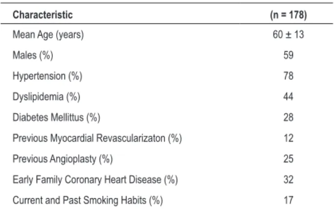
Table 1 - Patients’ Clinical Data
Characteristic (n = 178)
Mean Age (years) 60 ± 13
Males (%) 59
Hypertension (%) 78 Dyslipidemia (%) 44 Diabetes Mellittus (%) 28 Previous Myocardial Revascularizaton (%) 12 Previous Angioplasty (%) 25 Early Family Coronary Heart Disease (%) 32 Current and Past Smoking Habits (%) 17
n - number of patients.
individuals (25%) were diagnosed with non-cardiac pain (NC). Patients’ clinical data from the different diagnosis groups can be found in Table 2.
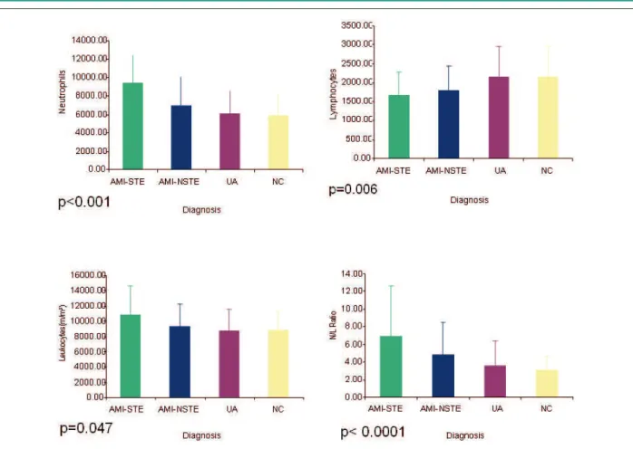
Leukocyte count and Subtypes Following Final Diagnosis Total leukocyte count showed gradual incerase of leukocytes, lower in the NC (8,825 ± 2,443) and higher among patients diagnosed with AMI-STE (10,804 ± 3,777); p<0.005. Similarly to total count, the number of neutrophils was lower among NC (5,921 ± 2,304), intermediate in UA patients (6,096 ± 2,448) and AMI-NSTE (6,889 ± 3,187) and higher among AMI-STE patients (9,391 ± 2,960). However, lower levels of lymphocytes could be observed in the AMI-STE group (1,684± 592), followed by AMI-NSTE (1,794 ± 647). Highest levels could be observed in the NC group (2,156 ± 782) (Graphic 1).
N/L ratio was directly associated to final diagnosis. That ratio was the highest among AMI-STE patients: 6.9 ± 5.7. Among AMI-NSTE patients the ratio was 4.8 ± 3.7, among those with UA it was 3.6 ± 2.9. The lowest ratio was found whenever a cardiologic cause was ruled out (NC) - 3.0 ± 1.6.
Assessing Diagnostic Value of N/L Test
Above 5.7 (highest quartile), the N/L ratio reported a 31% sensitivity (CI 95%: 23-38%) with specificity and 91.1% PPV (CI 95%: 83-99%) for the diagnosis of ACS. Test accuracy was 53%. ROC curve was constructed to assess the ability of N/L ratio to predict the presence of ACS. The under ROC curve area obtained was 63%.
Total count of leukocytes, neutrophils, lymphocytes, as well as N/L ratio were also calculated through Odds Ratio (OR) for ACS diagnosis by comparing patients in the highest quartile to all the others. OR for total leukocyte count for ACS and NC patients was 1.23 (CI 95% 0.55-2.75). Neutrophils and lymphocytes count: 2.14 (CI 95% 0.88-5.23) and 0.47 (CI 95% 0.22-1.003) respectively. As for N/L ratio, OR was 4.51 (CI 95% 1.51-13.45) (Graphic 2).
Considering that AMI-STE diagnosis is essentially electrocardiographic, 35 patients who were diagnosed were excluded. Specificity and sensitivity were also calculated for
the diagnosis of ACS with no ST elevation (UA and AMI-NSTE). Sensitivity was kept low: 23% (CI 95% 15 – 32%). The N/L ratio, however, also reported high specificity - 91% (CI 95% 83 - 99%)- and high positive predictive value – 85% (CI 95% 72 – 99%) for the diagnosis of ACS with no ST elevation.
N/L Correlation and Myocardial Necrosis Marker
With the purpose to assess whether the N/L ratio was directly associated to myocardial necrosis, the study correlated N/L and the highest CKMB obtained at the Chest Pain Unit. The correlation was not strong (Pearson r=0.48) but significant (p<0.0001) between the two variables. However, for the 68 patients who presented CKMB above the upper limit of normal levels the two variables presented poor correlation (Pearson r=0.40; p=ns).
Discussion
The findings of the present study demonstrate that the ratio obtained from a universally known, simple, low-cost test provides relevant information regarding the risk of patients who are admitted with chest pain to also be presenting ACS. In a country where the lack of resources keeps the access of so many to the best diagnostic methods, the N/L ratio may be turned into an additional parameter for the preliminary approach of patients with suspicion of ACS.
It has been estimated that from 5% to 10% of all emergency room assistance in the United States results from chest pain or other symptoms suggesting acute myocardial ischemia every year10. In Brazil no statistical estimate is available.
Although chest pain may have a wide range of causes, those resulting from cardiocirculatory system are of highest concern to patients and health professionals. In an attempt to both improve diagnosis accuracy and optimize expenses, the Chest Pain Units have come to the scene as a new approach for emergency assistance. Low-cost and highly predictive diagnostic methods for ACS are the very objective of CPUs.
High leukocyte count showed to be an independent predictor for ACS at the emergency room setting for individuals presenting chest pain suggestive of coronary heart disease, thus
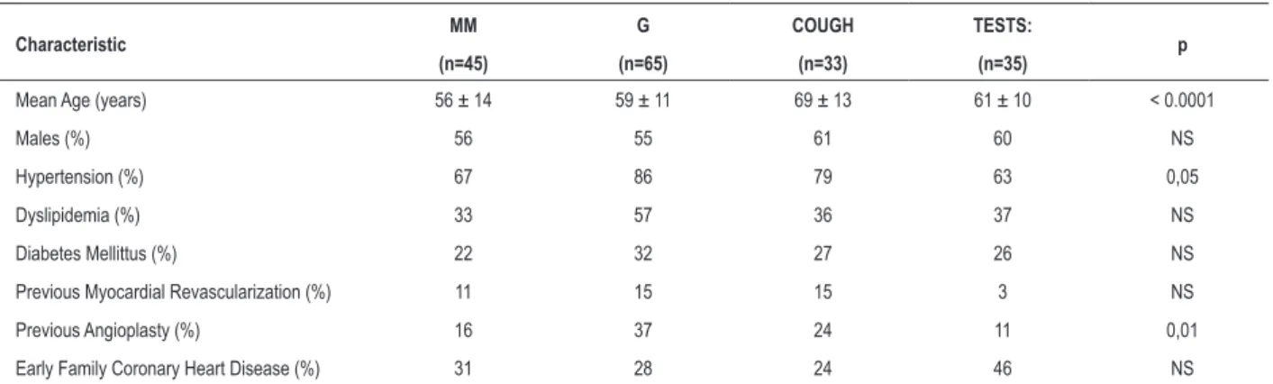
Table 2 -Clinical Data and Risk Factors in Diagnostic Groups
Characteristic MM G COUGH TESTS: p
(n=45) (n=65) (n=33) (n=35)
Mean Age (years) 56 ± 14 59 ± 11 69 ± 13 61 ± 10 < 0.0001
Males (%) 56 55 61 60 NS
Hypertension (%) 67 86 79 63 0,05
Dyslipidemia (%) 33 57 36 37 NS
Diabetes Mellittus (%) 22 32 27 26 NS
Previous Myocardial Revascularization (%) 11 15 15 3 NS
Previous Angioplasty (%) 16 37 24 11 0,01
Early Family Coronary Heart Disease (%) 31 28 24 46 NS
highest predictive cells12, whereas others have demonstrated
that neutrophils are the highest preditors13,14.
Post-AMI healing processes suggest the adaptive characteristic of neutrophils. More recently, it was proposed that increased vasculogenesis may be the response to ischemia conditions, therefore explaining a chronic adaptation process with higher number of circulating neutrophils. On the other hand, in the ACS scenario, neutrophils may be associated to the formation of aggregates between platelets and leukocytes in the intravascular lumen, thus even determining the increase of infarction extension areas15. Recent studies with animal models
have shown that neutrophilic invasion of the atherosclerotic plaque had direct visualization. Neutrophils may make plaque rupture easier through the release of protheolytic enzymes, arachidonic acid derivatives, and superoxide radicals16.
Therefore, the higher count of neutrophils may not only mirror the exacerbated inflammatory condition found in atherosclerotic patients but also be associated to the role played by those cells in atherosclerotic plaque instability.
Considering that the atherosclerotic plaque contains macrophage infiltrates and lymphocytes in its subendothelial layer, it could be postulated that the increase of polymorphonuclear cells is associated to the increase of mononuclear cells for the stability of the different plaque components. However, it has been observed that AMI-showing evidence that leukocytes would act as markers for
exacerbated inflammatory conditions11. However, few studies
have evaluated the role played by leukocytes subtypes in determining cardiovascular risk, and results are somewhat conflicting. One study demonstrated that monocytes are the
Fig. 1 - Neutrophils, lymphocyte, leukocyte count, as well as N/L ratio in the different diagnoses . AMI-STE (Acute Myocardial Infarction with Segment T Elevation; AMI-NSTE (Acute Myocardial Infarction with No Segment T Elevation; UA - Unstable Angina; NC - non-cardiologic - patients with non-cardiac pain; N/L - Neutrophils/ Lymphocytes.
STE patients presented lower lymphocyte concentration as compared to AMI-NSTE patients, followed by those presenting UA and NC. Results from other prospective studies have demonstrated that absolute and relative lymphocyte concentrations are significantly lower in patients with cardiac events. They have also shown those patients to be exposed to higher risk of future cardiac events17-19. Such reduction seems
to be related to the physiological stress of such conditions, thus determining cortisol production increase, which in its turn results in lymphocyte count decrease in peripheral blood20.
It has been demonstrated that increased total leukocyte count after coronary angioplasty is associated to higher mortality rate7. More recently, the relevance of N/L ratio
has been investigated in the 3-year follow-up of coronary angioplasty patients8. The present study showed an
independent, strong association between N/L ratio and mortality rate of patients submitted to percutaneous intervention (PI), irrespective of indication, thus showing that N/L ratio (but not total leukocyte count) increased mortality rate in the long run. Those findings support the evidence that neutrophils are associated to ischemic processes, particularly in acute phases.
The present study has not evaluated N/L ratio increase mechanisms. However, the N/L ratio does not seem to be associated to myocardial necrosis per se, since in patients presenting increased CKMB its correlation with N/L was poor and had no statistical significance. Studies on chronic condition patients may demonstrate whether there is any correlation between this ratio and the already established coronary heart disease. Afiune et al21 have demonstrated that
among all leukocytes subtypes, the monocytes are the ones to be associated to the highest rate of coronary disease. One possibility is that the N/L ratio acts as a better marker for ACS than leukocytes isolated subtypes. Such possibility is based on two distinctive mechanisms: neutrophilia would reflect systemic inflammatory status, and as a consequence, higher cardiovascular risk, and lymphopenia would reflect the acute stress presented by ACS.
Low test sensitivity (31%) makes clear that the N/L ratio will not play a diagnostic role if used isolatedly. On the other hand, high specificity and high positive predictive value may be most invaluable at centers where myocardial necrosis evaluation through laboratory exams is still carried out exclusively through CKMB dosing. Our data
are suggestive that individuals admitted with acute chest pain presenting significant N/L increase may need more detailed investigation to avoid inadverted patient discharge and consequent future complications. Cost-effectiveness studies are to confirm such hypothesis.
The present study does pose some limitations. Although it was a known fact that their diagnosis and therapeutic decision essentially depended on ECG, AMI-STE patients were included in the analysis. However, it was important for the authors to be aware of N/L ratio in all ACS spectrums. Another limitation was the fact that troponin dosing was not calculated in those patients. Therefore, diagnosis was obtained without that high specificity and sensitivity marker. Troponin dosing would also allow a more accurate evaluation of UA and AMI-NSTE groups. As study sample was calculated to detect the difference between ACS patients and those with non-cardiac pain, the role played by N/L in the differentiation of those patients was also limited by sample size. The detection of smaller differences – such as the one between those groups – obviously requires larger samples.
Non-dosing of troponin is a common limitation at tertiary services providing assistance to the National Health System (SUS) only. And that impossibility is exactly what shows the relevance of the present study: the proposal of a new ACS marker that is low cost, widely available and not new technology dependent. Therefore, the present study should be seen as a generator for new hypotheses only. Future mechanism studies may ellucidate the correct pathophysiology of neutrophil increase, and particularly lymphocytes reduction in these patients. Likewise, new studies should elucidate whether N/L ratio may play a role in diagnostic flowcharts currently in use at Chest Pain Units.
Potential Conflict of Interest
No potential conflict of interest relevant to this article was reported.
Sources of Funding
This study was partially funded by Fundação Araucária.
Study Association
This study is not associated with any graduation program.
References
1. Ross R. Atherosclerosis –an inflammatory disease. N Engl J Med. 1999; 340: 115-26.
2. Libby P. Vascular biology of atherosclerosis: overview and state of the art. Am J Cardiol. 2003; 91: 3A – 6A.
3. Libby P. Molecular basis of the acute coronary syndromes. Circulation. 1995; 91: 2844-52.
4. Falk E, Shah P, Fuster V. Coronary plaque disruption. Circulation. 1995; 92: 657-71.
5. Pearson T, Mensah G, Alexander R, Anderson JL, Cannon RO3rd, Criqui M,
et al. Markers of inflammation and cardiovascular disease: application to clinical and public health practice: a statement for healthcare professionals from the Centers for Disease Control and Prevention of the American Heart Association. Circulation. 2003; 107: 499-511.
6. Tall AR. C- reactive protein reassesed. N Eng J Med. 2004; 350: 1350-2.
7. Horne BD, Anderson JL, John JM, Weaver A, Bair TL, Jensen KR, et al. Which white blood cells subtypes predict increased cardiovascular risk? J Am Coll Cardiol. 2005; 45: 1638-43.
elevated neutrophil to lymphocyte ratio in predicting long term mortality after percutaneous coronary intervention. Am J Cardiol. 2006; 97 (7):993-6.
9. Sociedade Brasileira de Cardiologia. I Diretriz de dor torácica na sala de emergência. Arq Bras Cardiol. 2002; 79 (supl. 2): 1-22.
10. Nourjah, P. National Hospital Ambulatory Medical Care Survey: 1997 emergency department summary. Adv Data. 1999; 304: 1-24.
11. Menon, V; Lessard, D; Yarzbski, J. Leukocytosis and adverse hospital outcomes after acute myocardial infarction. Am J Cardiol. 2003; 92: 368-72.
12. Sweetnam P, Thomas H, Yarnell J, Baker I, Elwood P. Total and differential leukocyte counts as predictors of ischemic heart disease: the Caerphilly and Speedwell studies. Am J Epidemiol. 1997; 145: 416-21.
13. Huang Z-C, Chien K, Yang C, Wang C-H, Chang T, Chen C. Peripheral differential leukocyte counts and subsequent mortality for all diseases, cancers, and cardiovascular diseases in Taiwanese. J Formos Med Assoc. 2003; 102: 775-81.
14. Kawaguchi H, Mori T, Kawano T, Kono S, Sasaki J, Arakawa K. Band neutrophil count and the presence and severity of coronary atherosclerosis. Am Heart J. 1996; 132 (1 Pt 1): 9-12.
15. Siminiak T, Flores N, Sheridan D. Neutrophil interactions with endothelium and platelets: possible role in the development of cardiovascular injury. Eur Heart J. 1995; 16: 160-70.
16. Naruko T, Ueda M, Haze K, van der Wal AC, van der Loos CM, Itoh A, et al. Neutrophil infiltration of culprit lesions in acute coronary syndromes. Circulation. 2002; 106: 2894-900.
17. Roy D, Quiles J, Avanzas P, Arroyo-Espliguero R, Sinha M, Kaski JC. A comparative study of markers of inflammation for the assessment of cardiovascular risk in patients presenting to the emergency department with acute chest pain suggestive of acute coronary syndrome. Int J Cardiol. 2006; 109 (3): 317-21.
18. Zouridakis EG, Garcia-Moll X, Kaski JC. Usefulness of the blood lymphocyte count in predicting recurrent instability and death in patients with unstable angina pectoris. Am J Cardiol. 2000; 86: 449-51.
19. Ommen SR, Gibbons RJ, Hodge DO, Thomson SP. Uselfulness of the lymphocyte concentration as a prognostic marker in coronary heart disease. Am J Cardiol. 1997; 79: 812-4.
20. Thompson, SP; Mcmahon, LJ; Nugent, CA. Endogenous cortisol: a regulator of the number of lymphocytes in peripheral blood. Clin Immunol Immunopathol. 1980; 17: 506-14.