The Importance of the Interaction between Leukocyte Integrin Mac-1
DQG3ODWHOHW*O\FRSURWHLQ,EƠIRU/HXNRF\WH5HFUXLWPHQWE\3ODWHOHWV
and for the Inflammatory Response to Vascular Injury.
Alexandre C. Zago, Daniel I. Simon, Yunmei Wang, Masashi Sakuma, Zhiping Chen, Kevin Croce, Valentin Ustinov, Can Shi, Eulógio E. Martinez Filho
Instituto do Coração (InCor) - Universidade de São Paulo, Brigham and Women`s Hospital/Harvard University, Boston, Massachussets, EUA, Hospital de Clínicas de Porto Alegre (HCPA)/Universidade Federal do Rio Grande do Sul, The Cleveland Clinic Foundation of Medicine, Houston, Texas, EUA
Summary
Objective: To assess the importance of the interaction between leukocyte integrin Mac-1 (DME2) and platelet glycoprotein (GP) Ib-D for leukocyte recruitment after vascular injury and the effect of the neutralization of the Mac-1-GPIbD interaction on cell proliferation and the neointimal hyperplasia triggered by the vascular injury.
Methods: A peptide called M2 or anti-M2 antibody was developed to block the Mac-1-GPIbD interaction. This peptide was injected and compared to a control-peptide in C57B1/6J mice submitted to vascular injury of the femoral artery with a guide wire. One, five or 28 days after the vascular injury, the femoral arteries were removed for morphometric and immunohistochemical analyses.
Results: The blocking of the Mac-1-GPIbD interaction promoted a statistically significant reduction in the number of leukocytes in the neointimal layer on the first day after the vascular injury (control: 7.9±5.0% of the cell total versus anti-M2: 2.0±1.6%, p=0.021), as well as determined a statistically significant decrease in leukocyte accumulation in the neointimal layer on days 5 and 28 (control: 42.3±12.9% versus anti-M2: 24.6±10.8%, p=0.047 and control: 7.9±3.0% versus anti-M2: 3.3±1.3%, p=0.012; respectively). Cell proliferation in the neointimal layer of the vessel five days post-injury was reduced with the blocking of the Mac-1-GPIbD interaction (control: 5.0±2.9% of the cell total versus anti-M2: 1.8±0.5%; p=0.043), along with a significant decrease in cell proliferation in the vessel neointimal layer 28 days post-injury (control: 3.8±1.7% versus anti-M2: 2.0±1.2%; p=0.047). The blocking of the Mac-1-GPIbD interaction also determined a statistically significant decrease of the intimal thickening 28 days post-injury (control: 10,395±3,549 Pm2 versus anti-M2: 4,561±4,915Pm2; p=0.012).
Conclusion: Leukocyte recruitment after a vascular injury depends on the Mac-1-GPIbD interaction and the neutralization of this interaction inhibits cell proliferation and neointimal formation. (Arq Bras Cardiol 2008;90(1):52-60)
Key words: Cell adhesion molecules; inflammation; leukocytes; blood platelets; coronary restenosis.
Mailing address: Alexandre do Canto Zago •
Rua Ramiro Barcelos, 2350, 2º andar - Hemodinâmica – 90035-003 - Porto Alegre, RS - Brazil
E-mail: [email protected] / [email protected]
Manuscript received on 12/20/06; revised manuscript received on 03/28/07; accepted on 05/09/07.
Introduction
Increasing evidence of experimental and clinical studies supports the perception of atherosclerosis and re-stenosis as an inflammatory process. In this context, the inflammation is characterized by the interaction between platelets, leukocytes and endothelial cells1.
Experimental and clinical studies suggest a central role of leukocytes in neointimal proliferation due to vascular injury. In experimental models of vascular injury, the recruited leukocytes are pointed out as the precursors of the neointimal proliferation2,3.
Once adhered to the vascular wall, the platelets form an adhesive surface for the circulating leukocyte recruitment, which involves the following phases: adhesion mediated by selectins and rolling over the platelet surface, leukocyte activation, firm adhesion mediated by integrins and diapedesis4-6.
The phase of leukocyte firm adhesion to activated platelet surface is mediated by integrins from the E2family, of which integrin Mac-1 is emphasized, also known as macrophage-1 antigen,DME2or CD11b/CD18.
A study in mice with integrin Mac-1 deficiency and submitted to vascular injury showed a decreased migration and accumulation of leukocytes on the arterial wall as well as attenuation of the neointimal proliferation7. Another study,
Simon and cols.9 recently located the binding site of integrin
Mac-1 of the leukocytes with the IbD glycoprotein (GP) of platelets inside the segment DM(P201-K217) of the D
MI
9 domain, which
allowed the development of a peptide called M2 or anti-M2.
In vitro studies with the M2 peptide showed a significant adhesion reduction of 293 cells, which express integrin Mac-1, to sGPIbD, as well as a significant reduction of human leukocyte adhesion to platelets under continuous flow10.
The present study aims at evaluating the importance of the interaction between leukocyte integrin Mac-1 and platelet IbD glycoprotein for leukocyte recruitment after the vascular injury and the effect of the interaction neutralization between integrin Mac-1 and IbD glycoprotein on cell proliferation and neointimal hyperplasia triggered by vascular injury.
Methods
Materials
Peptides corresponding to the binding site of integrin Mac-1 on GPIbD receptors of mice (SGSG-214LYFRHWLQENANNVYL229-C)
and control peptides (SGSG-VEAFHLNYYRNNVWLQ-C) were obtained from W.M. Keck Biotechnology Resource Center – Yale University (New Haven, CT, EUA). The peptides were diluted in dimetilsulphoxide (DMSO) and stored at -80°C.
Fifty-three male C57BI/6J mice, aged 8 to 10 weeks, were obtained from the Animal Facility of Harvard University (Boston, MA, USA).
The common antigen leukocyte marker (CD45) –anti-CD45 antibody – was obtained from BD Biosciences (San Diego, CA, EUA); the specific marker for mouse neutrophils – monoclonal antibody 7/4 was supplied by Serotec (Indianapolis, IN, EUA); the Mac-3 marker, specific for mouse macrophages –monoclonal antibody M3/84 – was obtained from BD Biosciences and the bromodeoxyuridine (BrdU) was supplied byDAKO (Carpinteria, CA, USA).
Antibody development
A peptide-specific polyclonal antibody called peptide M2 or anti-M2 antibody, with affinity to the binding site of integrin Mac-1 to GPIbD receptor, was developed through the immunization of rabbits with the C-201
PITQLLGRTHTATGIRK217 peptide coupled to KLH (Zymed
Laboratories, South San Francisco, CA, USA), corresponding to the human sequence Dm (P
201-K217).
Subsequently, blood tests were conducted, which demonstrated high titers of anti-serum bound to the immobilized solid phase containing the C-P201-K217amino
acid sequence.
The last phase was the purification of the peptide-specific polyclonal antibody – anti-M2 antibody or M2 peptide – by affinity chromatography (Zymed Laboratories).
IgG immunoglobulin from non-immunized rabbits was used as control antibody.
Antibody treatment
The C57BI/6J mice were randomly divided into two groups
of treatment:
1- Rabbit M2 peptide and;
2- IgG immunoglobulin from non-immunized rabbits. The M2 peptide or anti-M2 antibody was injected with the objective of blocking the interaction of integrin Mac-1 of the leukocytes with the platelet GPIbD, aiming at inhibiting the leukocyte adhesion to the vessel surface covered by platelets. The IgG immunoglobulin from non-immunized rabbits was injected to serve as control, as it does not interact with Mac-1 integrin of the leukocytes or the platelet GPIbD and does not present any inhibitory effect on the adhesion between leukocytes and platelets.
The antibodies were injected intraperitoneally in a single dose of 250 µg, 4 hours before the femoral artery injury (day 0) and subsequently in a single dose of 100 µg intraperitoneally on days 2, 4, 6, 8, 10 and 12 after the femoral artery injury.
Femoral artery injury
Male C57B1/6J mice aged 8 to 10 weeks were anesthetized on day 0 with ketamine (80 mg/kg SC) and xylazine (5 mg/kg SC). Subsequently, an incision in the groin was made, followed by dissection and exposure of the femoral artery, temporary clamping of the femoral artery at the inguinal ligament level and arteriotomy distal to the epigastric artery under visualization with the surgical microscope.
A 0.010” guide wire was inserted into the femoral artery through the arteriotomy orifice, the clamp was removed and the guide wire was advanced until the bifurcation with the aorta and retreated to the anterior position three times (total of three insertions), aiming at causing an endoluminal injury to the common femoral artery. After the guide wire removal, the arteriotomy site was ligated.
This experimental model, described by Roque and cols.11
and used in the present study by a surgeon blinded to the treatment groups, is very similar to the process of vascular injury repair observed in humans after a percutaneous coronary intervention with stent implantation. The vascular injury technique by guide wire determines the stripping of the endothelium, deposition of platelets and fibrin and accentuated vascular inflammation, which makes this model useful to demonstrate the function of inflammatory cells (leukocytes) recruited at the neointimal formation modulation12-14.
All animals survived until the moment of the planned euthanasia without bleeding or infection.
All animals received humane care throughout the experiment and the procedures were revised and approved by the Harvard Medical School Standing Committee on Animals, which follows the criteria established by the American Association for Accreditation of Laboratory Animal Care and by the National Institutes of Health.
Removal of the femoral artery and tissue collection
9; M2: n= 9) or 28 days (control antibody: n= 9; anti-M2: n= 9) after the vascular injury.
A 22-gauge butterfly catheter was inserted into the left ventricle for in situ perfusion of 0.9% saline solution followed by paraformaldehyde 4 % fixation in 0.1 M phosphate buffer (pH=7.3) for 10 minutes at a pressure of 100 mmHg.
The right and left femoral arteries were removed and immersed in buffered paraformaldehyde for posterior morphometric and immunohistochemical analysis.
All animals received BrdU at a dose of 50 mg/kg intraperitoneally (IP), 18 hours and one hour before the euthanasia, for the assessment of cell proliferation on the femoral artery wall.
The spleen and small intestine of three animals were used as controls for the immunohistochemical reactions due to the presence of abundant lymphoid tissue (leukocytes) in these organs.
Histology and morphometry
The removed femoral arteries were initially formalin-fixed and later processed and paraffin-embedded. Two 1-mm distant cross-sectional cuts were performed, which were stained by the hematoxylin-eosin and Verhoeff techniques.
A researcher blinded to the treatment groups measured the luminal, intimal and medial areas of each transversal plane, using a microscope equipped with a CCD camera coupled to a computer running the NIH Image software, v1.60.
The luminal area was defined as the internal area surrounded by the layer of endothelial cells; the intimal area was defined as the area between the lumen and the internal elastic lamina; and the medial area was defined as the area between the internal and external elastic laminas.
The ratio between the intimal and the medial areas (intimal area/medial area) was also calculated to assess the effect of the inhibition of the Mac-1-GPIbD interaction on this ratio, as well as to allow future comparisons with experimental models using larger animals.
The area delimitated by the external elastic lamina was measured to evaluate the vessel size behavior in the two groups throughout time, i.e., to determine whether there was positive or negative remodeling of the vessel.
The results of the two cross-sectional cuts of each artery were used to calculate the mean, which was considered in the evaluation and statistical analysis of the data.
To calculate the medial area and the assessment of the external elastic lamina in basal conditions, i.e., before the injury or treatment with anti-M2 antibody, a group of mice (n=5) was euthanized to perform the quantitative morphometry.
Immunohistochemistry
The immunohistochemical analysis was performed using the immunoperoxidase technique by the avidin-biotin system, with specific antibodies for each cell.
The anti-CD45 antibody (common leukocyte antigen) of mice was used as a leukocyte marker; the monoclonal antibody 7/4 was used for neutrophil identification and the
monoclonal antibody M3/84 or anti-CD163 was used as a macrophage marker.
The BrdU uses the substitution of nucleotides to replace thymidine by uridine in the DNA structure of the dividing cells,in vitro as well as in vivo. The identification of the cells marked by this technique allows proliferation assessment and quantification. Hence, this technique constitutes an important tool in the assessment of smooth muscle cell proliferation, which predominates in the composition of neointimal hyperplasia due to the inflammatory response to the vascular injury.
Cross-sectional cuts of the femoral artery, immunologically stained, were assessed to quantify the number of cells positively immunostained in relation to the total number of cells.
Statistical analysis
The data are presented as mean ± standard deviation (SD); the comparisons between the groups were carried out by non-pairedt test; and the probability values were considered to be statistically significant when p< 0.05.
Results
Quantitative Assessment of leukocytes, neutrophils and macrophages in the femoral artery of mice post-vascular injury after blocking the DME2-GPIbD interaction
Initially, the post-injury leukocyte recruitment was assessed in relation to time. The function of the DME2-GPIbD interaction
in this process was established, as there was a difference in the accumulation of leukocytes on the vessel wall of mice treated with M2 peptide or anti-M2 in comparison to the control group.
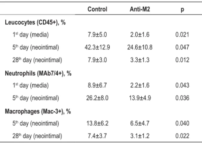
The CD45-positive inflammatory cells (leucocytes), which invaded the medial layer of the vessel on the first day post-injury were reduced by 75%, that is, from 7.9±5.0% of the total cells in the medial layer in the group of mice treated with non-immune IgG (control) to 2.0±1.6% in the group treated with anti-M2 (p=0.021) (Table 1) (Chart 1) (Fig. 1).
Table 1 - Quantitative analysis of leucocytes, neutrophils and macrophages in the femoral artery of mice after vascular injury
Control Anti-M2 p
Leucocytes (CD45+), %
1st day (media) 7.9±5.0 2.0±1.6 0.021
5th day (neointimal) 42.3±12.9 24.6±10.8 0.047
28th day (neointimal) 7.9±3.0 3.3±1.3 0.012
Neutrophils (MAb7/4+), %
1st day (media) 8.9±6.7 2.2±1.6 0.043
5th day (neointimal) 26.2±8.0 13.9±4.9 0.036
Macrophages (Mac-3+), %
5th day (neointimal) 13.8±6.2 6.5±4.7 0.040
The accumulation of leukocytes in the developing neointimal was also significantly reduced by the anti-M2 treatment, as there was a reduction of 42% in five days (control: 42.3±12.9% of the total cells in the neointimal versus
anti-M2: 24.6 ± 10.8%, p=0.047) and of 58% in 28 days (control: 7.9±3.0% versus anti-M2: 3.3±1.3%, p=0.012) in the group of mice treated with anti-M2 in comparison with the control group (Table 1) (Chart 1) (fig. 1).
The analysis of CD45-positive cells was extended through immunological staining techniques using specific cell markers, with the objective of quantifying the presence of neutrophils and macrophages.
The accumulation of neutrophils (cells that are positive for the monoclonal antibody 7/4) on the medial layer of the vessel on the first day post-injury was reduced by 75% in the group of mice treated with anti-M2, in comparison with the control group (control: 8.9±6.7% versus anti-M2: 2.2±1.6%; p=0.043). On the 5th day post-injury, the accumulation of
neutrophils in the developing neointimal layer was 47% lower in the group of mice treated with anti-M2, in comparison with the control group (control: 26.2±8.0% versus anti-M2: 13.9±4.9%; p=0.036).
No neutrophils were detected on the intimal and medial layers on the 28th day post-injury, in either treatment groups
(Table 1).
The macrophages (Mac-3-positive cells) were practically undetectable on the 1st day post-injury (<0.5% of the total
cells on the medial layer). On the 5th day post-injury, the
macrophage accumulation in the neointimal layer was reduced by 53% in the group of mice treated with anti-M2, in relation to the control group (control: 13.8±6.2% versus
anti-M2: 6.5±4.7%; p=0.040), whereas on the 28th day
post-injury, there was a reduction of 58% of macrophage
accumulation in the neointimal layer of mice treated with anti-M2 in relation with the control group (control: 7.4±3.7%
versus anti-M2: 3.1±1.2%; p=0.022) (Table 1).
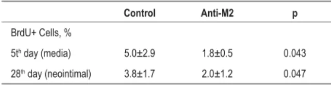
Effect of blocking the DME2–GPIbD interaction on cell proliferation after vascular injury
Significant cell proliferation was observed 5 days after the vascular injury in the group of mice treated with the control antibody and the cell proliferation was still evident on the 28th day post-injury.
The treatment of mice with the anti-M2 antibody determined, however, a statistically significant decrease of 64% in cell proliferation in the medial layer of the vessel five days post-injury (control: 5.0±2.9% of the total cells in the medial layer of the vessel versus anti-M2: 1.8±0.5%; p=0.043) and of 47% of cell proliferation in the intimal layer of the vessel 28 days post-injury (control: 3.8±1.7% of the total cells in the intimal layer of the vessel versus anti-M2: 2.0±1.2%; p=0.047) (Table 2).
Effect of blocking the DME2-GPIbD interaction on the quantitative morphometry of the intimal and medial areas after the vascular injury.
In mice treated with the control antibody, the intimal thickening started 5 days post-injury and progressed significantly between 5 and 28 days post-injury. The blocking of the
DME2-GPIbD interaction, promoted by the treatment with anti-M2 antibody or M2 peptide, resulted in the decrease of the intimal (neointimal) thickening 5 days post-injury, in relation to the control group (control: 921±534 µm2
versus anti-M2: 505±357 µm2; p=0.077), as well as in the
statistically significant reduction of the neointimal layer 28 post-injury in comparison to the control group (control:
10,395±3,549 µm2 versus anti-M2: 4,561±4,915 µm2;
p=0.012) (Table 3) (Figure 1).
The blocking of the DME2-GPIbD interaction in mice
treated with the anti-M2 antibody or M2 peptide determined, therefore, a reduction of 56% in the intimal thickening 28 days post-injury in relation to the control-group (Chart2).
The medial layer area was not affected by the treatment with the anti-M2 antibody. Hence, the relation between the intimal and medial layer areas expressed by the intimal
area/medial area ratio (I:M) 28 days post-injury was 56% lower in mice treated with the M-2 antibody in comparison with the control group (control: 1.20 ± 0.62 versus anti-M2: 0.53±0.62; p=0.036) (Table 3) (Chart 3).
Quantitative morphometry of the external elastic lamina of the femoral artery of mice treated with anti-M2 antibody after the vascular injury
The thickening of the intimal and medial layers was followed by progressive increase in vessel size (positive
Fig. 1 - Microphotographs of femoral arteries of mice after vascular injury. (a) Control-antibody (38X); (b) anti-M2 (38X); (c) control-antibody (150X); (d) anti-M2 (150X). The long arrows indicate the internal elastic lamina and the short arrows the external elastic lamina. The neointimal layer separates the internal elastic lamina from the lumen. CD45+ cells in brown (150X): (e) control-antibody – 1st day; (f) anti-M2 – 1st day; (g) control-antibody – 5th day; (h) anti-M2 – 5th day; (i) control-antibody – 28th
Table 2 - Quantitative analysis of cell proliferation (BrdU+ cells) in the femoral artery of mice after vascular injury
Control Anti-M2 p
BrdU+ Cells, %
5th day (media) 5.0±2.9 1.8±0.5 0.043
28th day (neointimal) 3.8±1.7 2.0±1.2 0.047
Table 3 - Morphometric analysis of the intimal and media areas of the femoral artery of mice after vascular injury
Control Anti-M2 p
Intimal Layer Area (µm2)
5th day 921±534 505±357 0.077
28th day 10,395±3,549 4,561±4,915 0.012
Media Layer Area (µm2)
0 day 9,583±1,203 ND
5th day 10,839±1,297 11,211±1,642 0.62
28th day 9,342±1,762 9,172±1,925 0.85
Intimal/Media Area
5th day 0.08±0.05 0.05±0.03 0.079
28th day 1.20±0.62 0.53±0.62 0.036
Chart 2 - Intimal area of the femoral artery of mice 28 days after vascular injury.
Chart 3 - Ratio between the intimal layer area and the media layer area (I:M) 28 days after vascular injury.
remodeling), assessed by the measurement of the area of the external elastic lamina in different periods of time (Table 4).
The positive remodeling of the vessel was similar in the two groups of treatment, 5 days (p=0.12) as well as 28 days after the vascular injury (p=0.99).
Discussion
The present study demonstrated that the antibody, which has as target the segment P201-K217 of the ID
M domain,
reduces leukocyte accumulation, cell proliferation and neointimal thickening after vascular injury in vivo. These observations demonstrate that the platelet GPIbD is a ligand that is physiologically relevant for the DME2 receptor (Mac-1) of leukocytes and that the DME2-GPIbD interaction is crucial for the pro-inflammatory leukocyte activity in the process of the vascular injury repair.
TheDME2 integrin regulates important leukocyte functions such as adhesion, migration, coagulation, proteolysis, phagocytosis, oxidative stress and signaling as it binds to several ligands such as fibrinogen15,16, factor X17, C3bi18, GPIbD9,19
and JAM-320.
The accurate identification of the ligand responsible for the accumulation of leukocytes at the vascular injury site, covered by adhered platelets, was, to date, still unknown. Although previous studies had shown that DME2 integrin directly promotes leukocyte recruitment at the sites of platelet and fibrin deposition6, the actual biological importance of
platelet contra-receptors for DME2, such as GPIbD, JAM-3 and fibrinogen bound to GPIIb/IIIa, is still quite unknown.
A previous study by Rogers and cols.8 ,in which the blocking
ofDME2 integrin with the monoclonal antibody M1/70 was carried out, was not conclusive regarding the identification of the physiological contra-receptor for DME2 integrin, because
M1/70 is a pan ligand antibody, i.e., it blocks all of the Mac-1(DME2) receptor.8Therefore, it is not possible to use M1/70
in clinical studies, despite the satisfactory results in this experimental study, considering that DME2integrin regulates important leukocyte functions and its total blocking would determine severe and life-threatening adverse effects.
Another study by Simon and cols.7 identified D
ME2 as a
molecular determinant of neointimal thickening after vascular injury. It was observed that the selective absence of DME2
in mice deficient for integrin Mac-1 impaired transplatelet Table 4 - External elastic lamina area of the femoral artery of mice after vascular injury
Control Anti-M2 p
External Elastic
/DPLQDȝP2
0 day* 31,792±11,293 NM
5th day 35,491±4,915 29,258±9,217 0.12
28th day 34,692±8,955 34,651±8,351 0.99
leukocyte migration to the inner vessel wall, decreasing the accumulation of leukocytes in the medial layer and neointimal thickening after experimental angioplasty. In the same study,
DME2deficiency was associated with a reduction of 67% in the early accumulation of leukocytes7.
It is noteworthy the fact that the magnitude of the reduction in the early leukocyte accumulation in the previously described study was similar to that obtained with the blocking of the
DME2-GPIbD interaction through the M2 peptide in the present study (75% of inhibition of CD45-positive cells), suggesting that leukocyte recruitment is largely dependent on the interaction betweenDME2 and GPIbD. This finding is also consistent with another, observed in a previous in vitro study, carried out by Ehlers and cols.9, in which the predominant interaction
between neutrophils and platelets adhered to a surface, after vigorous washing, seems to be mediated by the DME2-GPIbD interaction, considering that the M2 peptide caused inhibition superior to 80% of the neutrophil adhesion9.
The relative specificity of the inhibitory action of anti-M2 in relation to GPIbD, that is, it does not react with ICAM-1, fibrinogen and JAM-3, suggests a small contribution of other ligands for DME2 integrin in the context of vascular injury, with stripped endothelium and platelet deposition10.
Other potential ligands for DME2 integrin present in the platelet membrane include fibrinogen, which binds to GPIIb/ IIIa15,16, ICAM-221 and JAM-320.
The leukocyte-platelet interaction, mediated by fibrinogen bridges between DME2 and GPIIb/IIIa, was studied by Ostrovsky
and cols.22, who demonstrated in an experimental in vitro
study, that RGDS peptides as well as the substitution of normal platelets by thrombastenic ones (absence of GPIIb/IIIa) do not affect leukocyte accumulation on a surface of platelets22. The
deficiency of E3 (subunit of GPIIIa) affected neither leukocyte recruitment nor neointimal formation in mice submitted to an experimental model of femoral artery injury, identical to that of the present study13.
Although DME2 integrin binds to ICAM-1, this receptor is not
found in platelets, which express a similar receptor – ICAM-2. Diacovo and cols. demonstrated that the blocking of ICAM-2 has no effect on the firm adhesion of neutrophils over a surface of activated platelets, under continuous flow6.
Finally, Santoso and cols.20 recently reported that D ME2
integrin can also bind to JAM-3 of platelets, cooperating with GPIbD for the adhesion between neutrophils and platelets in vitro20. However, it was demonstrated in a previous study that
anti-M2 had a minimal inhibitory effect (13% of inhibition) in the interaction between the receptors DME2and JAM-310.
Anin vitro study, carried out by Wang and cols.10, using
the M2 peptide, demonstrated that leukocyte recruitment by platelets is largely mediated by the interaction between Mac-1 integrin (DME2) of the leucocytes and GPIbD of the platelets, in humans as well as in mice, as the biological model is very similar and the percentage of inhibition resulting from adhesion studies were statistically significant and comparable (80% in humans and 70% in mice). Even under flow conditions, the results of the adhesion inhibition were also statistically significant and similar (92% in humans and 81% in mice)10
In the present study, the data of the vascular injury experiment in mice allow us to conclude that leukocyte recruitment, after the vascular injury, depends on the interaction between Mac-1 integrin and GPIbD, as a specific antibody (anti-M2 or M2 peptide) directed at blocking the P201-K217 segment of the ID
M domain, which is responsible
for the interaction of leukocyte Mac-1 integrin and platelet GPIbD, promoted a statistically significant reduction of 75% in the leukocyte accumulation in the medial layer of the vessel, on the first day post-injury and reduced by 42% and 58% the leukocyte accumulation in the intimal layer of the vessel, 5 and 28 days after the vascular injury, respectively.
Similarly, the morphometric data of this vascular injury experiment showed, through the neutralization of the Mac-1-GPIbD interaction, that the latter modulates cell proliferation and neointimal formation. The anti-M2 antibody directed at blocking the Mac-1-GPIbD interaction was able to significantly reduce cell proliferation in the medial layer of the vessel wall by 64% on the 5th day post-injury and cell proliferation in the
intimal layer by 47%, on the 28th day post-injury, as well as
decrease neointimal formation by 56% on the 28th day after
the vascular injury (p=0.012).
Although the treatment with anti-M2 has reduced neointimal thickening by 56%, a less robust inhibitory effect when compared to the 80% of neointimal thickening inhibition with complete DME2 deficiency in genetically modified mice
in comparison to the wild-type ones, the anti-M2 outcome, in this first in vitro study, was better or at least similar, to the effect of rapamycin in experimental studies.
A study in rats submitted to carotid artery injury with balloon-catheter showed a reduction of 45% of neointimal thickening in the group of rats treated with rapamycin, after 14 days, when compared to the control group23. Another study,
carried out by Gallo and cols.24, showed that the administration
of rapamycin i.m., 3 days before and 14 days after angioplasty with over-dimensioned balloon-catheter in the coronary artery, reduced neointimal thickening by 50% in relation to the control group in the period of 28 days24.
Similarly, the outcome of anti-M2 was better, or at least comparable, to most studies carried out with paclitaxel. Drachman and cols.25 showed a reduction of 50% in the
neointimal thickening in iliac arteries of rabbits treated with paclitaxel and submitted to stent implantation, in relation to the control group25.
A study carried out in rats, using a model of carotid artery injury, showed a reduction of 70% in the neointimal thickening in the group treated with paclitaxel26. Two studies in swine
treated with super-dimensioned stents, in the coronary artery, showed that paclitaxel-eluting covered stents reduced neointimal thickening by 40%, in a period of 4 weeks, in comparison with the control group27,28.
It is noteworthy that rapamycin and paclitaxel are the anti-cell-proliferation drugs largely used in drug-eluting stents, also called drug-coated stents or medicated stents.
The first study with rapamycin-eluting stent in humans showed an angiographic restenosis rate equal to 0% at 4 and 12 months29,30. The randomized studies showed a statistically
months of 35.4%-41.7% for the conventional stent versus 3.2-3.9% for the rapamycin-eluting stent31,32.
Similarly, the RESEARCH study, which evaluated the performance of the rapamycin-eluting stent in the real world, i.e., in patients that were not selected by inclusion or exclusion criteria, confirmed these promising results in Intervention Cardiology practice, with an intra-stent restenosis rate of 6.3% at 6 months33.
The first study with paclitaxel-eluting stent in humans also showed an angiographic restenosis rate at 6 months of 0%34, whereas the randomized studies showed a reduction
of the angiographic restenosis rate at 6-9 months from 17.9-24.4% for the conventional stent to 2.3-5.5% for the paclitaxel-eluting stent35,36. The T-SEARCH study evaluated
the results of the paclitaxel-eluting stent in the real world and showed the need for target-vessel revascularization in patients selected by clinical criteria in only 5.4% of them, within a one-year period.37.
Considering the results of the experimental and clinical studies with rapamycin and paclitaxel, as well as the initial outcome of the blocking of the Mac-1-GPIbD interaction with anti-M2 antibody in this experimental study, it can inferred that there is a potential angiographic and clinical benefit for anti-M2 antibody or M2 peptide. It is clear that differences in methodology, the drug administration pathway and the species of animals used in the mentioned experimental studies with rapamycin and paclitaxel must be taken into account in the assessment of results when compared to those obtained in the present study.
The neutralization of the Mac-1-GPIbD interaction through the blocking of the DM(P201-K217) segment with anti-M2 antibody,
seems, therefore, to be a promising new therapeutic approach to prevent restenosis after percutaneous coronary intervention. However, additional studies with the local release of anti-M2 at different doses are strongly recommended, considering that the local release showed to have a crucial role for the successful use of anti-cell-proliferation drug-eluting stents.
Additionally, anti-M2 reduces firm adhesion and transmigration of leukocytes in sites of platelet deposition, one of the first steps of vascular inflammation. Hence, anti-M2 has a potential effect against vascular inflammation and seems to be promising in the control and/or prevention of coronary artery disease and aggressive/progressive vascular disease.
One of the limitations of the present study is that one cannot rule out the contribution of other ligands of the DME2 receptor and its functions, as well as exclude the participation
of other cell adhesion molecules in the biological response to the vascular injury. Likewise, the function of the adventitial layer of the vessel wall was not assessed as a source of infiltrating inflammatory cells, which constitutes another study limitation.
Leucocytes can infiltrate the wall through the lumen as well as through the adventitia; however, leukocyte recruitment through the adventitia layer is difficult to reliably assess in this experimental model due to the need for dissection and isolation of the artery from the adjacent tissues to cause the injury with guide wire.
Additionally, although the thickening of the intimal and media layers was followed by the progressive increase in vessel size or positive remodeling, which was similar in the mice treated with anti-M2 antibody or control, the cells from adventitia layer influenced vascular remodeling5, suggesting
that alternative experimental models of vascular injury will be necessary to investigate the responses of the adventitia layer to the injury.
Finally, the experimental model used does not contemplate the presence of the pre-existing atherosclerotic plaque in the inflammatory process site and the stent implantation, factors that might be considered additional limitations to this study, as it differs from the clinical treatment of atherosclerotic lesions with stent implantation.
Conclusions
Leukocyte recruitment after a vascular injury depends on the interaction between the leukocyte Mac-1-integrin (DME2)
and the platelet IbD glycoprotein and the neutralization of the Mac-1-GPIba interaction inhibits cell proliferation and neointimal formation.
Potential Conflict of Interest
No potential conflict of interest relevant to this article was reported.
Sources of Funding
This study was funded by Harvard University.
Study Association
This article is part of the thesis of doctoral submitted by Alexandre do Canto Zago, from Universidade de São Paulo-USP and Harvard University.
References
1. Gawaz M, Langer H, May AE. Platelets in inflammation and atherogenesis. J Clin Invest. 2005; 115: 3378-84.
2. Tanaka H, Sukhova GK, Swanson SJ, Clinton SK, Ganz P, Cybulsky MI, et al. Sustained activation of vascular cells and leukocytes in the rabbit aorta after balloon injury. Circulation. 1993; 88: 1788-803.
3. Rogers C, Welt FG, Karnovsky MJ, Edelman ER. Monocyte recruitment and
neointimal hyperplasia in rabbits: coupled inhibitory effects of heparin. Arterioscler Thromb Vasc Biol. 1996; 16: 1312-8.
4. Springer TA. Traffic signals for lymphocyte recirculation and leukocyte emigration: the multistep paradigm. Cell. 1994; 76: 301-14.
6. Diacovo TG, Roth SJ, Buccola JM, Bainton DF, Springer TA. Neutrophil rolling, arrest, and transmigration across activated, surface-adherent platelets via sequential action of P-selectin and the D2-integrin CD11b/CD18. Blood. 1996; 88: 146-57.
7. Simon DI, Chen Z, Seifert P, Edelman E, Ballantyne CM, Rogers C. Decreased neointimal formation in Mac-1-/- mice reveals a role for inflammation in vascular repair after angioplasty. J Clin Invest. 2000; 105: 293-300. 8. Rogers C, Edelman E, Simon DI. A mAb to the E2-leukocyte integrin
Mac-1 (CDMac-1Mac-1b/CDMac-18) reduces intimal thickening after angioplasty or stent implantation in rabbits. Proc Natl Acad Sci USA. 1998; 95: 10134-9. 9. Ehlers R, Ustinov V, Chen Z, Zhang X, Rao R, Luscinskas FW, et al. Targeting
platelet-leukocyte interactions: identification of the integrin Mac-1 binding site for the platelet counter receptor Glycoprotein IBD. J Exp Med. 2003; 198: 1077-88.
10. Wang Y, Sakuma M, Chen Z, Ustinov V, Shi C, Croce K, et al. Leukocyte engagement of platelet glycoprotein IbD via the integrin Mac-1 is critical for the biological response to vascular injury. Circulation. 2005; 112: 2993-3000.
11. Roque M, Fallon JT, Badimon JJ, Zhang WX, Taubman MB, Reis ED. Mouse model of femoral artery denudation injury associated with the rapid accumulation of adhesion molecules on the luminal surface and recruitment of neutrophils. Arterioscler Thromb Vasc Biol. 2000; 20: 335-42. 12. Chen Z, Keaney JF Jr., Schulz E, Levison B, Shan L, Sakuma M, et al. Decreased
neointimal formation in Nox2-deficient mice reveals a direct role for NADPH oxidase in the response to arterial injury. Proc Natl Acad Sci USA. 2004; 101: 13014-9.
13. Smyth SS, Reis ED, Zhang W, Fallon JT, Gordon RE, Coller BS. Beta(3)-integrin-deficient mice but not P-selectin-Beta(3)-integrin-deficient mice develop intimal hyperplasia after vascular injury: correlation with leukocyte recruitment to adherent platelets 1 hour after injury. Circulation. 2001; 103: 2501-7.
14. Roque M, Kim WJ, Gazdoin M, Malik A, Reis ED, Fallon JT, et al. CCR2 deficiency decreases intimal hyperplasia after arterial injury. Arterioscler Thromb Vasc Biol. 2002; 22: 554-9.
15. Wright SD, Weitz JS, Huang AJ, Levin SM, Silverstein SC, Loike JD. Complement receptor type three (CD11b/CD18) of human polymorphonuclear leukocytes recognizes fibrinogen. Proc Natl Acad Sci USA. 1988; 85: 7734-8. 16. Altieri DC, Bader R, Mannucci PM, Edgington TS. Oligospecificity of the
cellular adhesion receptor Mac-1 encompasses an inducible recognition specificity for fibrinogen. J Cell Biol. 1988; 107: 1893-900.
17. Diamond MS, Staunton DE, Marlin SD, Springer TA. Binding of the integrin Mac-1 (CD11b/CD18) to the third immunoglobulin-like domain of ICAM-1 (CD54) and its regulation by glycosylation. Cell. 1991; 65: 961-71. 18. Altieri DC, Morrissey JH, Edgington TS. Adhesive receptor Mac-1 coordinates
the activation of factor X on stimulated cells of monocytic and myeloid differentiation: an alternative initiation of the coagulation cascade. Proc Natl Acad Sci USA. 1988; 85: 7426-66.
19. Simon DI, Chen Z, Xu H, Li CQ, Dong J, McIntire LV, et al. Platelet glycoprotein IbD is a counterreceptor for the leukocyte integrin Mac-1 (CD11b/CD18). J Exp Med. 2000; 192: 193-204.
20. Santoso S, Sachs UJ, Kroll H, Linder M, Ruf A, Preissner KT, et al. The junctional adhesion molecule 3 (JAM-3) on human platelets is a counterreceptor for the leukocyte integrin Mac-1. J Exp Med. 2002; 196: 679-91.
21. Diacovo TG, de Fougerolles AR, Bainton DF, Springer TA. A functional integrin ligand on the surface of platelets: intercellular adhesion molecule-2. J Clin Invest. 1994; 94: 1243-51.
22. Ostrovsky L, King AJ, Bond S, Mitchell D, Lorant DE, Zimmerman GA, et al. A
juxtacrine mechanism for neutrophil adhesion on platelets involves platelet-activating factor and a selectin-dependent activation process. Blood. 1998; 91: 3028-36.
23. Gregory CR, Huie P, Billingham ME, Morris RE. Rapamycin inhibits arterial intimal thickening caused by both alloimmune and mechanical injury: its effect on cellular, growth factor, and cytokine response in injured vessels. Transplantation. 1993; 55: 1409-18.
24. Gallo R, Padurean A, Jayaraman T, Marx S, Roque M, Adelman S, et al. Inhibition of intimal thickening after balloon angioplasty in porcine coronary arteries by targeting regulators of the cell cycle. Circulation. 1999; 99: 2164-70. 25. Drachman DE, Edelman ER, Seifert P, Groothuis AR, Bornstein DA, Kamath
KR, et al. Neointimal thickening after stent delivery of paclitaxel: change in composition and arrest of growth over six months. J Am Coll Cardiol. 2000; 36: 2325-32.
26. Sollott SJ, Cheng L, Pauly RR, Jenkins GM, Monticone RE, Kuzuya M, et al. Taxol inhibits neointimal smooth muscle cell accumulation after angioplasty in the rat. J Clin Invest. 1995: 95: 1869-76.
27. Heldman AW, Cheng L, Jenkins GM, Heller PF, Kim DW, Ware M, et al. Paclitaxel stent coating inhibits neointimal hyperplasia at 4 weeks in a porcine model of coronary restenosis. Circulation. 2001; 103: 2289-95.
28. Hong MK, Kornowski R, Bramwell O, Ragheb AO, Leon MB. Paclitaxel-coated Gianturco-Roubin II (GRII) stents reduce neointimal hyperplasia in a porcine coronary in-stent restenosis model. Coron Artery Dis. 2001; 12: 513-5. 29. Sousa JE, Costa MA, Abizaid A, Abizaid AS, Feres F, Pinto IMF, et al. Lack
of neointimal proliferation after implantation of sirolimus-coated stents in human coronary arteries. Circulation. 2001; 103: 192-5.
30. Sousa JE, Costa MA, Abizaid A, Rensing BJ, Abizaid AS, Tanajura LF, et al. Sustained suppression of neointimal proliferation by sirolimus-eluting stents. Circulation. 2001; 104: 2007-11.
31. Moses JW, Leon MB, Popma JJ, Fitzgerald PJ, Holmes DR, O’Shaughnessy C, et al. Sirolimus-eluting stents versus standard stents in patients with stenosis in a native coronary artery. N Engl J Med. 2003; 349: 1315-23.
32. Schofer J, Schlüter M, Gershlick AH, Wijns W, Garcia E, Schampaert E, et al. Sirolimus-eluting stents for treatment of patients with long atherosclerotic lesions in small coronary arteries: double-blind, randomized controlled trial (E-SIRIUS). Lancet. 2003; 362: 1093-9.
33. Lemos PA, Hoye A, Goedhart D, Arampatzis CA, Saia F, van der Giessen WJ, et al. Clinical, angiographic, and procedural predictors of angiographic restenosis after sirolimus-eluting stent implantation in complex patients – an evaluation from the rapamycin-eluting stent evaluated at Rotterdam Cardiology Hospital (RESEARCH) Study. Circulation. 2004; 109: 1366-70. 34. Grube E, Silber S, Hauptmann KE, Mueller R, Buellesfeld L, Gerckens U, et
al. Six- and twelve-month results from a randomized, double-blind trial on a slow-release paclitaxel-eluting stent for de novo coronary lesions. Circulation. 2003; 107: 38-42.
35. Colombo A, Drzewiecki J, Banning A, Grube E, Hauptmann K, Silber S, et al. Randomized study to assess the effectiveness of slow- and moderate-release polymer-based paclitaxel-eluting stents for coronary artery lesions. Circulation. 2003; 108: 788-94.
36. Stone GW, Ellis SG, Cox DA, Hermiller J, O’Shaughnessy C, Mann JT, et al. A polymer-based, paclitaxel-eluting stent in patients with coronary artery disease. N Engl J Med. 2004; 350: 221-31.