Arq Bras Oftalmol. 2006;69(2):251-4
Perda da camada de fibras nervosas retiniana documentada por meio do Stratus OCT
TMem pacientes com adenoma hipofisário: relato de caso
Department of Ophthalmology. Faculdade de Medicina da Universidade de São Paulo - USP - São Paulo (SP) - Brasil.
1Postgraduate student of Department of Ophthalmology. Faculdade de Medicina da Universidade de São Paulo - USP - São Paulo (SP) - Brasil.
2Associate Professor of Department of Ophthalmology. Faculdade de Medicina da USP - São Paulo (SP) - Brasil. 3Associate Professor of Department of Ophthalmology. Faculdade de Medicina da USP - São Paulo (SP) - Brasil.
Endereço para correspondência: Mário Luiz Ribeiro Monteiro. Av. Angélica, 1757 - Conj. 61 - São Paulo (SP) CEP 01227-200
E-mail: [email protected] Recebido para publicação em 03.05.2005 Versão revisada recebida em 13.11.2005 Aprovação em 23.11.2005
Os autores não têm interesse comercial no equipamento utilizado neste estudo.
Nota Editorial: Depois de concluída a análise do artigo sob sigilo editorial e com a anuência do Dr. Eric Pinhei-ro de Andrade sobre a divulgação de seu nome como revisor, agradecemos sua participação neste processo. Bruno Campelo Leal1
Frederico Castelo Moura2
Mário Luiz Ribeiro Monteiro3
Keywords: Pituitary neoplasms; Retinal ganglion cells/cytology; Tomography, optical coherence; Optic chiasm/abnormalities; Nerve fibers/pathology; Vision disorders; Case reports [publication type]
INTRODUCTION
Pituitary adenomas are among the most important causes of lesions in optic pathways and produce bitemporal visual field loss due to chiasmal compression. Abnormalities in the retinal nerve fiber layer (RNFL) in long-standing lesions are also characteristic(1). In such cases, the crossed nerve
fibers are lost with preservation of the uncrossed nerve fibers. Therefore, RNFL loss occurs predominantly in the nasal and temporal side of the optic disc, a pattern identified on ophthalmoscopy as band atrophy (BA) that is important in diagnosis and in estimating the chances of visual improvement after optic pathway decompression(2-4).
Optical coherence tomography (OCT) is an in vivo technique for acquisi-tion of cross-secacquisi-tional images of retinal structures from which estimates of the thickness of retinal layers can be made. Recent studies showed that OCT-1 was able to distinguish mean values of RNFL of eyes with BA from those of normal controls in two series of patients(5-6). However, due to large variation
of normal values, very often it is difficult to define RNFL as abnormal in an individual patient. The newer version of OCT (Stratus OCTTM) has
incorpora-ted advances in image acquisition and provides a classification (within nor-mal limits, borderline, or outside nornor-mal limits) for each studied parameter based on comparison with an internal normative database. We had the opportunity of documenting the usefulness of such program in two patients with pituitary adenomas that demonstrated good correlation with the expected pattern of RNFL loss.
Retinal nerve fiber layer loss documented by Stratus OCT
TMin patients with pituitary adenoma: case report
Purpose: To report abnormalities of retinal nerve fiber layer (RNFL) thickness using optical coherence tomography (Stratus OCTTM) in patients
with pituitary adenoma. Methods: Two patients with long-standing bitemporal visual field defects and optic nerve band atrophy were submitted to optical coherence tomography examination (Stratus OCTTM).
Results: Both patients with band atrophy revealed diffuse loss of the retinal nerve fiber layer on Stratus OCTTM, with severe reduction in the
nasal and temporal areas of the optic disc. Retinal nerve fiber layer loss correlated well with visual field loss and with previous histological studies of band atrophy of the optic nerve. Conclusions: Stratus optical coherence tomography can provide useful information in the diagnosis of band atrophy from chiasmal lesions such as pituitary adenomas.
ABSTRACT
Arq Bras Oftalmol. 2006;69(2):251-4
252Retinal nerve fiber layer loss documented by optical coherence tomography (Stratus OCTTM) in patients with pituitary adenoma: case report
CASE REPORTS
Case 1
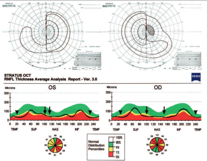
A 49-year-old man complained of visual loss 20 years pre-viously when a large pituitary adenoma was diagnosed. Sur-gical removal and postoperative use of bromocriptine resulted in visual improvement, however, a severe temporal visual loss remained in both eyes. Ophthalmic examination revealed a visu-al acuity of 20/20 in each eye. Fundus examination showed diffuse band atrophy of the optic disc in each eye and visual field testing revealed severe bitemporal hemianopia (Figure 1). Stratus OCTTM showed diffuse reduction of RNFL in both eyes
including the nasal and temporal portions of the disc (Figure 1).
Case 2
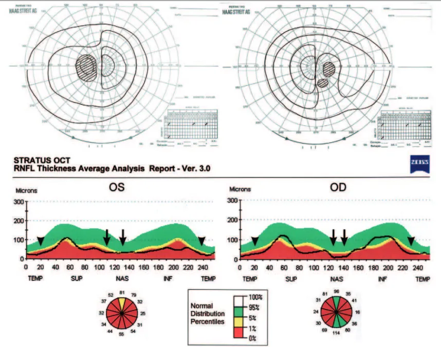
A 36-year-old woman presented headache and visual loss 10 years previously and was diagnosed as having a large pituitary
adenoma that was surgically removed. On examination her best corrected visual acuity was 20/20 in the right eye (OD) and 20/25 in the left eye (OS). She had bitemporal hemianopic scotomas with I/4e target and complete temporal hemianopia with I/2e target in both eyes (Figure 2). Fundus examination revealed band atrophy of the optic disc in both eyes. Stratus OCTTM
examina-tion showed diffuse reducexamina-tion of RNFL in both eyes, including the nasal and temporal portions of the disc (Figure 2).
DISCUSSION
In both cases Stratus OCTTM demonstrated RNFL loss
involving predominantly the nasal and temporal portions of the disc, with relative preservation of the superior and inferior quadrants. Such findings are in agreement with Mikelberg and Yidegiligne’s(3) histological analysis of a patient with band
Figure 1 - Case 1: Goldmann visual fields showing bitemporal severe loss. Stratus OCTTM showing diffuse retinal nerve fiber layer loss in both
Arq Bras Oftalmol. 2006;69(2):251-4
Retinal nerve fiber layer loss documented by optical coherence tomography (Stratus OCTTM) in patients with pituitary adenoma: case report 253
atrophy and seems to confirm a better potential of OCT when compared to the scanning laser polarimetry (SLP) in evalua-ting RNFL in the nasal and temporal areas of the disc(4-6). In a
previous study using an early commercial version of OCT (OCT-1), we have documented that RNFL measurements in patients with BA were significantly different from normal ones in each quadrant as well as in each of twelve, 30-degree seg-ments of the disc(5), a finding confirmed by the study of
Kanamori et al.(6). Large normal range and absence of a
norma-tive database in OCT-1, however, many times makes it difficult to define as abnormal an individual case. In this paper we have documented good correlation of the normative database of Stratus OCTTM and the expected RNFL loss. Further studies
with a larger number of patients, however, are necessary in order to better understand the real value of such normative database in diagnosis and management of diseases of the anterior visual pathways.
RESUMO
Objetivo: Relatar alterações na camada de fibras nervosas retiniana (CFNR) com o uso da tomografia de coerência óptica (Stratus OCTTM) em pacientes com adenoma hipofisário.
Mé-todos: Dois pacientes com defeito perimétrico bitemporal de longa duração e atrofia em banda do nervo óptico foram exami-nados com tomografia de coerência óptica (Stratus OCTTM).
Resultados: Ambos pacientes demonstraram perda difusa da camada de fibras nervosas retiniana com redução acentuada nas regiões nasal e temporal do disco óptico. A perda da camada de fibras nervosas retiniana se correlacionou com o defeito de campo visual e com os estudos histológicos de atrofia em banda do nervo óptico. Conclusões: A tomografia de coerência óptica (Stratus OCTTM) pode apresentar grande
utilidade no diagnóstico da atrofia em banda decorrente de lesões quiasmáticas tais como os adenomas pituitários.
Figure 2 - Case 2: Goldmann visual fields showing bitemporal hemianopic scotomas. Stratus OCTTM showing diffuse reduction of RNFL in both
Arq Bras Oftalmol. 2006;69(2):251-4
254Retinal nerve fiber layer loss documented by optical coherence tomography (Stratus OCTTM) in patients with pituitary adenoma: case report
Descritores: Neoplasias hipofisárias; Células do glângio reti-niano/citologia; Tomografia de coerência óptica; Quiasma óptico/abnormalidades; Fibras nervosas/patologia; Transtor-nos da visão; Relato de casos [tipo de publicação]
REFERENCES
1. Tanito M, Itai N, Goto T, Ohira A, Chihara E. Abnormalities of scanning laser polarimetry associated with pituitary adenoma. Am J Ophthalmol. 2003;135 (4):565-7.
2. Unsöld R, Hoyt WF. Band atrophy of the optic nerve. The histology of temporal hemianopia. Arch Ophthalmol. 1980;98(9):1637-8.
3. Mikelberg FS, Yidegiligne HM. Axonal loss in band atrophy of the optic nerve in craniopharyngioma: a quantitative analysis. Can J Ophthalmol. 1993;28(2): 69-71. 4. Monteiro MLR, Medeiros FA, Ostroscki MR. Quantitative analysis of axonal loss in band atrophy of the optic nerve using scanning laser polarimetry. Br J Ophthalmol. 2003;87(1):32-7.
5. Monteiro ML, Leal BC, Rosa AA, Bronstein MD. Optical coherence analysis of axonal loss in band atrophy of the optic nerve. Br J Ophthalmol. 2004;88(7): 896-9.
6. Kanamori A, Nakamura M, Matsui N, Nagai A, Nakanishi Y, Kusuhara S, et al. Optical coherence tomography detects characteristic retinal nerve fiber layer thickness corresponding to band atrophy of the optic discs. Ophthalmology. 2004;111(12):2278-83.