ABSTRACT
Evaluation of opening pattern and bone
neoformation at median palatal suture area in
patients submitted to surgically assisted rapid
maxillary expansion (SARME) through cone beam
computed tomography
Daniel Gomes SALGUEIRO1, Vitor Hugo Leite de Oliveira RODRIGUES1, Victor TIEGHI NETO1, Carolina Carmo de MENEZES2, Eduardo Sanches GONÇALES3, Osny FERREIRA JÚNIOR3
1- Master’s Student, Department of Stomatology, Bauru School of Dentistry, University of São Paulo, Bauru, Brazil.
2- Department of Pediatric Dentistry, Orthodontics and Community Health, Bauru School of Dentistry, University of São Paulo, Bauru, Brazil. 3- Department of Stomatology, Discipline of Oral Surgery, Bauru School of Dentistry, University of São Paulo, Bauru, Brazil.
Corresponding address: Osny Ferreira Júnior - Universidade de São Paulo, Faculdade de Odontologia de Bauru - Departamento de Estomatologia - Al. Octávio Pinheiro Brisola, 9-75 - 17012-901 - Bauru - SP - Brazil - Phone: +551432358258 - e-mail: [email protected]
6XEPLWWHG'HFHPEHU0RGL¿FDWLRQ0D\$FFHSWHG0D\
S
urgically assisted rapid maxillary expansion (SARME) is the treatment of choice to adult patients even with severe transversal maxillary discrepancies. However, the adequate retention period to achieve the bone remodeling, thus assuring treatment stability, is controversial. Objective: To evaluate the opening pattern and bone neoformation process at the midpalatal suture in patients submitted to surgically assisted (SARME) through cone beam computed tomography (CBCT). Material and Methods: Fourteen patients were submitted to SARME through subtotal Le Fort I osteotomy. Both the opening pattern and the mean bone density at midpalatal suture area to evaluate bone formation were assessed pre- and post-operatively (15, 60 and 180 days) through CBCT. Results: Type I opening pattern (from anterior to posterior nasal spine) occurred in 12 subjects while type II opening pattern (from anterior nasal spine to transverse palatine suture) occurred in 2 individuals. The 180-day postoperative mean (PO 180) of bone density value was 49.9% of the preoperative mean (Pre) value. Conclusions: The opening pattern of midpalatal suture is more related to patients’ age (23.9 years in type I and 33.5 years in type II) and surgical technique. It was not possible to observe complete bone formation at midpalatal suture area at the ending of the retention period studied (180 days).Keywords:
Cone beam computed tomography
INTRODUCTION
Aging results in both bone density increasing and alterations on the biological behavior of the craniofacial complex sutures due to skeletal maturity. Although the increasing of bone rigidity is variable among individuals, it offers greater resistance to the forces applied to midpalatal suture during maxillary expansion3.
Surgically assisted rapid maxillary expansion (SARME) or surgically assisted maxillary expansion
(SAME) has been indicated both for patients reaching skeletal maturity and those previously submitted to rapid maxillary expansion (RME) whose treatment failed5,22.
SARME varied from 19 to 29. The groups studied were quite small and mostly contained not more than 20 patients. The period of retention after expansion varies from 2 to 12 months. Generally, a period of 3 months is used13.
Still, the biological behavior of midpalatal suture of adult patients submitted to SARME has not amount required to achieve bone neoformation/ mineralization and the adequate retention period to achieve bone remodeling, thus assuring treatment stability9.
Cone beam computed tomography (CBCT) is a useful tool to asses quantitatively the effects of maxillary expansion30 and measure bone density at midpalatal suture area1. Moreover, CBCT provides tridimensional visualization of craniofacial structures and other advantages, such as: relatively conventional multislice tomography7.
The aim of this study was to evaluate the opening pattern and bone neoformation at the midpalatal suture area, immediately before and during orthodontic retention period, in patients submitted to surgically assisted rapid maxillary expansion through cone beam computed tomography.
MATERIAL AND METHODS
It was a prospective study and the sample comprised 56 digital volumetric tomographies of 14 adult patients (four images per patient), both genders, presenting posterior cross bite due to assisted rapid maxillary expansion (SARME), no systemic disease that contraindicated the procedure. The tomography device used in this research was !"#$ $ & Pennsylvania, USA). Expanders were intermittently activated. Patients wore either Hyrax (n=8) or Haas (n=6) appliances and did not have any contributory
disease contraindicating the surgical procedure. One surgeon performed all surgeries at hospital environment. The images used in this study came by the Ethics in Research Committee protocol number 401.136 and all participants signed an informed consent agreement.
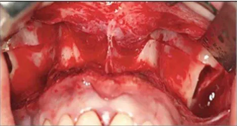
" ' Le Fort I with osteotomy of pterygoid pillars and midpalatal suture according to Betts, et al.5 (1995), but without osteotomy of the nasal septum (Figure 1). The four tomographies were obtained at pre-operative and 15-, 60-, and 180-day post-pre-operative periods for each patient. During the surgery, 1.0 g of cephalothin and 10 mg of intravenous dexamethasone were administered in addition to the drugs needed for the general anesthesia. Cephalothin was maintained for 24 hours and during this period they were under nursing care.
The following activation protocol was used: after 0.5 mm per day (¼turn at the morning and ¼turn at night) for 7 days followed by no activation for the next 7 days. The activation-latency cycles were interpolated until the palatal cusps of maxillary pre-molars and molars touched the buccal cusps of mandibular pre-molars and molars, achieving the desired expansion23.
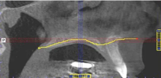
All tomographies were evaluated through i-CAT Vision software. Firstly irregular tool was used to mark an irregular line from posterior to anterior nasal spine, following the hard palate curvature at sagittal reformations (Figure 2). This tracing enabled to obtain the axial reformation of the marked area and to analyze the opening pattern and bone neoformation density at the midpalatal suture area (Figure 3).
The midpalatal suture opening pattern *+ i m a g e s a t 6 0 - d ay p o s t- o p e ra t i ve p e r i o d , corresponding to the ending of the expansion period, through visual analysis of the images. The
Figure 1- Subtotal Le Fort I osteotomy with zygomatic pillar step
; et al.19 (2010), as follows: Type I – opening from
$$– opening
from anterior nasal spine to transversal palatal suture (Figure 4).
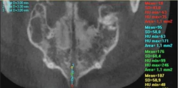
To measure the bone neoformation density, HU Statistics tool was used by marking rectangles (1 mm2) on all extension of the suture/disjunction of midpalatal suture at about 3 mm spacing among
them through Distance tool.
Once the rectangular area was marked, the & units (HU). Approximately 15 rectangular marks were performed along with the midpalatal suture on 56 sample images, divided into the following areas: anterior (from the anterior nasal spine to incisor foramen), median (from incisor foramen to transversal palatal suture) and posterior (from
Figure 2- Demarcation of hard palate curvature with the aid of “irregular” tool
Figure 3- New axial reformation obtained from hard palate demarcation
A B
transversal palatal suture to posterior transversal palatal suture) (Figures 5 and 6).
A single examiner assessed the images. Paired t-test was applied to verify the systematic error, at <>>?@ ’s formula was used to evaluate the casual error. ANOVA was applied for statistical analysis. All statistical tests were performed by Statistica 7.0 software.
RESULTS
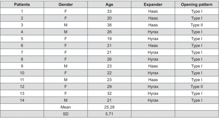
The studied sample comprised 56 tomographies of 14 individuals (9 females and 5 males) with mean age of 25.3 years (SD=5.71 years). Orthodontic appliances consisted of Hyrax (tooth born) and Haas (tooth and mucosa born), respectively installed in 6 and 8 individuals. Type I opening pattern of midpalatal suture was observed in 12 images, while Type II in 2 images (Figure 7 and 8).
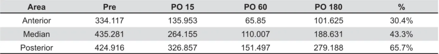
Table 1 displays the bone density mean of the
three evaluated maxillary areas: anterior, median and posterior, at each studied period. The bone density at these 3 periods: 15-day post-operative (PO 15), 60-day post-operative (PO 60) and 180-day post-operative (PO 180) was smaller than the Initial Period (Pre) in the three regions studied. The period allowed the evaluation of bone density after that the total activation (PO 60) demonstrated the smallest density values. The density values found after six months retention (PO 180) were lower than the Initial Period (Pre). The last column compares
Table 2 shows the density results at each studied period: pre-operative (Pre), 15-day post-operative (PO 15), 60-day post-operative (PO 60), and 180-day post-operative (PO 180) with the mean of each period and the percentage regarding to initial period J "KQX" differences were seen by Tukey test.
Figure 5- Density mean along with the area of interest
Figure 6- Marked anterior, median and posterior areas
DISCUSSION
Rapid maxillary expansion (RME) is considered to correct even severe transversal maxillary discrepancies. However, RME success rate decreases with aging24. Moreover, aging and the increasing of resistance against skeletal expansion is directly related because of cranial and face suture fusion12,13. Accordingly, RME is a treatment with high predictability in children and teenagers, but not in adults, commonly resulting in treatment failure and complications like tooth tipping and reduction in buccal cortical bone thickness23.
Ideally, the maxillary orthodontic expansion should preferably occur before 15 years old. However, expansion in individuals reaching skeletal maturity is possible, but without predictable and stable outcomes. RME after the pubertal peak tends to show more relapse, hence it is important to plan
6.
Usually, the orthopedic mechanics is capable of opening the midpalatal suture until 16-17 years old10. Considering individuals achieving skeletal maturity, RME does not provide stable expansion than 5 mm28.
Thus, currently, surgically assisted rapid
expansion (SARME) has been the treatment of choice, well accepted by the literature, to overcome the bone resistance to expansion5,22. This treatment modality consists of associating surgical and orthodontic procedures aiming at providing space on tooth arch for tooth alignment8.
A few studies demonstrated through finite element analysis the need of osteotomy of pterygoid pillars to decrease the stress concentration on this area, which was very high when the osteotomy was not performed2,12. Moreover, this procedure prevents damage to sphenoid bone and results in a more uniform expansion of the maxillary posterior area.
Other authors concluded that SARME associated with pterygomaxillary disjunction should be chosen in patients aged above 20 years15. The aforementioned study was conducted through computed tomography aiming to compare pre- and post-operative periods of patients submitted to SARME with and without pterygoid pillar disjunction.
SARME is considered a procedure with little risk of serious complications, although major complications are mentioned in literature, such as: life threatening epistaxis to a cerebrovascular accident, skull base fracture with reversible oculomotor nerve pareses and orbital compartment syndrome13. Less serious complications reported are post-operative hemorrhage, pain, sinusitis, palatal tissue irritation/ulceration, asymmetrical expansion, nasal septum deviation, periodontal problems and relapse23.
The mean age of this study’s patients was 25.3 years (Figure 1 and 2), which is in agreement with
Patients Gender Age Expander Opening pattern
1 F 33 Haas Type I
2 F 20 Haas Type I
3 M 38 Haas Type II
4 M 26 Hyrax Type I
5 F 19 Hyrax Type I
6 F 21 Haas Type I
7 F 21 Hyrax Type I
8 F 26 Hyrax Type I
9 M 23 Haas Type I
10 F 22 Hyrax Type I
11 M 23 Haas Type I
12 F 29 Hyrax Type II
13 F 32 Hyrax Type I
14 M 21 Hyrax Type I
Mean 25.28
SD 5.71
Figure 7- Gender, age, expander type and opening pattern of midpalatal suture
Type I Type II
Age 23.9 years 33.5 years
Density variables
Mean SD p
Pre 379.214C 148.193 0.000*
PO 15 234.732B 143.393 0.000*
PO 60 104.995A 152.092 0.000*
PO 180 189.287AB 100.968 0.000*
* p<0.05
Table 2- ANOVA comparative analysis of density variables before and after 15, 60, and 180 days in relation to the mean values and standard deviation (SD) of all areas. among means by Tukey test
the literature which reports that patients submitted to SARME showed an age range from 19-29 years15. The mean age of patients exhibiting type I opening pattern was 23.9 years, while the mean age of those presenting type II opening pattern was 33.5 years. This result corroborates the literature reporting the maxillary expansion in older individuals4. According to the opening pattern of the midpalatal suture. The were submitted to a single surgical technique, but nevertheless exhibited different opening patterns of the midpalatal suture. Thus, we can hypothesize that older individuals would show a tendency ' posterior area of maxilla, evidencing an opening pattern less parallel (V shaped). Consequently, these individuals would tend to present a type II opening pattern. Also, the results of this study regarding the expander type.
Although Hyrax and Haas appliances show similar effects, maxillary expansion is obtained through different mechanisms: dental-alveolar expansion by the former and more orthopedic expansion by the latter18. In this study, regardless of the expander type, the desired expansion of maxillary expansion was achieved with both appliances and the opening pattern of the midpalatal suture seems to be more related to the patient’s age and surgical technique than to the expander type used in the orthopedic mechanics, as aforementioned explained.
It is worth emphasizing the indispensable role of post-expansion orthodontic retention of the midpalatal suture because at this period both bone neoformation and remodeling take place after disjunction, decreasing relapse possibilities26. According to a study which compared the stability between rapid and slow maxillary expansion, the dental arch transverse dimensions showed a more pronounced relapse maintaining approximately 40% from control21. Progel, et al.22 (1992), Berger, et al.4(1998)and Koudstaal, et al.13 (2005), reported 0.88 mm (12%), 1.01 mm (17.5%) and 0.5 mm (55%) of relapse, respectively, in a 12-month study period. They concluded that expansion obtained with SARPE is stable, but all their patients were still in orthodontic treatment. Although a number of articles on the stability of surgically assisted rapid palatal expansion (SARPE) have been published, the reported stability varies considerably1,4,24.
After midpalatal suture disjunction, bone repair of the area is required to reach treatment stability. However, literature has reached no consensus regarding to the retention period necessary for bone repair29.
The midpalatal suture is the area in which the skeletal expansion is observed after RME or SARME and is commonly seen on conventional oclusal radiographs. On occlusal view, obtained through occlusal radiographs of the maxilla, midpalatal suture is visualized up to the middle of the palate. Consequently, the proper view of the posterior area of palate is not possible because of the superposition of the cranial base structures. Thus, the main advantage of computed tomography (CT) over oclusal radiograph of maxilla is the perfect visualization of the posterior area of midpalatal suture, without interference of the cranial structures16.
In one study through CBCT comprising 60 adult and 60 teenager patients, the authors concluded than that of teenagers, but the density of the anterior palatal area of teenagers was similar to that of posterior palatal area of adults11.
Similarly, a CT study with teenagers showed anterior to posterior nasal spine after a mean retention period of 8-9 months27. Nevertheless, in this present study, the complete bone neoformation
Area Pre PO 15 PO 60 PO 180 %
Anterior 334.117 135.953 65.85 101.625 30.4%
Median 435.281 264.155 110.007 188.631 43.3%
Posterior 424.916 326.857 151.497 279.188 65.7%
Table 1- 180) in relation to that of initial period (Pre)
of midpalatal suture did not take place within the studied period, that is, 180 days (6 months) of of the study conducted by Petrick, et al.20 (2011) through CT, who evidenced different density values between pre-operative and 7-month post-operative periods20. The bone density of the anterior, median and posterior areas was 48%, 53% and 75% of their initial values, respectively. The aforementioned "Z[\ should be longer. Probably the difference in the retention periods between these studies occurred because the former employed children submitted to
RME20,27. As shown by some studies5,12, RME tends
to result in incomplete opening of posterior palatal area and consequently bone repair is faster together with the fact that bone neoformation takes place faster in children27,20
.
In this study, similarly to that of Petrick, et al.20 (2011), the density values of the anterior area and posterior areas of maxilla. The bone density of anterior, median, and posterior areas was 30.4%, 43.3% and 65.7% of the initial values, respectively.
The authors from this research acknowledge the limitation on the small sample size. A study with a large number of patients, especially older individuals, is needed to conclude emphatically that the age affects the opening pattern and the bone neoformation. Another limitation was the assessment performed by a single examiner.
The importance of this study relies on the fact that the bone density at the ending of the retention period (180 days) was smaller than that before SARME. Accordingly, a retention period longer than *]> between pre- and post-operative periods.
CONCLUSIONS
Based on the methodology employed and the results obtained, it can be concluded:
1. The opening pattern of the palatal suture seems to be more related to patients’ age than to type of orthopedic mechanics employed.
2. The bone density values after 180 days of retention were smaller than those of the pre-operative period, demonstrating that this retention neoformation at the midpalatal suture area of the studied patients.
REFERENCES
1- Angelieri F, Cevidanes LH, Franchi L, Gonçalves JR, Benavides \ [K _" _ [ ` method for individual assessment before rapid maxillary "_Q@ Q{>*|*++`}?~~
2- Assis DS, Xavier TA, Noritomi PY, Gonçales AG, Ferreira O Jr, Carvalho PC, et al. Finite element analysis of stress distribution in anchor teeth in surgically assisted rapid palatal expansion. Int _Q[{>*|+{`*>~|~
3- Bell RA. A review of maxillary expansion in relation to rate of "_Q*~]{]*`|{} 4- Berger JL, Pangrazio-Kulbersh V, Borgula T, Kaczynski R. Stability of orthopedic and surgically assisted rapid palatal expansion over "_Q@ Q*~~]**+#`|]+? 5- Betts NJ, Vanarsdall RL, Barber HD, Higgins-Barber K, Fonseca Z_@ $_"QQ*~~?*>`}?~
6- Bishara SE, Staley RN. Maxillary expansion: clinical implications. "_Q@ Q*~]}~*`|*+
7- De Vos W, Casselman J, Swennen GR. Cone-beam computerized tomography (CBCT) imaging of the oral and maxillofacial region: a systematic review of the literature. Int J Oral Maxillofac Surg. {>>~|]`>~{?
8- Gonçales ES. Cirurgia ortognática: guia de orientação para portadores de deformidades faciais esqueléticas. São Paulo: \{>*>
~ _" [ [ ;JX !Z Q of the midpalatal suture after surgically assisted rapid maxillary \_Q{>*{|+`|~+|
10- Haas AJ. Palatal expansion: just the beginning of dentofacial "_Q*~}>?}`{*~??
11- Han S, Bayome M, Lee J, Lee YJ, Song HH, Kook YA. Evaluation of palatal bone density in adults and adolescents for application " Q{>*{]{`{?|* 12- Holberg C, Steinhäuser S, Rudzki I. Surgically assisted rapid maxillary expansion: midfacial and cranial stress distribution. Am _Q@ Q{>>}*|{`}}]{
13- Koudstaal MJ, Poort LJ, van der Wal KG, Wolvius EB, Prahl-Andersen B, Schulten AJ. Surgically assisted rapid maxillary expansion (SARME): a review of the literature. Int J Oral Maxillofac {>>?|+`}>~*+
*+ "" " " [ &" ! nasopharyngeal airway following orthopedic and surgically assisted _!{>*>{*`|*{} 15- Laudemann K, Petruchin O, Mack MG, Kopp S, Sader R, Landes CA. Evaluation of surgically assisted rapid maxillary expansion with or without pterygomaxillary disjunction based upon preoperative and post-expansion 3D computed tomography data. Oral Maxillofac {>>~*|`*?~~
16- Loddi PP, Pereira MD, Wolosker AB, Hino CT, Kreniski TM, Ferreira LM. Transverse effects after surgically assisted rapid maxillary expansion in the midpalatal suture using computed _!{>>]*~`+||]
17- Mitsuda ST, Pereira MD, Passos AP, Hino CT, Ferreira LM. Effects of surgically assisted rapid maxillary expansion on nasal dimensions using acoustic rhinometry. Oral Surg Oral Med Oral ;QZ\{>*>*>~`*~*
18- Oliveira NL, Silveira AC, Kusnoto B, Viana G. Three-dimensional assessment of morphologic changes of the maxilla: a comparison of 2 kinds of palatal expanders. Am J Orthod Dentofacial Orthop. {>>+*{`|?+{
19- Pereira MD, Prado GP, Abramoff MM, Aloise AC, Masako Ferreira ! assisted rapid maxillary expansion using computed tomography. QQ[ Q;QZ\{>*>**>`+*? 20- Petrick S, Hothan T, Hietschold V, Schneider M, Harzer W, Tausche E. Bone density of the midpalatal suture 7 months after surgically assisted rapid palatal expansion in adults. Am J Orthod @ Q{>***|~`*>~*
21- Pinheiro FH, Garib DG, Janson G, Bombonatti R, Freitas MR. Longitudinal stability of rapid and slow maxillary expansion. Dental ; _Q{>*+*~`}>}
23- Ribeiro PD Jr, Gonçales ES, Souza PC, Nary Filho H, Luz JGC. Clinical evaluation of surgically assisted maxillary expansion #"[\Z @ ; QQ{>>**`++?~ 24- Schauseil M, Ludwig B, Zorkun B, Hellak A, Korbmacher-Steiner H. Density of the midpalatal suture after RME treatment - a retrospective comparative low-dose CT-study. Head Face Med. {>*+*>`*]
25- Seeberger R, Kater W, Davids R, Thiele OC. Long term effects of surgically assisted rapid maxillary expansion without performing osteotomy of the pterygoid plates. J Craniomaxillofac {>*>|]`*}?]
26- Silva Filho OG, Capelozza Filho L, Fornazari RF, Cavassan AO. Rapid maxillary expansion: an essay on its instability. Rev Dental ; QQ{>>|]`*}|
27- Silva Filho OG, Lara TS, Silva HC, Bertoz FA. Behavior of the midpalatal suture in children submitted to rapid maxillary expansion: evaluation with computerized tomography. Rev Dent ; QQ{>>}*{`~+*>|
28- Silverstein K, Quinn PD. Surgically-assisted rapid palatal _ Q[*~~}??`}{?}
29- Suri L, Taneja P. Surgically assisted rapid palatal expansion: a "_Q@ Q{>>]*||`{~> 302.
30- Woller JL, Kim KB, Behrents RG, Buschang PH. An assessment of the maxila after rapid maxillary expansion using cone beam computed tomography in growing children. Dental Press J Orthod. {>*+*~`{|?