Rev Bras Anestesiol SCIENTIFIC ARTICLE 2011; 61: 3: 311-319
Revista Brasileira de Anestesiologia 311
Vol. 61, No 3, May-June, 2011
Received from Universidade Estadual de Campinas (Unicamp) and Faculdade de Ciências Médicas, Brazil.
1. Master’s Degree in Surgery Cirurgia, Post-graduate Student at the Departamento de Ciru-rgia of Faculdade de Ciências Médicas da Unicamp.
2. Head and Neck Surgeon of Hospital Centro Médico de Campinas; Chief of Oncologia Prefeitura de Paulínia
3. Assisting Physician of the Departamento de Clínica Médica, Faculdade de Ciências Médi-cas da Unicamp
4. Physiotherapist; Serviço de Doenças Pulmonares Unicamp
5. Professor, Physician of the Departamento de Anatomia Patológica of Faculdade de Ciên-cias Médicas da Unicamp
6. Medical Student at Faculdade de Ciências Médicas da Unicamp
7. Associate Professor of the Departamento de Cirurgia of Disciplina de Cirurgia de Cabeça e Pescoço of Faculdade de Ciências Médicas da Unicamp
Submitted on October 3, 2010. Approved on December 7, 2010. Correspondence to:
Dr. Alfio José Tincani
Departamento de Cirurgia Faculdade de Ciências Médicas Cidade Universitária “Zeferino Vaz”, s/nº
Barão Geraldo
13083970 – Campinas, SP, Brazil E-mail: [email protected]
SCIENTIFIC ARTICLE
Atraumatic Endotracheal Tube for Mechanical Ventilation
Silvio Oscar Noguera Servin
1, Gilson Barreto
2, Luiz Cláudio Martins
3, Marcos Mello Moreira
4,
Luciana Meirelles
5, José Alexandre Colli Neto
6, José Hélio Zen Júnior
6, Alfio José Tincani
7Summary: Servin SON, Barreto G, Martins LC, Moreira MM, Meirelles L, Colli Neto JA, Zen Júnior JH, Tincani AJ – Atraumatic Endotracheal Tube for Mechanical Ventilation.
Background and objectives: Patients who need to stay under endotracheal intubationfor long periods or when undergoing general anesthesia may develop tracheal lumen injuries due to pressure from distal cuff. In some cases, these injuries may evolve to stenosis or, occasionally, necro-sis. The objective of this study was to present a modified endotracheal tube (METT) in which the cuff pressure is variable according to the cycle of mechanical ventilation (MV), which was tested on a lung simulator and animal model.
Methods: Two models of endotracheal tubes, a modified (METT) and a conventional (CETT), number 7.5 mm and 8.0 mm, were connected to a lung simulator in a mechanical ventilator adjusted with two tidal volumes (TV) of 10 and 15 mL.kg-1 and a compliance of 60 mL.cmH
2O to evaluate the ventilatory efficiency of METT. Both models were also compared in Large-White pigs under general anesthesia and MV for 48 consecutive hours. Subsequently, animals were sacrificed for histopathological analysis of their tracheas.
Results: Both METTs (#7.5 and 8.0) presented air leaks in lung simulator. The smallest air leak(13%) was observed in METT #7.5 with TV = 15 mL.kg-1, while the largest air leak (32%) was observed in METT #8.0 with TV = 10 mL.kg-1. Nevertheless, both METTs showed good efficiency on the lung simulator. In animals, on histopathological analysis of their tracheas, it was found that METT caused less trauma to the epithelium when compared to CETT.
Conclusion: The use of a new model of ETT may decrease the risks of tracheal injury without hindering respiratory mechanics.
Keywords: Intubation, Intratracheal; Disposable equipament; Technology Assessment, Biomedical; Respiration, Artificial; Swine.
[Rev Bras Anestesiol 2011;61(3): 311-319] ©Elsevier Editora Ltda.
INTRODUCTION
Endotracheal tube (ETT) is used quite often in the medical field in patients requiring mechanical ventilation (MV). This might be for a short period for example in general anesthesia, or more prolonged periods such as in patients who need MV in intensive care units (ICU).
The endotracheal tube has a distal cuff, whose function is to seal and protect the airways, therefore preventing aspiration of secretions and allowing positive pressure ventilation. The
distal cuff exerts a certain amount of pressure on the tracheal wall, which should not be elevated. If this happens, it could prevent blood flow to the tracheal mucosa. High-compliance and low-pressure cuffs may minimize the risk of mucosal is-chemia, preventing injury. Therefore, the cuff inflation pressu-res should be adjusted between 20 and 30 cmH2O 1.
The most common endotracheal complications caused by the ETT cuff include laryngitis, glottic edema, mucosal ulcera-tion, laryngeal stenosis, tracheal stenosis or dilaulcera-tion, inadver-tent esophageal intubation, and innominate artery fistula 2,3.
Tracheal ischemia may occur when the tracheal cuff is infla-ted with high pressures or when large ETTs are used for long periods. This may cause an inflammatory reaction and, often, tracheal stenosis. The studies of Cooper & Grillo 4 indicated
that the pressure applied by the cuff on tracheal wall repre-sents the main factor of tracheal stenosis. This complication varies between 1.5% and 19.5%. Other studies demonstrated that patients may develop laryngeal stenosis when intubated for short periods between 24 and 48 hours 3,5,6, more often
affecting the glottis at the level of cricoid cartilage 5,7.
The pressure inside the cuff is the most important factor in the genesis of post-intubation tracheal injury 4,8. In an attempt
to decrease the effects of cuff’s pressure on the tracheal epi-thelium, Kamen & Wilkinson 9 recommended a cuffed tube
filled with foam. More than three decades ago, Arola & Ant-tinen 9 in an experimental study with tubes with double cuffs
REFERÊNCIAS / REFERENCES
01. Sole Ml, penoyer dA, Su X et al. – Assessment of endotracheal cuff pressure by continuous monitoring: a pilot study. Am J crit care, 2009;18:133-143.
02. Bain JA – late complications of tracheostomy and prolonged endotra-cheal intubation. int Anesthesiol clin, 1972;10:225-244.
03. Tornvall SS, Jackson KH, oyanedel E – Tracheal rupture, complica-tion of cuffed endotracheal tube. chest, 1971;59:237-239.
04. cooper Jd, Grillo Hc – Experimental production and prevention of inju-ry due to cuffed tracheal tubes. Surg Gynecol obstet, 1969;129:1235-1241.
05. Bishop MJ – Mechanisms of laryngotracheal injury following pro-longed tracheal intubation. chest, 1989;96:185-186.
06. Yang Kl – Tracheal stenosis after a brief intubation. Anesth Analg, 1995;80:625-627.
07. Mcculloch TM, Bishop MJ – complications of translaryngeal intuba-tion. clin chest Med, 1991;12:507-521.
08. Magovern GJ, Shively JG, Fecht d et al. – The clinical and experi-mental evaluation of a controlled-pressure intratracheal cuff. J Thorac cardiovasc Surg, 1972;64:747-756.
09. Kamen JM, Wilkinson cJ – A new low-pressure cuff for endotracheal tubes. Anesthesiology, 1971;34:482-485.
10. Arola MK, Anttinen J – post-mortem findings of tracheal injury after cuffed intubation and tracheostomy. A clinical and histopathological study. Acta Anaesthesiol Scand, 1979;23:57-68.
11. conti M, pougeoise M, Wurtz A et al. – Management of postintubation tracheobronchial ruptures. chest, 2006;130:412-418.
12. lindholm cE – Experience with a new orotracheal tube. Acta otolar-yngol (Stockh), 1973;75:389-390.
13. Nordin u, lindholm cE, Wolgast M – Blood flow in the rabbit tracheal mucosa under normal conditions and under the influence of tracheal intubation. Acta Anaesthesiol Scand, 1977;21:81-94.
14. Moro ET – prevenção da aspiração pulmonar do conteúdo gástrico.. Rev Bras Anestesiol, 2004;54:261-275.
15. American Association for Respiratory care – AARc (American Asso-ciation for Respiratory care) clinical practice guideline. Management of airway emergencies. Respir care, 1995;40:749-760.
16. Brichet A, Verkindre c, dupont J et al. – Multidisciplinary approach to management of postintubation tracheal stenoses. Eur Respir J, 1999;13:888-893.
17. lindholm cE - prolonged endotracheal intubation. Acta Anaesthesiol Scand, 1970;( Suppl 33):1-131.
18. Klainer AS, Turndorf H, Wu WH et al. – Surface alterations due to endotracheal intubation. Am J Med, 1975;58:674-683.
19. paegle Rd, Ayres SM, davis S – Rapid tracheal injury by cuffed airways and healing with loss of ciliated epithelium. Arch Surg, 1973;106:31-34.
20. Bryant lR, Trinkle JK, dubilier l – Reappraisal of tracheal in-jury from cuffed tracheostomy tubes. Experiments in dogs. JAMA, 1971;215:625-628.
21. Valles J, Artigas A, Rello J et al. – continuous aspiration of subglottic secretions in preventing ventilator-associated pneumonia. Ann intern Med, 1995;122:179-186.
Resumen: Servin SoN, Barreto G, Martins lc, Moreira MM, Meirel-les l, colli Neto JA, Zen Júnior JH, Tincani AJ – Fijación Atraumática de Tubo Endotraqueal para Ventilación Mecánica.
Justificativa y objetivos: pacientes que necesitan permanecer bajo intubación endotraqueal (ioT), durante largos períodos o que tienen que ser sometidos a la anestesia general, podrán tener lesiones en la luz de la tráquea debido a presiones ejercidas por el balón terminal. En algunos casos, esas lesiones podrán evolucionar para una este-nosis o a veces necrosis. El presente trabajo quiso presentar un tubo endotraqueal modificado (TETM), en que la presión del balón varía de acuerdo con el ciclo de la ventilación mecánica (VM), siendo el mismo testado en un simulador pulmonar y modelo animal.
Método: En un simulador pulmonar acoplado a un ventilador mecáni-co y ajustado mecáni-con dos volúmenes mecáni-corrientes (Vc) de 10 y 15 ml.kg-1 y complacencia de 60 ml.cmH2o-1, fueron utilizados dos modelos de tubos endotraqueales: uno modificado (TETM), y el otro convencio-nal (TETc), números (#) 7,5 mm y 8,0 mm, para evaluar la eficiencia de la ventilación con el TETM. También se hizo la comparación entre los dos modelos, en cerdos de la raza large-White, bajo anestesia general y VM por 48 horas consecutivas. posteriormente, los anima-les se sacrificaron para el análisis histopatológico de las tráqueas.
Resultados: los dos TETMs (#7,5 y 8,0) presentaron un escape de aire en el simulador pulmonar. El menor de los escapes de aire (13%), fue visto en el TETM #7,5 mm, con Vc = 15 ml.kg-1, y el mayor (32%) en el TETM #8,0 mm, con Vc = 10 ml.kg-1. A pesar de eso, los dos TETMs presentaron una buena eficacia en el simulador pulmonar. En la evaluación del uso de los TETs en animales, analizando la his-topatología de sus tráqueas, verificamos que el TETM causó menos áreas traumáticas en su epitelio en comparación con el TETc.
Conclusiones: El uso de un nuevo modelo de TET podrá disminuir los riesgos de lesión traqueal sin prejudicar la mecánica respiratoria.
SERVIN, BARRETO, MARTINS ET AL.
312 Revista Brasileira de Anestesiologia
Vol. 61, No 3, May-June, 2011 In an experimental study with rabbits, the authors
eva-luated tracheal blood flow. They demonstrated that tra-cheal mucosal ischemia is seen when the cuff pressure exceeds 30 mmHg 11,12. In another study also with rabbits
Nordin et al. 13 demonstrated that a cuff pressure above
40 cmH2O causes mucosal ischemia and recommended that
this pressure should be below 26 cmH2O.
To attenuate adverse effects caused by cuff pressure on tracheal mucosa during tracheal intubation a modified ETT (METT) was developed and patented, both nationally and in-ternationally (Patent deposit # INPI: 11/08/2007. Patent num-ber MU8702392-0U2, Classification A61M39/10). The main goal of the METT is to allow variation in the cuff internal pres-sure during respiratory cycles, according to MV.
Respiratory mechanics and occasional deleterious effects on the trachea were evaluated in a lung simulator (test lung). To demonstrate possible tracheal mucosal injuries, an animal model was ventilated mechanically with the conventional ETT (CETT) and compared to the METT.
METHODS
The new ETT model shows variation in the distal cuff pressu-reaccording to cycle of MV. The ETT model has three iden-tical perforations within its distal cuff, of 3 mm each, to allow the cuff inflation during the inspiratory cycle and deflation in the expiratory phase. The tube also allows the use of an ex-ternal tube to aspirate secretions that accumulates around the cuff.
An experimental study and a pilot study were performed to determine air leaks and tracheal injuries that the METT and CETT could cause, respectively.
A DX 3010® Dixtal mechanical ventilator was used cycled
by volume with a tidal volume of 10 and 15 mL.kg-1 and
po-sitive end-expiratory pressure (PEEP) of zero and 5 cmH2O,
respectively, with a respiratory rate of 12 cycles per minute, and an inspiratory/expiratory ratio of 1:2,.
The lung simulation equipment used was the Vent Aid®
TLLTM Training/Test Lung (Michigan Instruments
Incorpora-tion, Michigan, MC). The adjusted compliance used in this si-mulator was 60 mL.cmH2O-1.
Endotracheal tubes #7.5 and 8.0 mm were used. The pres-sure on the CETT cuff was adjusted for 25 cmH2O, which
differed from the METT, in which it varied during the respi-ratory cycle. A flow sensor of the respirespi-ratory profile monitor CO2MO PLUS DX-8100 (Dixtal/Novametrix, São Paulo,
Bra-zil) was connected to both ETTs (placed between the mecha-nical ventilator and the pulmonary simulator). Air leakage was calculated by the difference between the inspiratory (Vi) and expiratory (Ve) volumes. Data regarding respiratory mecha-nics were stored continuously on a computer, for three minu-tes, by the software Analysis Plus®. At the end of this period,
data were processed and analyzed.
An experimental pilot study was performed to compare both ETTs and the occasional occurrence of lesions. The same endotracheal tubes (METT and CETT, #7.5 mm) were used in two Large-White pigs with similar weights of 35 kg. The same mechanical ventilator of the first study was used cycled by volume (15 mL.kg-1), respiratory rate of 12 cycles per minute,
PEEP = 0 cmH2O, FiO2 = 0.21%. CETT cuff pressure was set
to 25 cmH2O. Both animals remained anesthetized and on
mechanical ventilation for 48 hours. Afterwards, the animals were sacrificed and both tracheas were analyzed for possible macroscopic and histological injuries.
RESULTS
Table I shows the mean values obtained in the first experiment with the # 7.5 mm CETT and METT. Greater air leaking (23%) was observed in METT with TV of 10 mL.kg-1 and PEEP of
0 cmH2O, and lower air leaking (13%) with TV of 15 mL.kg-1
and PEEP of 5 cmH2O.
There was less air leakage in CETT (3% and 6%).
The greater inspiratory pressure of METT cuff was 10.5 cmH2O, while the CETT cuff pressure was constant at
25 cmH2O.
Table II shows the mean values obtained with the #8 mm CETT and METT in which the greater air leaking (32%) was observed with TV of 10 mL.kg-1 and PEEP of 5 cmH
2O. The
lower air leaking (20%) was observed with TV of 15 mL.kg-1
and PEEP of 0 cmH2O.
The air leakage observed with the CETT was 8% and 13%. The greater inspiratory pressure in METT cuff was 10 cmH2O, while the pressure in CETT cuff was constant, at
25 cmH2O.
e
er
s a
e ry s
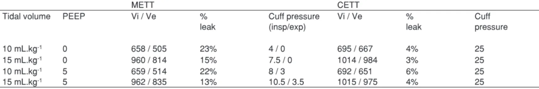
Table I – Mean Values Obtained with Both #7.5 mm Endotracheal Tubes
METT CETT
Tidal volume PEEP Vi / Ve %
leak
Cuff pressure (insp/exp)
Vi / Ve %
leak
Cuff pressure
10 mL.kg-1 0 658 / 505 23% 4 / 0 695 / 667 4% 25
15 mL.kg-1 0 960 / 814 15% 7.5 / 0 1014 / 984 3% 25
10 mL.kg-1 15 mL.kg-1
5 5
659 / 514 962 / 835
22% 13%
8 / 3 10.5 / 3.5
692 / 651 1015 / 975
6% 4%
25 25
CETT: conventional endotracheal tube; METT: modified endotracheal tube; PEEP: positive end-expiratory pressure (cmH2O); Vi/Ve: inspiratory and expiratory
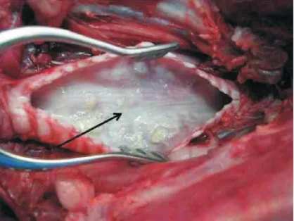
In the pilot study, the animal using METT showed macros-copically less severe tracheal lesions without visible lesions (Figure 1, arrow), while the animal using CETT showed areas of necrosis and ulcerations (Figure 2, arrow).
Microscopically, the animal using METT showed areas with preserved respiratory epithelium (Figure 3, arrow). On
Table II – Mean Values of Both # 8 mm Endotracheal Tubes (CETT and METT)
METT CETT
Tidal volume PEEP Vi / Ve %
modified leak Cuff pressure (insp/exp)
Vi / Ve %
leak
Cuff pressure
10 mL.kg-1 0 663 / 479 28% 3/0 697 / 630 10% 25
15 mL.kg-1 0 966 / 769 20% 6/0 1020 / 941 8% 25
10 mL.kg-1 5 660 / 451 32% 7/2 698 / 609 13% 25
15 mL.kg-1 5 964 / 749 22% 10/3 1024 / 923 10% 25
CETT: conventional endotracheal tube; METT: modified endotracheal tube; PEEP: positive end-expiratory pressure (cmH2O); Vi/Ve: inspiratory and expiratory
volu-mes (mL); % leak: difference between inspiratory and expiratory voluvolu-mes (mL); cuff pressure (cmH2O) during the expiratory and inspiratory phases.
Figure 1 – Animal Trachea Opened Longitudinally. The animal re-mained with the Modified Endotracheal Tube METT) for 48 Consecu-tive Hours. Note the Preservation of the Epithelium (arrow).
Figure 2 – The Arrows Demonstrate on the Trachea, Opened Lon-gitudinally, Damage to the Epithelium. Those are Greater on a Lower Segment, Indicated by the Ruler. This Animal Remained with the Conventional Endotracheal Tube (CETT) for 48 Consecutive Hours.
Figure 3 – Microscopic Tracheal Examination of the Animal that Re-mained with the METT for 48 Consecutive Hours. The Arrow Shows the Epithelium with Mild inflammatory Process and Preserved Muco-sa (H&E - 400X).
SERVIN, BARRETO, MARTINS ET AL.
314 Revista Brasileira de Anestesiologia
Vol. 61, No 3, May-June, 2011 histological evaluation the animal using CETT had an intense
inflammatory process with ulcerated areas and erosions on tracheal epithelium (Figure 4, arrow). Extensive areas of de-epithelization and foci of associated hemorrhage were also observed. In ulcerated areas neutrophilic perichondritis and acute inflammation extending up to the adventitia was obser-ved, contrary to what happened to the animal using METT.
DISCUSSION
When compared with CETT, the METT showed in the first ex-periment lower cuff pressure. This did not depend on the pha-se of the respiratory cycle or larger tidal volumes and PEEP used. On the other hand, # 8 mm METT showed greater air leaking (32% higher than the CETT). In the pilot study, we observed that METT was associated with less tracheal injury, both macro- and microscopically.
Compared with CETT, the METT is composed of an iden-tical endotracheal tube used for intubation. In order to have cycles during MV, the tube inside the cuff has millimetric ho-les. These holes cause the passage and exit of air during MV (both on inspiration and expiration). Since it is not constantly insufflated the contact with tracheal mucosa is smaller and, consequently, possible mucosal injuries are practicably ne-gligible. This comparative demonstration was possible in the pilot study with animals.
In general, the objective of ETTs is to allow the passage of air. The inflated cuff protects the airways from aspirating secretions, closing the trachea and allowing effective alve-olar ventilation. Aspiration of gastric contents is not com-mon during anesthesia, but it requires preventive measures such as the use of drugs that reduce its volume and acidity. However, tracheal intubation is the best method to prevent gastric aspiration in these situations 14. The cuff, when in
con-tact with the mucosa, will cause endotracheal injury due to the pressure exerted. Current cuffs close the trachea through a greater area of contact with the mucosa. This allows a lower pressure inside, reflecting on the mucosa minimizing the inci-dence of ischemia, and consequently fewer injuries. This less deleterious effect of the low pressure tubes will not be seen if a smaller diameter than necessary is used. In tubes of lower caliber, cuffs will require larger volumes to achieve adequate closure, causing an increase in pressure and ischemic chan-ges, which lead to complications especially if longer periods of intubation are required.
The most feared complications of tracheal intubation for longer periods continue to attract considerable attention in the literature. Among the most common, we could mention subglottic laryngeal and tracheal stenosis 11,15,16.
Lindholm 17 investigated 35 cases of prolonged intubation
with rubber, latex, and PVC tubes. In all cases, he observed damage in the area of contact with the cuff, with different
degrees of inflammation and/or necrosis. Klainer et al. 18
de-monstrated the presence of tracheal damage in the area of the low-pressure cuff after two hours of intubation, which was demonstrated through electron microscopy. Ciliary disorgani-zation with loss of the histological pattern was observed in some areas.
Magovern et al. 8 developed a cuff with an external
pres-sure regulator system. When excessive volumes of air are injected inside the cuff, another external cuff of an extremely elastic material distends. This will avoid an increase in pres-sure above 30 cmH2O in the internal cuff. This system has
some drawbacks, such as the price (approximately 10 times more than the conventional tube) and it can frequently show rupture of the system.
In another extreme, Peagle et al. 19 investigated 54 patients
in whom two models of PVC tube with latex cuff (red tube) were used and who died and underwent autopsy. Pressures were not systematically controlled, but they were between 150 and 300 mmHg. The histological study of the tracheas showed loss of ciliary epithelium with less than 12 hours of in-tubation. The inflammatory reaction extended to the margins of cartilage with signs of early necrosis.
Therefore, the METT represents an alternative to the si-tuation presented and indicated by the pilot study presented here, since it causes little damage to the tracheal mucosa, as observed in the studied animal.
In the first phase of the study, the METT showed greater air leaking than the CETT. In an attempt to minimize this situ-ation, a PEEP of 5 cmH2O was used. This measure was not
effective in reducing the percentage of air leaking (Tables I and II).
Note that with the smaller diameter METT (# 7.5 mm) the percentage of air leaking was also smaller. The smaller dia-meter cuff of METT has a greater volume capacity when com-pared to the METT of greater diameter during the inspiratory phase, which could explain this reduction in air leaking. This will result in delayed emptying during the expiratory phase, therefore allowing better seal and less air leaking.
Bryant et al. 20 concluded, in their findings on ETT-related
damage, that temporarily emptying the cuff or changing its position was not enough to prevent tracheal mucosa necrosis and/or cartilage damage.
An ETT with a pressure regulating valve of distal cuff is available on the market. This decreases tracheal trauma; ho-wever, the damages remain and its cost is very high 21.
REFERÊNCIAS / REFERENCES
01. Sole Ml, penoyer dA, Su X et al. – Assessment of endotracheal cuff pressure by continuous monitoring: a pilot study. Am J crit care, 2009;18:133-143.
02. Bain JA – late complications of tracheostomy and prolonged endotra-cheal intubation. int Anesthesiol clin, 1972;10:225-244.
03. Tornvall SS, Jackson KH, oyanedel E – Tracheal rupture, complica-tion of cuffed endotracheal tube. chest, 1971;59:237-239.
04. cooper Jd, Grillo Hc – Experimental production and prevention of inju-ry due to cuffed tracheal tubes. Surg Gynecol obstet, 1969;129:1235-1241.
05. Bishop MJ – Mechanisms of laryngotracheal injury following pro-longed tracheal intubation. chest, 1989;96:185-186.
06. Yang Kl – Tracheal stenosis after a brief intubation. Anesth Analg, 1995;80:625-627.
07. Mcculloch TM, Bishop MJ – complications of translaryngeal intuba-tion. clin chest Med, 1991;12:507-521.
08. Magovern GJ, Shively JG, Fecht d et al. – The clinical and experi-mental evaluation of a controlled-pressure intratracheal cuff. J Thorac cardiovasc Surg, 1972;64:747-756.
09. Kamen JM, Wilkinson cJ – A new low-pressure cuff for endotracheal tubes. Anesthesiology, 1971;34:482-485.
10. Arola MK, Anttinen J – post-mortem findings of tracheal injury after cuffed intubation and tracheostomy. A clinical and histopathological study. Acta Anaesthesiol Scand, 1979;23:57-68.
11. conti M, pougeoise M, Wurtz A et al. – Management of postintubation tracheobronchial ruptures. chest, 2006;130:412-418.
12. lindholm cE – Experience with a new orotracheal tube. Acta otolar-yngol (Stockh), 1973;75:389-390.
13. Nordin u, lindholm cE, Wolgast M – Blood flow in the rabbit tracheal mucosa under normal conditions and under the influence of tracheal intubation. Acta Anaesthesiol Scand, 1977;21:81-94.
14. Moro ET – prevenção da aspiração pulmonar do conteúdo gástrico.. Rev Bras Anestesiol, 2004;54:261-275.
15. American Association for Respiratory care – AARc (American Asso-ciation for Respiratory care) clinical practice guideline. Management of airway emergencies. Respir care, 1995;40:749-760.
16. Brichet A, Verkindre c, dupont J et al. – Multidisciplinary approach to management of postintubation tracheal stenoses. Eur Respir J, 1999;13:888-893.
17. lindholm cE - prolonged endotracheal intubation. Acta Anaesthesiol Scand, 1970;( Suppl 33):1-131.
18. Klainer AS, Turndorf H, Wu WH et al. – Surface alterations due to endotracheal intubation. Am J Med, 1975;58:674-683.
19. paegle Rd, Ayres SM, davis S – Rapid tracheal injury by cuffed airways and healing with loss of ciliated epithelium. Arch Surg, 1973;106:31-34.
20. Bryant lR, Trinkle JK, dubilier l – Reappraisal of tracheal in-jury from cuffed tracheostomy tubes. Experiments in dogs. JAMA, 1971;215:625-628.
21. Valles J, Artigas A, Rello J et al. – continuous aspiration of subglottic secretions in preventing ventilator-associated pneumonia. Ann intern Med, 1995;122:179-186.
Resumen: Servin SoN, Barreto G, Martins lc, Moreira MM, Meirel-les l, colli Neto JA, Zen Júnior JH, Tincani AJ – Fijación Atraumática de Tubo Endotraqueal para Ventilación Mecánica.
Justificativa y objetivos: pacientes que necesitan permanecer bajo intubación endotraqueal (ioT), durante largos períodos o que tienen que ser sometidos a la anestesia general, podrán tener lesiones en la luz de la tráquea debido a presiones ejercidas por el balón terminal. En algunos casos, esas lesiones podrán evolucionar para una este-nosis o a veces necrosis. El presente trabajo quiso presentar un tubo endotraqueal modificado (TETM), en que la presión del balón varía de acuerdo con el ciclo de la ventilación mecánica (VM), siendo el mismo testado en un simulador pulmonar y modelo animal.
Método: En un simulador pulmonar acoplado a un ventilador mecáni-co y ajustado mecáni-con dos volúmenes mecáni-corrientes (Vc) de 10 y 15 ml.kg-1 y complacencia de 60 ml.cmH2o-1, fueron utilizados dos modelos de tubos endotraqueales: uno modificado (TETM), y el otro convencio-nal (TETc), números (#) 7,5 mm y 8,0 mm, para evaluar la eficiencia de la ventilación con el TETM. También se hizo la comparación entre los dos modelos, en cerdos de la raza large-White, bajo anestesia general y VM por 48 horas consecutivas. posteriormente, los anima-les se sacrificaron para el análisis histopatológico de las tráqueas.
Resultados: los dos TETMs (#7,5 y 8,0) presentaron un escape de aire en el simulador pulmonar. El menor de los escapes de aire (13%), fue visto en el TETM #7,5 mm, con Vc = 15 ml.kg-1, y el mayor (32%) en el TETM #8,0 mm, con Vc = 10 ml.kg-1. A pesar de eso, los dos TETMs presentaron una buena eficacia en el simulador pulmonar. En la evaluación del uso de los TETs en animales, analizando la his-topatología de sus tráqueas, verificamos que el TETM causó menos áreas traumáticas en su epitelio en comparación con el TETc.
Conclusiones: El uso de un nuevo modelo de TET podrá disminuir los riesgos de lesión traqueal sin prejudicar la mecánica respiratoria.