Article
ISSN 0102-695X
http://dx.doi.org/10.1590/S0102-695X2011005000190 Received 16 Dec 2010 Accepted 28 Jul 2011 Available online 14 Oct 2011
sucuriju
Frederico A. Vanderlinde,
*,1Fabio F. Rocha,
1David C. Malvar,
1Raquel T. Ferreira,
1Elson A. Costa,
2Iziara F. Florentino,
2Giselle M. S. P. Guilhon,
3Thereza C. M. de Lima
41Departamento de Ciências Fisiológicas, Instituto de Biologia, Universidade Federal Rural do Rio de Janeiro, Seropédica, Brazil,
2Departamento de Ciências Fisiológicas, Instituto de Ciências Biológicas, Universidade Federal de Goiás, Goiânia, Brazil,
3Departamento de Química, Centro de Ciências Exatas e Naturais, Universidade Federal do Pará, Brazil,
4Departamento de Farmacologia, Universidade Federal de Santa Catarina, Brazil.
Abstract: Mikania lindleyana DC., Asteraceae (sucuriju), grows in the Amazon region, where is frequently used to treat pain, inflammatory diseases and scarring. This study was carried out to investigate phytochemical profile accompanied by in vivo antinociceptive and anti-inflammatory screening of n-hexane (HE), dichloromethane (DME) and methanol (ME) extracts obtained from the aerial parts of the plant. The oral administration of ME (0.1, 0.3, 1 g/kg) caused a dose-related reduction (16.2, 42.1 e 70.2%) of acetic acid-induced abdominal writhing while HE and DME (1 g/kg, p.o.) were ineffective. In the hot plate test, ME (300 mg/kg,
p.o.) increased the latency of heat stimulus between 30 and 120 min and inhibited the first (45%) and second (60%) phases of nociception in the formalin test. The antinociception induced by ME or positive control fentanyl (150 µg/kg, s.c.) in hot plate and formalin tests was prevented by naloxone (3 mg/kg, s.c.). When submitted to the carrageenan-induced peritonitis test, ME (0.5, 1.0, 2.0 g/kg, p.o.) impaired leukocyte migration into the peritoneal cavity by 46.8, 59.4 and 64.8% respectively, while positive control dexamethasone (2 mg/kg, s.c.), inhibited leukocyte migration by 71.1%. These results indicate that the antinociception obtained after oral administration of methanol extract of M. lindleyana involves anti-inflammatory mechanisms accompanied with opioid-like activity which could explain the use of the specie for pain and inflammatory diseases.
Keywords:
Mikania lindleyana
anti-inflammatory antinociceptive Asteraceae sucuriju phytomedicine
Introduction
Mikania lindleyana DC., Asteraceae, is a creeper plant found in the Amazon region, known as sucuriju. The infusion of its leaves is empirically used as an anti-inl ammatory, analgesic or cicatrizing and in the treatments of hepatitis, chronic gastric ulcers, varicose veins, acne and various types of cutaneous eruptions. Also is used as a diuretic and anti-hypertensive medication through orally administration of the tea leaves. The dry leaves of sucuriju are sold in some of Belém do Pará markets for medicinal purposes (Berg, 1993; Martins et al., 2005). The genus Mikania Willd belongs to the subtribe Mikaniinae and to the tribe Eupatorieae, from the family Asteraceae, and includes among 430 species across tropical regions.
Nowadays, several anti-inl ammatory agents are used to treat different types of pain associated or not
to the inl ammatory process. These agents are efi cient in most cases, but the collateral effects are common, especially when they are used in chronic treatments. The most common collateral effect is gastrointestinal disturb (Peura & Goldkind, 2005). Considering the research of medicinal plants can propitiate the discovery of new molecules with innovative mechanisms or less adverse side effects (McCurdy & Scully, 2005), the ethnopharmacological proi le of Mikania lindleyana
Material and Methods
Extraction
Mikania lindleyana DC., Asteraceae, aerial parts were collected in Benevides-PA (Brazil) and samples were identiied by Dr. M. E. van den Berg, botanist from Emílio Goeldi Paraense Museum (MPEG). The voucher specimens were deposited at the João Murça Pires Herbarium of MPEG under MG-01607412 number.
Botanical material were dried and submitted to extraction with hexane at room temperature, followed by dichloromethane and methanol extraction. The solutions obtained were concentrated dry under reduced pressure in a rotary evaporator, below 40 ºC, to obtain the n-hexane (HE), dichloromethane (DME) and methanol (ME) extracts.
Phytochemical characterization
In the phytochemical study of M. lindleyana
extracts, the substances were isolated by chromatographic methods and identiied by usual spectroscopic methods (NMR, IR and MS) being detected in the hexane and dichloromethane extracts: stigmasterol (0.27% of the extracts), stigmasta-4,24-dien-3-one (0.07%), methylene-cycloartan-3-ol (0.60%), a mixture of 24-methylene-cycloartan-3-ol and cycloart-24-en-3-ol (0.51%), a mixture of 24-methylene-cycloartan-3-one and cycloart-24-en-3-one (0.29%), trans-phytol (0.29%) and also long chain esters of stigmasterol (0.57%) and fatty acids (0.25%). A mixture of the inorganic salts KNO3 and KCl (0.50%) was obtained from the methanol extracts (Guilhon et al., 1998a,b,c, 1999).
When partitioned with water and chloroform, methanol extract (1 g) formed the aqueous (A-F) and chloroform (CH-F) fractions. Analyses by comparative thin layer chromatography (CCDC) on plates of silica gel (Merck F254) were eluted with butanol/acetic acid/ water (BAW, 4:1:5) and chloroform-methanol 9:1 (CHCl3:MeOH) and visualized in UV chromatograms at 254 nm and 366 nm. The reagent NP/PEG (BAW) revealed the presence of lavonoids in CH-F (Rf 0.70 and
0.57) and A-F (Rf 0.20) for rutin and quercetin Rf 0.55/Rf 77 (Merck) and triterpenes (Liberman-Burchard) AF (Rf 0.21 and 0.27). The Drangendorff reagent did not detect alkaloids (Moreira et al., 2008).
Animals
Adult male Swiss mice (25-35 g) were housed in plastic cages, with food and tap water available ad libitum in the colony room. Mice were acclimatized to the laboratory for at least 60 min prior to the test procedure and were fasted for 12-18 h before the gavage. All
experiments were carried out in accordance with current guidelines for the care of laboratory animals and the ethical guidelines on the use of animals in pain research (Zimmermann, 1986). The experimental protocols were approved by the local Animal Care and Use Committee (006/2006/CEPEB/UFRRJ). The minimum number of animals and duration of observation required to obtain consistent data were employed.
Drugs
Formalin, acetic acid, acetone (Merck AG, Darmstadt, Germany), indomethacin, carrageenan, naloxone hydrochloride (Sigma Chemical Co., St. Louis, MO, USA), fentanyl (Janssen Pharmaceutical), dexamethasone (Prodome, Brazil). The n-hexane (HE), dichloromethane (DME) and methanol (ME) extracts were prepared and provided by Dr. Giselle M. S. P. Guilhon (Federal University of Pará, Brazil). Test solutions of extracts and drugs were diluted in water (p.o.) or saline (s.c. or i.p.) in such a concentration as to allow the administration of constant volumes of 10 mL/kg for each dose employed. The oral pre-treatment (p.o.) was always done 1 h and subcutaneous (s.c.) 30 min before tests.
Acetic acid-induced writhing test
In an initial screening of antinociceptive activity, group of six mice were orally treated with vehicle (water),
n-hexane (HE), dichloromethane (DME), methanol (ME) extracts (1.0 g/kg) or positive control indomethacin (10 mg/kg), 60 min before acetic acid injection (1.2%, 0.1 mL/10 g) and the number of writhes was counted for the following 30 min (Koster et al., 1959). The absence of activity with the HE and DME directed the study to obtain in this method the dose effects of ME (0.1, 0.3 and 1.0 g/kg).
Hot-plate test
The latency (s) of heat stimulus (55.0±0.5 oC)
was measured every 30 min, starting 30 min before and up to 150 min after pretreatment of mice (n=8) with vehicle (water), ME (300 mg/kg, p.o.), fentanyl (positive control - 100 µg/kg, s.c.), ME+naloxone (3 mg/kg, s.c.) or fentanyl+naloxone. The animals whose basal lick responses were bigger than 9 s were discharged and a cut-off time of 30 s was maintained through the experiment, to prevent tissue damage. (D’Amour & Smith, 1941).
Formalin-induced nociception
injection of the formalin solution (3%, 20 µL/paw, s.c.) into the plantar surface of the hind paw. To investigate the mechanism of antinociception, naloxone (300 mg/kg,
s.c.) treatment preceded (15 min) the administration of ME or fentanyl. The time that animals spent licking the injected paw was measured with a chronometer and was considered as an index of pain (licking time) in seconds. From the formalin injection, the initial nociceptive response peaked at about 5 min (irst phase) and was followed by a second peak (second phase) that occurred at 15-30 min post injection (Hunskaar et al., 1986; Hunskaar & Hole, 1987).
Peritonitis induced by carrageenan
Mice (n=10) were orally treated with vehicle (water), ME (0.5, 1.0 and 2.0 g/kg) 60 min prior to an injection of carrageenan (1% in saline; 250 µL/mouse) into the peritoneal cavity. The positive control group was pre-treated (30 min) with dexamethasone (2 mg/ kg, s.c.). The number of leukocytes that had migrated to the peritoneum was counted in a Neubauer chamber and results were expressed as cells×106/mL and % of
inhibition of leukocyte migration compared to control groups (Ferrándiz & Alcaraz, 1991).
Statistical analysis of data
Data were statistically analyzed by one-way ANOVA followed by the Dunnet multicomparison test. In the hot-plate test, the two-way ANOVA followed by the Bonferroni’s test analysis was used. The values are reported as mean±standard error of the mean (SEM). P values less than 0.05 (p<0.05) were considered to be signiicant (Sokal & Rohlf, 1981).
Results
Acetic acid-induced writhing test
The initial screening of antinociceptive activity demonstrated that the oral treatments with hexane (HE) or dichloromethane (DME) extracts (1.0 g/kg) did not alter the number of acetic acid-induced abdominal writhing (48.3±3.9 and 54.1±4.0, respectively) compared to the control group (51.3±3.8 writhes) while as shown in Figure 1, methanol extract (ME - 0.1, 0.3 and 1 g/kg) produced a dose-related inhibition of acetic acid-induced abdominal writhing (16.2±1.1, 42.1±3.8 and 70.2±4.8 %, respectively; IC50=431.5 mg/kg),. The group treated with indomethacin showed a decrease in accumulated abdominal writhes (14±2.5 writes).
Hot-plate test
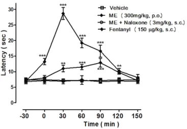
In the hot-plate test Figure 2 shows that the basal latency of control group was 7.1±0.6 s and orally administration of ME (300 mg/kg) increased the pain latency by 51.7±4.8, 62.4±4.3, 79.5±8.6 and 33.8±1.7% after 30, 60, 90 and 120 min respectively. The positive control fentanyl (150 µg/kg, s.c.) produced a signiicant
increased of reactivity time to heat stimulus in the intervals between zero and 120 min with a maximum latency of 3.9-fold (30 min). Pre-treatment (15 min) with naloxone (3 mg/kg, s.c.) inhibited antinociceptive effect of ME (igure 2) and fentanyl (results no show).
Figure 1. Effect of methanol extract (ME) obtained from the aerial parts of Mikania lindleyana or indomethacin on the acetic acid-induced writhing in mice. The vertical bars and lines express means±SEM of the number of writhing registered
for 30 min after the acetic acid injection. Signiicantly different
from vehicle group: **p<0.01; ***p<0.001 (n=6).
Figure 2. Latency of response to thermal stimulus (hot plate at 55 ºC) in group of mice (n=8) before and after treatment with water (Vehicle), methanol extract (ME) obtained from the aerial parts of Mikania lindleyana, fentanyl or ME+naloxone. The symbols and vertical lines express means±SEM of the latency for the nociceptive behavior. **p<0.01, ***p<0.001 when compared with the vehicle group. Symbols represent the different between the ME and fentanyl.
Formalin-induced nociception
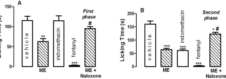
licking time of animals previously treated with vehicle presented 115.8±10.3 s in the irst phase and 160.5±11.4 s in the second phase. ME (300 mg/kg, p.o.) treatment produced reduction in pain reaction to 63.0±7.4 s (Figure 3A) and 64.1±5.5 s (Figure 3B) in the irst and second phases respectively. The licking times of animals treated with fentanyl (100 µg/kg, s.c.) were reduced in both phases (around 97%), while indomethacin treatment (10 mg/kg, p.o.), a positive control of second phase, inhibited 62.3% this phase without inluencing the irst phase of nociception. The naloxone pre-treatment (3.0 mg/kg, s.c.) reversed totally the antinociceptive effects of ME in the irst phase (95.3±5.1 s) and partially in the second phase (122.6±7.2 s) of test (Figures 3A and 3B). The fentanyl analgesic effect also were blocked by naloxone (results no show).
Table 1. Effect of ME of M. lindleyana on carrageenan-induced peritonitis in mice.
Treatment Dose (g/kg)
Number of leukocytes (x 106/mL)
Inhibition (%)
Vehicle (p.o.) - 12.8±1.7
-ME (p.o.) 0.5 6.8±1.0** 46.8
ME (p.o.) 1.0 5.2± 0.8*** 59.4
ME (p.o.) 2.0 4.5±0.9*** 64.8 Dexamethasone (s.c.) 0.002 3.7±0.3*** 71.1
Values represent the means±SEM of ten mice. Signiicantly different
from vehicle group. p<0.01, p<0.001.
Peritonitis induced by carrageenan
Using carrageenan as a stimulus, it was possible to produce an acute inlammatory response after 4 h
in the peritoneal cavity of mice, with a large number of leukocytes in the exudates. With the aim of evaluating a possible inhibitory effect of ME on the cell migration into the peritoneal cavity, the carrageenan-induced peritonitis test was used. As can be seen in Table 1 in comparison with vehicle group (12.8±1.7×10⁶ leukocytes/mL), ME (0.5, 1.0
and 2.0 g/kg, p.o.) exhibited a dose-related reduction of leukocyte migration by 46.8, 59.4 and 64.8%, respectively. The positive control dexamethasone (2 mg/kg, s.c.) also was effective, inhibiting leukocyte migration by 71.1%.
Discussion
In the antinociceptive screening using acetic acid-induced abdominal writhing model and high doses of hexane (HE), dichloromethane (DME) or methanol (ME) extracts obtained from the aerial parts of the Mikania lindleyana DC., Asteraceae, only the ME was effective. The writhing induced by acetic acid in mice result from chemical-induced a peripheral acute inlammatory reaction (Zakaria et al., 2006a). The treatment with ME promoted a dose-related antinociceptive effect in this model, normally used for screening of synthetic and natural compounds to determinate the presence of central and/or peripheral antinociceptive activity, because it is sensitive to nonsteroidal anti-inlammatory drugs (NSAID) and opioids (Zakaria et al., 2006b; Fischer et al., 2008). Moreover, the nociception induced in writhing method has been associated with some prostanoids, like as PGE2, PGF2α and PGI2 (Deraedt et al., 1980) as well as lipoxygenase products (Ballou et al., 2000; Parveen et al., 2007), neurokinin A (Julia & Buéno, 1997) and CGRP (Friese et al., 1997), for example. The consequence of the involvement of different mediators in the nociception,
Figure 3. Effect of methanol extract of Mikania lindleyana (ME-300 mg/kg, p.o.) on the licking time (s) in the first phase (0-5 min/A) and second phase (15-30 min/B) of the formalin test in mice (n=6-8). Fentanyl (100 µg/kg, s.c.) and indomethacin (10 mg/kg, p.o.) were used as positive controls of the first and second phases respectively. The administration of naloxone (3 mg/ kg, p.o.) preceded ME treatment. The vertical bars and lines express means±SEM of licking time. *p<0.05, **p<0.01; ***
phases suggest that ME antinociception is produced by anti-inlammatory and opioid-like mechanisms. The antinociception of ME is not related to nonspeciic central effects, since no detectable effect was observed after oral administration of ME high dose in the rota-rod test (results not showed).
Considering the popular use of specie to treat inlammatory diseases and antinociception obtained in the second phase of formalin test accompanied by partial reversal with naloxone pre-treatment, the anti-inlammatory hypothesis was tested in leukocyte migration model. The carrageenan-induced peritonitis was used to investigate possible alterations in the leukocyte migration. ME reduced in a dose-related manner the total leukocyte migration to the peritoneum induced by carrageenan with a highest inhibition value by 64.8%. This process is dependent of the synthesis and release of chemo-attractants such as lipoxygenase products and chemokines being constituted by three phase: leukocyte rolling, attachment and diapedesis. Drugs can reduce the leukocyte migration by inhibit the synthesis and release of chemo-attractants or by block of some leukocyte migration phase (Muller, 2002). Several studies have been reported that leukocytes migration would not be directly related to cyclooxygenase products, but the process could be inhibited by some, indicating that many mechanisms may be implicated in its control (Higgs et al., 1980; Mikami & Miyasaka, 1983; Brooks & Day, 1991). A recent study showed that PGF2α was still able to induce leukocyte migration, suggesting that the inhibitory property of NSAID appears to be primarily related to the reduced production of the PGF2α (De Menezes et al., 2005). Since several mechanisms may be involved in the leukocytes migration more studies must be done to elucidate the mechanism of the anti-inlammatory effect of ME.
These results showing that the oral administration of methanol extract of Mikania lindleyana produced a potent antinociceptive activity in the acetic acid-induced abdominal writhing, inhibitory effect on the reactivity of heat stimulus in the hot-plate test and the first phase (neurogenic pain) and second phase (inflammatory pain) of the formalin test with effects reversed by naloxone, are indicative that ME antinociception involves opioid-like mechanisms. The fact of ME also reduced the leukocyte migration, a characteristic activity present of anti-inflammatory drugs, allow to state that antinociception of ME is also associated with anti-inflammatory mechanisms.
The anti-inflammatory and antinociceptive effects of stigmasterol isolated from different vegetal species have been demonstrated in pharmacological models (Santos et al., 1995; Garcia et al., 1999) as flavonoids have long been recognized to possess a wide variety of biological activities such as antioxidant which means that based only on this model, is not possible
to suggest any mechanism to the antinociceptive effect of ME.
and anti-inflammatory (Middleton et al., 2000), besides the blockade of phospholipase A2, inhibit the cyclooxygenase and/or the lipoxygenase pathways of arachidonate metabolism (Lindahl & Tagesson, 1997; Middleton et al., 2000; Kwak et al., 2003). Although the phytochemical study of hexane and dichloromethane extracts has detected stigmasterol compound (Guilhon et al., 1998a,b,c, 1999), high doses of these extractions were ineffective in acetic acid-induced writhing model, however the antinociceptive and anti-inflammatory activities of ME can be attributed to flavonoids compounds, such quercetin and rutin, present in this extraction (Moreira et al., 2008), without excluding the possibility of stigmasterol, and other chemical compounds present in the aerial parts of plant also participate in the analgesic and anti-inflammatory effectiveness of specie. These results could explain the use of the plant for pain and inflammatory diseases and a bioassay-guided fractioning of the extracts is now in progress to identify a greater number bioactive substances in aerial parts of M. lindleyana as well as the mechanisms of action involved in the effects described before.
Acknowledgments
The authors thank Carlos Chagas Filho Foundation for Research Support of Rio de Janeiro and Brazilian National Research Council for fellowships and inancial support.
References
Ballou LR, Botting RM, Goorha S, Zhang J, Vane, JR 2000.
Nociception in cyclooxygenase isozyme-deicient mice. Proc Natl Acad Sci USA 97: 10272-10276.
Berg MEVDE 1993. Plantas medicinais na Amazônia. Pará: Coleção Adolfho Ducke.
Brooks PM, Day RO 1991. Nonsteroidal anti-inlammatory
drugs - differences and similarities. New Engl J Med 324: 1716-1725.
D’Amour FE, Smith DL 1941. A method for determining loss of pain sensation. J Pharmacol Exp Ther 72: 74-79. Deraedt R, Jouquey S, Delevallee F, Flahaut M 1980. Release
of prostaglandins E and F in an algogenic reaction and its inhibition. Eur J Pharmacol 61: 17-24.
De Menezes GB, Dos Reis WG, Santos JM, Duarte ID, De Francischi JN 2005. Inhibition of prostaglandin F(2alpha) by selective cyclooxygenase 2 inhibitors accounts for reduced rat leukocyte migration.
Inlammation 29: 163-169.
Ferrándiz ML, Alcaraz MJ 1991. Antiinlammatory activity and inhibition of arachidonic acidmetabolism by lavonoids.
Inlamm Res 32: 283-288.
Fischer LG, Santos D, Serain C, Malheiros A, Monache FD,
Monache GD, Cechinel FV, Souza MM 2008. Further antinociceptive properties of extracts and phenolic compounds from Plinia glomerata (Myrtaceae) leaves.
Biol Pharm Bull 32: 235-239.
Friese N, Diop L, Chevalier E, Angel F, Riviere PJ, Dahl SG 1997. Involvement of prostaglandins and CGRP dependent sensory afferents in peritoneal irritationinduced visceral pain. Regul Peptides 70: 1-7.
Garcia MD, Saenz MT, Gomez MA, Fernández MA 1999.
Topical antiinlammatory activity of phytosterols
isolated from Eryngium foetidum on chronic and acute
inlammation models. Phytother Res 13: 78-80. Gonçalves JCR, Oliveira FS, Benedito RB, Souza DP, Almeida
RN, Araujo DAM 2008. Antinociceptive activity of carvone: Evidence of association decrease peripheral nerve excitability. Biol Pharm Bull 31: 1017-1020. Guilhon GMSP, Miranda ACO, Muller AH, Brasil DSB 1998a.
Derivados cicloartânicos de Mikania lindleyana
(Compositae). 21a Reunião Anual da SBQ. Poços de
Caldas, Brasil.
Guilhon GMSP, Teixeira UBA, Muller AH 1998b. Constituintes químicos dos extratos diclorometânico e metanólico de
Mikania lindleyana (Compositae). Semana da Química 98 e I Encontro Paraense do Ensino de Química. Belém, Brasil.
Guilhon GMSP, Teixeira UBA, Brasil DSB, Maia JGS, Andrade EHA, Zoghbi MGB 1998c. Composição Química do Óleo Essencial de Mikania lindleyana DC. XXXVIII Congresso Brasileiro de Química. São Luiz, Brasil. Guilhon GMSP, Brasil DSB, Muller AH, Marinho AMR 1999.
Constituintes químicos dos extratos diclorometânico e metanólico de Mikania lindleyana A. DC. (Asteraceae).
22a Reunião Anual da SBQ. Poços de Caldas, Brasil.
Higgs GA, Eakins KE, Mugridge KG, Moncada S, Vane JR
1980. The effects on non-steroid anti-inlammatory
drugs on leukocyte migration in carrageenin-induced
inlammation. Eur J Pharmacol 66: 81-86.
Hunskaar S, Berger OG, Hole K 1986. Dissociation between
antinociceptive and antiinlammatory effects of
acetylsalicylic acid and indomethacin in the formalin test. Pain 25: 125-132.
Hunskaar S, Hole K 1987. The formalin test in mice: dissociation
between inlammatory and non-inlammatory pain. Pain 30: 103-114.
Julia V, Buéno L 1997. Tachykininergic mediation of
viscerosensitive responses to acute inlammation in rats:
evidence for a CGRP-induced release of tachykinins. Am J Physiol 272: 141-146.
Koster R, Anderson M, De Beer EJ 1959. Acetic acid for analgesic screening. Fed Proc 18: 412.
Kwak WJ, Moon TC, Lin CX, Rhyn HG, Jung HJ, Lee E, Kwon DY, Son KH, Kim HP, Kang SS, Murakami M, Kudo I,
A(2)-inhibitory activity. Biol Pharm Bull 26: 299-302. Lindahl M, Tagesson C 1997. Flavonoids as phospholipase A2
inhibitors: importance of their structures for selective inhibition of group II phospholipase A2. Inlammation
21: 347-356.
McCurdy CR, Scully SS 2005. Analgesic substances derived from natural products (natureceuticals). Life Sci 78: 476-484.
Martins AG, Rosário DL, Barros MN, Jardim MAG 2005. Levantamento etnobotânico de plantas medicinais, alimentares e tóxicas da Ilha do Combu, Município de Belém, Estado do Pará, Brasil. Rev Bras Farm 86: 21-30.
Middleton JrE, Kandaswami C, Theoharides TC 2000. The
effects of plant lavonoids on mammalian cells: implications for inlammation, heart disease, and cancer. Pharmacol Rev 52: 673-751.
Mikami T, Miyasaka K 1983. Effects of several
anti-inlammatory drugs on the various parameters involved in the inlammatory response in rat carrageenin-induced
pleurisy. Eur J Pharmacol 95: 1-12.
Moreira SF, Guilhon GMSP, Müller AH, Rocha FF, Vanderlinde FA, Torres LMB 2008. Mikania lindleyana A. Dc.: Estudo Químico e Avaliação de Atividade biológica.
XX Simpósio de Plantas Medicinais do Brasil - X International Congress of Ethnopharmacology. São Paulo, Brasil.
Muller WA 2002. Leukocyte-endothelial cell interactions in the
inlammatory response. Lab Invest 82: 521-533. Oliveira FS, Sousa DP, Almeida RN 2008. Antinociceptive
effect of hydroxydihydrocarvone. Biol Pharm Bull 31: 588-591.
Parveen Z, Deng Y, Saeed MK, Daí R, Ahamad W, Yu YH 2007.
Antiinlammatory and analgesic activities of Thesium chinese Turcz extracts and its major lavonoids,
kaampferol and kaempferol-3-O-glucoside. Yakugaku Zasshi 127: 1275-1279.
Peura DA, Goldkind L 2005. Balancing the gastrointestinal
beneits and risks of nonselective NSAIDs. Arthritis
Res Ther 7 (Suppl 4): S7-S13.
Santos ARS, Niero R, Cechinel FV, Yunes RA, Pizzolatti MG, Delle Monache F, Calixto JB 1995. Antinociceptive properties of steroids isolated from Phyllanthus corcovadensis in mice. Planta Med 61: 329-332. Sokal RR, Rohlf FJ 1981. Biometry: The Principle and Practice
of Statistics 2. ed. New York: W.H. Freeman.
Wang JR, Zhou H, Jiang ZH, Wong YF, Liu L 2008. In vivo antiinlammatory and analgesic activities of a puriied
saponin fraction derived from the root of Ilex pubescens. Biol Pharm Bull 31: 643-650.
Zakaria ZA, Gopalan HK, Zainal H, Pojan NHM, Morsid NA, Aris A, Sulaiman MR, 2006a. Antinociceptive,
antiinlammatory and antipyretic effects of Solanum nigrum Chloroform extract in animal models. Yakugaku Zasshi 126: 1171-1178.
Zakaria ZA, Abdul Ghan DF, Raden Mohd RNS, Gopalan HK, Sulaiman MR, Abdullah FC 2006b. Antinociceptive
and antiinlamatory activities of Dicranopteris linearis
leaves clhoroform extract in experimental animals.
Yakugaku Zasshi 126: 1197-1203.
Zakaria ZA, Sulaiman MR, Gopalan HK, Abdul Ghani ZDF, Raden Mohd RNS, Mat Jais AM, Abdullah FC 2007.
Antinociceptive and antiinlamatory properties of Corchorus capsularis leaves clhoroform extract in experimental animals models. Yakugaku Zasshi 127: 359-365.
Zimmermann M 1986. Ethical considerations in relation to pain in animal experimentation. Acta Physiol Scand 554: 221-223.
*Correspondence
Frederico A. Vanderlinde
Laboratório de Farmacologia, Sala 31, Pavilhão de Química, Universidade Federal Rural do Rio de Janeiro
BR 465, km 07, 23890-000, Seropédica, RJ, Brazil [email protected]