Radiol Bras. 2016 Jul/Ago;49(4):234–240 234
Study of scattered radiation during fluoroscopy in hip surgery
*
Estudo da radiação espalhada em fluoroscopia durante procedimentos cirúrgicos no quadrilLesyuk O, Sousa PE, Rodrigues SIES, Abrantes AF, Almeida RPP, Pinheiro JP, Azevedo KB, Ribeiro LPV. Study of scattered radiation during fluoroscopy in hip surgery. Radiol Bras. 2016 Jul/Ago;49(4):234–240.
Abstract
R e s u m o
Objective: To measure the scattered radiation dose at different positions simulating hip surgery.
Materials and Methods: We simulated fluoroscopy-assisted hip surgery in order to study the distribution of scattered radiation in the
operating room. To simulate the patient, we used a anthropomorphic whole-body phantom, and we used an X-ray-specific detector to quantify the radiation. Radiographs were obtained with a mobile C-arm X-ray system in continuous scan mode, with the tube at 0° (configuration 1) or 90° (configuration 2). The operating parameters employed (voltage, current, and exposure time) were determined by a statistical analysis based on the observation of orthopedic surgical procedures involving the hip.
Results: For all measurements, higher exposures were observed in configuration 2. In the measurements obtained as a function of
height, the maximum dose rates observed were 1.167 (± 0.023) µSv/s and 2.278 (± 0.023) µSv/s in configurations 1 and 2, respectively, corresponding to the chest level of health care professionals within the operating room. Proximal to the patient, the maximum values were recorded in the position occupied by the surgeon.
Conclusion: We can conclude that, in the scenario under study, health care professionals workers are exposed to low levels of radiation,
and that those levels can be reduced through the use of personal protective equipment.
Keywords: Radiation, ionizing; Operating rooms; Scattering, radiation; Radiation protection; Radiology, interventional.
Objetivo: Medir a intensidade da dose de radiação espalhada em diferentes posições simulando uma intervenção cirúrgica no quadril.
Materiais e Métodos: Simulou-se uma intervenção cirúrgica no quadril com apoio da fluoroscopia para estudar a distribuição da radiação
espalhada no bloco operatório. Para simular o paciente foi utilizado um simulador antropomórfico de corpo inteiro e para medir a radiação utilizou-se um detector específico para medir raios X. Realizaram-se incidências com um equipamento de raios X tipo arco em C móvel, em modo de escopia contínua, com a ampola a 0° (configuração 1) e a 90° (configuração 2). Os parâmetros operacionais utilizados (voltagem, corrente, tempo de exposição) foram determinados por meio de um estudo estatístico resultante da observação de cirurgias ortopédicas de quadril.
Resultados: Em todas as medições observaram-se exposições mais elevadas na configuração 2. Nas medições em função da altura,
observaram-se os valores máximos da taxa de dose de 1,167 (± 0,023) µSv/s e 2,278 (± 0,023) µSv/s nas configurações 1 e 2, respectivamente, correspondendo à altura do tórax dos profissionais. No estudo em torno do paciente os valores máximos registraram-se na posição ocupada pelo médico cirurgião.
Conclusão: Concluiu-se que a exposição à radiação dos profissionais é baixa, podendo ainda ser reduzida mediante o uso de
equipa-mentos de proteção individual.
Unitermos: Radiação ionizante; Salas cirúrgicas; Espalhamento de radiação; Proteção radiológica; Radiologia intervencionista.
* Study conducted in the Department of Medical Imaging and Radiotherapy of the Escola Superior de Saúde da Universidade do Algarve (ESSUAlg), Faro, Portugal. 1. Radiology Technician, Professor in the Department of Medical Imaging and Radiotherapy of the Escola Superior de Saúde da Universidade do Algarve (ESSUAlg), Faro, Portugal.
2. PhD, Physics Engineer, Professor in the Department of Medical Imaging and Radiotherapy of the Escola Superior de Saúde da Universidade do Algarve (ESSUAlg), Faro, Portugal.
3. MSc, Radiology Technician, Professor in the Department of Medical Imaging and Radiotherapy of the Escola Superior de Saúde da Universidade do Algarve (ESSUAlg), Faro, Portugal.
4. PhD, Professor and Head of the Department of Medical Imaging and Radio-therapy of the Escola Superior de Saúde da Universidade do Algarve (ESSUAlg), Faro, Portugal.
5. MsC, Radiology Technician, Graduate Student at the Universidad de Murcia, Murcia, Spain, Professor in the Department of Medical Imaging and Radiotherapy of the Escola Superior de Saúde da Universidade do Algarve (ESSUAlg), Faro, Portugal.
Oksana Lesyuk1, Patrick Emmanuel Sousa2, Sónia Isabel do Espirito Santo Rodrigues3, António Fernando Abrantes4, Rui Pedro Pereira de Almeida5, João Pedro Pinheiro6, Kevin Barros Azevedo7, Luís Pedro Vieira Ribeiro7
6. MSc, Doctoral Student at the Universidade de Coimbra, Coimbra, Portugal, Professor in the Department of Medical Imaging and Radiotherapy of the Escola Superior de Saúde da Universidade do Algarve (ESSUAlg), Faro, Portugal.
7. PhD, Professor in the Department of Medical Imaging and Radiotherapy of the Escola Superior de Saúde da Universidade do Algarve (ESSUAlg), Faro, Portugal.
Mailing address: Oksana Lesyuk. Universidade do Algarve, Área Departamental de Imagem Médica e Radioterapia – Escola Superior de Saúde. Avenida Dr. Adelino da Palma Carlos, 8000-510 Faro, Portugal. E-mail: [email protected].
Received December 18, 2014. Accepted after revision August 3, 2015.
INTRODUCTION
The use of ionizing radiation for diagnostic and treat-ment purposes has increased due to the developtreat-ment of new
equipment and easier access to radiologic exams(1). Medical
patients and health care professionals to radiation, and ra-diation protection is therefore necessary in order to reduce the levels of that exposure.
The involvement of professionals from various areas, without specific training in the field of radiation protection, can lead to excessive exposure to ionizing radiation in the
operating room(2,3). Previous studies have indicated that
non-radiologist physicians possess heterogeneous, inadequate knowledge of ionizing radiation, suggesting that there is
room for improvement(4).
Ionizing radiation produces lesions in cells and can have
deterministic or stochastic effects(5,6). To minimize
radia-tion exposure, there are laws stipulating dose limits for work-ers who are exposed while exercising their professions. The average annual effective dose received by a worker should not exceed 20 mSv (100 mSv in a period of five years) and may not surpass 50 mSv in any given year. The annual equiva-lent dose should not exceed 500 mSv for the skin and ex-tremities and 15 mSv for the lens of the eye. According to
Portuguese law(7), effective doses above 1.5 mSv/month
should be investigated.
There are many limitations that make proper dose moni-toring difficult. Such limitations include failure to use per-sonal dosimeters and the incorrect use of such dosimeters, as well as their inherent limitations, such as detecting radia-tion at a single angle, which depends on the posiradia-tion of the
device in relation to the source of the radiation(5).
Exposure to radiation has been given attention at gen-eral radiology centers. However, work conditions involving ionizing radiation exposure are not routinely monitored
during diagnostic or therapeutic orthopedic interventions(5).
According to information published on the International
Atomic Energy Agency website(8), there have been
numer-ous studies investigating the levels of ionizing radiation re-ceived by medical professionals during procedures that carry a high risk of such exposure, including those related to he-modynamics, angiography, or gastroenterology. However, there is still a need for studies of other, low-risk, procedures, such as orthopedic interventions, specifically those involv-ing the backbone and hip, where there is greater exposure
to ionizing radiation(9).
It is pertinent to study the distribution of scattered ra-diation in the operating room during a simulated fluoros-copy-guided orthopedic intervention, to evaluate the inten-sity of the scattered radiation in different zones of the oper-ating room, and to identify factors which influence profes-sionals’ exposure during interventions, thus establishing ra-diation protection recommendations to apply the “as low as reasonably achievable” principles with greater efficiency.
MATERIALS AND METHODS
Between January 1 and April 30 of 2014, a study of interventional radiology procedures in orthopedics was con-ducted at the Faro Branch of the Algarve Hospital Center, in the city of Faro, Portugal. We evaluated the respective
operating parameters (voltage, current, and fluoroscopy time) of the dose-area product received by the patient, the data related to the positions occupied by the professionals, and the configuration of the C-arm around the table, in order to determine which procedure produces the most radiation and to evaluate the image acquisition conditions.
After the statistical study described above, the scattered radiation dose rate was measured as a function of height, dis-tance, and the angle between the simulator and the detector in configuration 1 (tube at 0°) and configuration 2 (tube at 90°), during a simulation of fluoroscopy-guided hip surgery. An AR10A whole-body phantom (Adam,Rouilly Limited,
Kent, England) was used as a surrogate for the patient(10).
We employed a radiation monitor AT1123 (Atomtex; Minsk, Belarus). The monitor was used in order to measure the background dose rate, referred to throughout the text as the dose rate, with a maximum intrinsic uncertainty of ± 15%,
in continuous mode(11). The fluoroscopy equipment used in
the study was a Philips model BV300 (Philips Medical Sys-tems; Best, the Netherlands), with the voltage set at 80 kV and quality controlled, the maximum deviation being ± 0.6%,
which is well within the ± 10% tolerance defined by law(12).
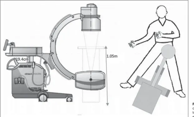
The phantom was positioned to simulate a surgical pro-cedure involving the left hip, with the lower left member ex-tended and lower right member in maximum abduction. The table was placed at a height of 1.05 m above the operating room floor, and the fluoroscopy equipment was placed with its longitudinal axis parallel to the longest axis of the lower right member, centered over the left hip joint (Figure 1).
The operating parameters for voltage and current were in accordance with the results of the statistical study, in two configurations of the C-arm: 67 kV and 2.4 mA, respectively, with the tube at 0° (configuration 1), and 76 kV and 2.8 mA, respectively, with the tube at 90° (configuration 2). The ra-diation reading was registered after the rara-diation beam had stabilized, typically after it had been on for 5 s.
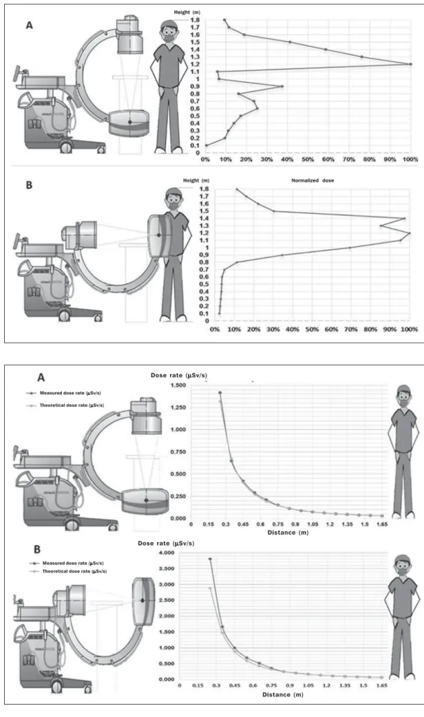
Variation in the dose rate as a function of height
For the study of the scattered radiation dose rate as a function of the dosimeter height, the initial settings for the table, equipment, and phantom were maintained, and the detector was placed at a fixed distance of 25 cm from the center of the exposure field, the approximate position of the lead surgeon. Readings were taken for both configurations, at a 90° angle to the median sagittal line of the phantom, dose readings being taken between 0.10 m and 1.80 m, chang-ing the position of the detector in increments of 10 cm.
Variation in the dose rate as a function of distance
sagittal line, only the distance between the phantom and the detector varying in both configurations. The doses were measured between 0.25 m and 1.65 m, the detector being repositioned in increments of 10 cm.
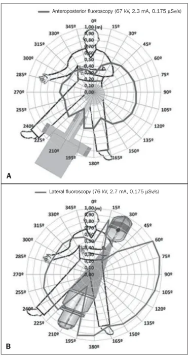
Variation in the dose rate around the phantom
For the study of the scattered radiation readings around the phantom, the initial positioning was maintained, and the radiation monitor was placed at 1.0 m from the center of the exposure field, at a height of 1.25 m in the plane of in-cidence of the radiation beam in configuration 1. The posi-tion of the detector was changed in 15° increments, the 0° angle corresponding to the median sagittal line in the direc-tion of the head.
RESULTS
The statistical study conducted prior to the dose rate readings involved a sample of 55 orthopedic interventions and showed that the procedure that emits the most scattered radiation is hip surgery, because it is the most common in-tervention and produces the highest dose values. The mean voltage and current were 67 kV and 2.4 mA, respectively, for configuration 1, compared with 76 kV and 2.8 mA, re-spectively, for configuration 2. The mean fluoroscopy time per intervention was 27 s.
Variation in the dose rate as a function of height above the operating room floor
The scattered radiation dose rate readings as a function of height are shown for configurations 1 and 2 in Figures 2A and 2B, respectively. At chest level, the maximum dose rates were 1.167 ± 0.023 µSv/s and 2.278 ± 0.023 µSv/s in configurations 1 and 2, respectively. At the thyroid level, the
mean dose rates registered were 0.481 ± 0.010 µSv/s and 0.692 ± 0.007 µSv/s in configurations 1 and 2, respectively, compared with 0.133 ± 0.0013 µSv/s and 0.367 ± 0.011 µSv/s, respectively, at the level of the lens of the eye.
Variation in the dose rate as a function of distance
For configurations 1 and 2, the scattered radiation dose rate readings as a function of the distance from the center of the exposure field are shown in Figures 3A and 3B, respec-tively. We also compared the experimental and theoretical distance values obtained by the inverse square law.
According to the general rule of irradiance, an extended source may be considered a point source if the distance from
the source is greater than five times its diameter(9).
There-fore, to calculate the theoretical values, the value measured at the greatest distance was used, allowing the application of the inverse square law.
We observed differences between the measured values and the theoretical values, those differences being more pro-nounced in configuration 2 and for distances less than 1.0 m.
Variation in the dose rate around the phantom
Considering the scattered radiation dose rate readings around the phantom (Table 1), we used the inverse square law formula to estimate, for each angle, the distance at which the detector should be to receive the maximum scattered ra-diation dose rate registered (0.175 µSv/s), thus tracing the isodose curves for configurations 1 and 2, as shown in Fig-ures 4A and 4B, respectively.
Figure 4 shows an anisotropic dose distribution around the phantom, indicated by the line that connects the points of equal doses at different distances. In both configurations, the highest doses registered were to the left of the patient and
Figure 2. Graphic illustra-tions of the variation in the dose rate as a function of height, with the tube at 0° (A) and at 90° (B) at a distance of 25 cm from the center of the exposure field, at a 90° angle to the median sagittal line of the phantom.
Height (m)
Normalized dose Height (m)
Figure 3. Graphic illustra-tions of the variation in the dose rate as a function of dis-tance, at a height of 1.25 m and at a 90° angle to the median sagittal line of the phantom, for configuration 1 (A) and configuration 2 (B).
Dose rate (µµµµµSv/s)
Dose rate (µµµµµSv/s)
Measured dose rate (µµµµµSv/s) Theoretical dose rate (µµµµµSv/s) Measured dose rate (µµµµµSv/s)
Theoretical dose rate (µµµµµSv/s)
Distance (m)
Table 1—Rates of scattered radiation doses around the phantom.
Configuration 1 (0°) Configuration 2 (90°)
Position 0° 15° 30° 45° 60° 75° 90° 105° 120° 135° 150° 165° 180° 195° 210° 225° 240° 255° 270° 285° 300° 315° 330° 345° Dose rate (µSv/s)
0.012 0.031 0.048 0.057 0.062 0.067 0.070 0.074 0.073 0.068 0.054 0.029 0.053 0.056 0.015 0.050 0.040 0.036 0.045 0.043 0.033 0.034 0.029 0.021 Distance (m) 0.26 0.42 0.52 0.57 0.59 0.62 0.63 0.65 0.65 0.62 0.56 0.41 0.55 0.57 0.29 0.53 0.48 0.45 0.51 0.49 0.43 0.44 0.41 0.35 Dose rate (µSv/s)
0.041 0.128 0.009 0.041 0.175 0.175 0.161 0.150 0.139 0.133 0.131 0.114 0.147 0.158 0.007 0.097 0.097 0.103 0.092 0.079 0.071 0.073 0.069 0.059 Distance (m) 0.48 0.85 0.23 0.48 1.00 1.00 0.96 0.93 0.89 0.87 0.86 0.81 0.92 0.95 0.20 0.75 0.75 0.77 0.72 0.67 0.64 0.65 0.63 0.58
the maximum dose rate was 0.175 µSv/s, registered for the incidence in profile, at a distance of 1.0 m, at 60° and 75°.
Estimate of the effective dose received by professionals
On the basis of the dose rates measured as a function of the angle and of the distance at which where professionals were from the center of exposure, we estimated the effective dose received at the position of each professional, assuming that the members of the team maintain the same positions throughout the surgical procedure.
In calculating the effective doses, we assumed that the overall duration of an intervention was 27 s. The interven-tions evaluated were distributed equally between configura-tions 1 and 2 (Table 2).
On the basis of previous studies, it is estimated that ap-proximately 282 surgical interventions involving the hip are performed per year in the Orthopedics Department of the Faro Branch of the Algarve Hospital Center. Assuming that there are five surgical teams performing these interventions, each team therefore carrying out approximately 57 fluoros-copy-guided hip interventions procedures per year, we esti-mated that the lead surgeon receives a cumulative annual scattered radiation dose of 1.974 mSv, compared with 0.653 mSv for the attending physician.
DISCUSSION
Fluoroscopy is frequently used by medical profession-als. Therefore, it is necessary to raise awareness in relation to the risks of ionizing radiation, as well as to encourage the use of personal protective equipment and greater attention to radiation protection recommendations in order to reduce
the doses received during medical procedures(1).
A previous study carried out by our group indicated that the medical field in which fluoroscopy is most frequently re-quested is orthopedics, primarily the subspecialty of hip surgery. Therefore, we decided to study the distribution of scattered radiation during those procedures and estimate the effective doses of radiation received by the different profes-sionals involved.
Figure 4. Isodose curve around the phantom traced for configuration 1 (A) and configuration 2 (B).
B
Anteroposterior fluoroscopy (67 kV, 2.3 mA, 0.175 µSv/s)
Lateral fluoroscopy (76 kV, 2.7 mA, 0.175 µSv/s)
In relation to the parameters used in this study during the radiation beam simulation and exposure time, the mean exposure time observed in the present study was similar to
the 26 s reported by Alonso et al.(13). In addition, our values
for current and voltage were similar to those reported by
Fuchs et al.(14).
The readings for the dose rate as a function of height in relation to the floor of the operating room showed that the radiation intensity was greatest at the level of the chest of the lead surgeon. That was true for both configurations.
Assuming that the exposure duration at the level of the lens of the eye is 30 s, we estimated that the equivalent dose to the eyes is 7.5 µSv per intervention, which is below the 11.2–45.5 µSv range of values indicated in the study
con-ducted by Fuchs et al.(14). It should be borne in mind that
the annual equivalent dose for the lens of the eye is 15 mSv
per year(15).
At the thyroid level, the estimated dose was 17.58 µSv per intervention under the same conditions described above. That is within the 16.7–67.9 µSv dose range indicated in
the study conducted by Fuchs et al.(14).
For the dose rate as a function of distance, there was a difference between the experimental and theoretical values for short distances from the exposure field. Therefore, the inverse square law underestimates the true dose rate in that simulation.
In relation to the dose rate around the phantom indi-cated by the isodose curves, we observed a 210° gap in the dose, corresponding to the space occupied by the C-arm fluo-roscopy equipment, probably due to the absorption of scat-tered radiation by the equipment. There was also a drop in the intensity of the dose at the positions corresponding to the location of the head and lower members of the patient, due to the absorption of scattered radiation by the patient.
The dose rates were higher for configuration 2 than for configuration 1. That was due to the fact that the detector was in the same plane of incidence of the primary X-ray beam, meaning that there was a higher concentration of
backscat-tered radiation(16).
In this study, it was estimated that the lead surgeon re-ceives an approximate effective dose of 34.6 µSv per proce-dure, which is within the range of dose values reported in
the study conducted by Fuchs et al.(14). Alonso et al.(13)
re-ported a dose value of 37 µSv, which is quite comparable to the value registered in the present study.
Even though the dose rate values obtained in this study are relatively low, the use of personal protective equipment
is recommended(17). The use of such equipment can
substan-tially reduce radiation exposure.
During surgical interventions involving the use of ra-diation, most health professionals wear lead aprons and thy-roid collars, although eye protection (with goggles) is rarely used.
According to the International Atomic Energy Agency, the effective dose per hip procedure received by the lead sur-geon, assuming a fluoroscopy time of 25 s and the use of a 0.5-mm lead apron, should be no more than approximately 5 µSv. Considering that an X-ray beam with energy between 60 keV and 100 keV transmits 1–7% of that energy through a 0.5-mm lead apron, we can conclude that, under the con-ditions presented in this study and assuming that the physi-cian is wearing a 0.5-mm lead apron, the effective dose re-ceived would be 2.5 µSv, which is below the reference
value(18).
On the basis of the doses estimated in this study, we can state that the use of 0.25-mm lead aprons would be suffi-cient to ensure safety and protection during surgical inter-ventions involving the use of radiation. That would afford health professionals greater comfort during such procedures.
CONCLUSION
In this study, we have shown that the radiation doses re-ceived by health professionals during fluoroscopy-guided hip surgery are low. Nevertheless, given that there are no safe levels of radiation, it is advisable to wear lead aprons, thy-roid collars, and protective goggles, which can substantially reduce radiation exposure during such procedures.
REFERENCES
1. Santana PC, Oliveira PMC, Mamede M, et al. Ambient radiation levels in positron emission tomography/computed tomography (PET/CT) imaging center. Radiol Bras. 2015;48:21–5.
2. Le Heron J, Padovani R, Smith I, et al. Radiation protection of medi-cal staff. Eur J Radiol. 2010;76:20–3.
3. Romano RFT, Salvadori PS, Torres LR, et al. Readjustment of ab-dominal computed tomography protocols in a university hospital: impact on radiation dose. Radiol Bras. 2015;48:292–7.
Table 2— Estimate of the effective dose received by health professionals based on the dose rate measured at 1.25 m in relation to the patient plane.
Professional
Lead surgeon Attending physician Instrument nurse Nurse anesthetist Anesthesiologist Circulating nurse Radiology technician
Distance (m)
0.3 0.5 1.6 1.2 1.2 2.4 1.9
Dose rate in configuration 1 (µSv/s)
0.775 0.293 0.029 0.015 0.015 0.009 0.004
Dose rate in configuration 2 (µSv/s)
1.790 0.556 0.059 0.041 0.041 0.026 0.002
Dose per intervention (µSv)
4. Madrigano RR, Abrão KC, Puchnick A, et al. Evaluation of non-radiologist physicians’ knowledge on aspects related to ionizing ra-diation in imaging. Radiol Bras. 2014;47:210–6.
5. Oliveira AD, Jesus J, Leite E, et al. Caracterização do feixe de radia-ção X num bloco operatório em cirurgia ortopédica. Rev Port Saúde Pública. 2009;27:59–70.
6. Navarro VCC, Navarro MVT, Maia AF, et al. Evaluation of medi-cal radiation exposure in pediatric interventional radiology proce-dures. Radiol Bras. 2012;45:210–4.
7. Portugal. Ministério da Saúde. Decreto-Lei nº 222/2008. Diário da República, 223 Série I, de 17 de novembro de 2008.
8. International Atomic Energy Agency. Patient and staff dose in fluo-roscopy. [cited 2015 Apr 8]. Available from: https://rpop.iaea.org/ RPOP/RPoP/Content/InformationFor/HealthProfessionals/ 4_InterventionalRadiology/patient-staff-dose-fluoroscopy.htm. 9. International Atomic Energy Agency. Orthopedic surgery. [cited
2014 Jul 12]. Available from: https://rpop.iaea.org/RPOP/RPoP/ C o n t e n t / I n f o r m a t i o n F o r / H e a l t h P r o f e s s i o n a l s / 6 _ OtherClinicalSpecialities/Orthopedic/index.htm.
10. Adam,Rouilly. AR10A X-ray/radiographic positioning doll. [cited 2014 May 29]. Available from: http://www.adam-rouilly.co.uk/ productdetails.aspx?pid=2792&cid=411.
11. Atomtex. AT1121, AT1123 X-ray and gamma radiation dosimeters.
[cited 2013 Dec 18]. Available from: http://www.atomtex.com/en/ products/portable-dosimeters/at1121-at1123-x-ray-and-gamma-radiation-dosimeters.
12. Soma Technology. Philips BV 300. [cited 2013 Dec 18]. Available from: http://www.somatechnology.com/MedicalProducts/philips-bv300-c-arms.asp.
13. Alonso JA, Shaw DL, Maxwell A, et al. Scattered radiation during fixation of hip fractures. Is distance alone enough protection? J Bone Joint Surg. 2001;83:815–8.
14. Fuchs M, Schmid A, Eiteljörge T, et al. Exposure of the surgeon to radiation during surgery. Int Orthop. 1998;22:153–6.
15. Conselho da União Europeia. Diretiva 2013/59/Euratom do Con-selho de 5 de dezembro de 2013. Jornal Oficial da União Europeia. 2014;13:1–73.
16. Bushong SC. Radiologic science for technologists: physics, biol-ogy, and protection. 7th ed. St. Louis, MO: Mosby; 2001. 17. Osman H, Sulieman A, Sam AK. Orthopedist’s thyroid radiation
dose during surgery. Journal of Advanced Medical Research. 2011;1: 55–60.