Radiol Bras. 2016 Mai/Jun;49(3):144–149 144
Imaging assessment of glenohumeral dysplasia secondary
to brachial plexus birth palsy
*
Avaliação de imagens de displasia glenoumeral secundária a paralisia obstétrica do plexo braquial
Chagas-Neto FA, Dalto VF, Crema MD, Waters PM, Gregio-Junior E, Mazzer N, Nogueira-Barbosa MH. Imaging assessment of glenohumeral dysplasia secondary to brachial plexus birth palsy. Radiol Bras. 2016 Mai/Jun;49(3):144–149.
Abstract
R e s u m o
Objective: To assess imaging parameters related to the morphology of the glenohumeral joint in children with unilateral brachial plexus birth palsy (BPBP), in comparison with those obtained for healthy shoulders.
Materials and Methods: We conducted a retrospective search for cases of unilateral BPBP diagnosed at our facility. Only patients with a clinical diagnosis of unilateral BPBP were included, and the final study sample consisted of 10 consecutive patients who were assessed with cross-sectional imaging. The glenoid version, the translation of the humeral head, and the degrees of glenohumeral dysplasia were assessed.
Results: The mean diameter of the affected humeral heads was 1.93 cm, compared with 2.33 cm for those of the normal limbs. In two cases, there was no significant posterior displacement of the humeral head, five cases showed posterior subluxation of the humeral head, and the remaining three cases showed total luxation of the humeral head. The mean glenoid version angle of the affected limbs (90–α) was –9.6°, versus +1.6° for the normal, contralateral limbs.
Conclusion: The main deformities found in this study were BPBP-associated retroversion of the glenoid cavity, developmental delay of the humeral head, and posterior translation of the humeral head.
Keywords: Birth injuries/complications; Joint diseases/diagnosis; Brachial plexus neuropathies/complications; Humeral head/abnormalities; Shoulder dislocation/diagnosis; Tomography.
Objetivo: Avaliar os parâmetros de imagem relacionados com a morfologia da articulação glenoumeral em crianças com paralisia obs-tétrica do plexo braquial (POPB) unilateral, comparando-os com os observados em ombros saudáveis.
Materiais e Métodos: Foi realizada uma busca retrospectiva de casos de POPB unilateral diagnosticados em nossa instituição. Somente foram incluídos os pacientes com diagnóstico clínico de POPB unilateral, e a amostra final do estudo consistiu em 10 pacientes conse-cutivos avaliados por meio de imagens transversais. Foram avaliados a retroversão da cavidade glenoide, a translação da cabeça do úmero e o grau de displasia glenoumeral.
Resultados: A média do diâmetro da cabeça do úmero foi 1,93 cm nos membros afetados e 2,33 cm nos membros normais. Em dois casos, não houve deslocamento posterior significativo da cabeça do úmero, cinco casos apresentaram subluxação posterior da cabeça do úmero, e os três casos restantes apresentaram luxação total da cabeça do úmero. A média do ângulo de retroversão glenoide dos membros afetados (90–α) foi –9,6°, ao passo que a dos membros contralaterais normais foi +1,6°.
Conclusão: As principais deformidades encontradas neste estudo foram retroversão da cavidade glenoide relacionada com POPB, atraso no desenvolvimento da cabeça do úmero e translação posterior da cabeça do úmero.
Unitermos: Traumatismos do nascimento/complicações; Artropatias/diagnóstico; Neuropatias do plexo braquial/complicações; Cabeça do úmero/anormalidades; Luxação do ombro/diagnóstico; Tomografia computadorizada.
* Study conducted in the Division of Radiology, Internal Medicine Department, Faculdade de Medicina de Ribeirão Preto da Universidade de São Paulo (FMRP-USP), Ribeirão Preto, SP, Brazil.
1. Radiology Professor, Division of Radiology, Universidade de Fortaleza (Unifor) and Centro Universitário Christus, Fortaleza, CE, Brazil.
2. PhD Student, Division of Radiology, Internal Medicine Department, Faculdade de Medicina de Ribeirão Preto da Universidade de São Paulo (FMRP-USP), Ribeirão Preto, SP, Brazil.
3. MD, Radiologist, Radiology Department, Hôpital Saint-Antoine, Université Paris VI, Paris, France; Department of Radiology, Quantitative Imaging Center, Boston University School of Medicine, Boston, MA, USA; Department of Radiology and Tele-Imaging, Hospital do Coração (HCor), São Paulo, SP, Brazil.
4. Orthopedic Surgeon-in-Chief, Brachial Plexus Program Director, Orthopedic Center, Boston Children’s Hospital, Harvard Medical School, Boston, MA, USA.
5. PhD, MD, Radiologist, União Médica Radiológica Catanduva (UMERC), Catan-duva, SP, Brazil.
INTRODUCTION
Brachial plexus birth palsy (BPBP) most commonly af-fects the upper trunk nerve components of the brachial plexus
Francisco Abaete Chagas-Neto1, Vitor Faeda Dalto2, Michel Daoud Crema3, Peter M. Waters4, Everaldo Gregio-Junior5, Nilton Mazzer6, Marcello Henrique Nogueira-Barbosa7
6. Full Professor of Orthopedics, Department of Biomechanics, Medicine, and Rehabilitation of the Locomotor Apparatus, Faculdade de Medicina de Ribeirão Preto da Universidade de São Paulo (FMRP-USP), Ribeirão Preto, SP, Brazil.
7. Associate Professor of Radiology, Division of Radiology, Internal Medicine De-partment, Faculdade de Medicina de Ribeirão Preto da Universidade de São Paulo (FMRP-USP), Ribeirão Preto, SP, Brazil.
Mailing Address: Dr. Vitor Faeda Dalto. Divisão de Radiologia, FMRP-USP. Avenida Bandeirantes, 3900, Monte Alegre. Ribeirão Preto, SP, Brazil, 14048-900. E-mail: [email protected].
(C5–T1) and may lead to severe dysfunction of the shoul-der. BPBP represents a significant source of motor disabil-ity, causing morbidity due to the limited active functional range of motion and joint contractures of the upper limbs, with an incidence of approximately 1.5 per 1000 live births in the United States(1).
Approximately 70% of obstetric injuries to the brachial plexus show spontaneous regression in the first months of life, although there is limited spontaneous recovery of mo-tor function in the remaining 30%(1). In cases of incomplete recovery during the first two to three years of life, changes in the glenoid cavity and in the humeral head can appear as early as the fifth month of life, due to contractures and muscle imbalance(2). Initial deformities result from glenohumeral joint contractures and an imbalance in the muscles of the shoulder girdle, caused by paralysis of the external abduc-tors and rotaabduc-tors, combined with relative hyperactivity of the internal adductors and internal rotators, which are partially spared from neurological involvement(1–3).
The main bone alterations secondary to birth palsy of the brachial plexus are as follows: progressive retroversion of the glenoid cavity; thinning and loss of bone at the poste-rior border of the glenoid; posteposte-rior subluxation or luxation of the humeral head; hypoplasia of the scapula; flattening or absence of the glenoid cavity; inferior deviation of the cora-coid process; conic deformation of the acromion; deformity and hypoplasia of the humeral head; delayed bone develop-ment in the proximal humerus; and shortening of the clavicle in comparison with that of the contralateral side.
Because of the risk of rapid progression of BPBP, early diagnosis and therapeutic interventions are critical. Several studies have evaluated different ways to quantify the degrees of glenoid anteversion and retroversion, as well as to measure the different degrees of translation of the humeral head(4–7). The aim of our study was to assess imaging parameters related to the morphology of the glenohumeral joint in chil-dren with unilateral BPBP, comparing these parameters be-tween pathological and healthy shoulders.
MATERIALS AND METHODS Subject selection and clinical data
This study was approved by the local institutional re-view board, which conceded an exemption from the require-ment to obtain informed consent. We conducted a retrospec-tive search for cases of unilateral BPBP diagnosed at our ser-vice. The identification of cases was based on searches of reports from computed tomography (CT) and magnetic resonance imaging (MRI) studies of the shoulders of chil-dren, conducted between January 1, 2005, and December 31, 2010, in radiology information systems. We used the following search terms: “glenoid retroversion”; “glenoid dysplasia”; “posterior subluxation or luxation of the humeral head”; “brachial plexus lesion”; and “hypoplasia of the hu-meral head”. Reports and images were reviewed and corre-lated with additional imaging reports, as well as with the
clinical and surgical history. Only patients with a clinical diagnosis of unilateral BPBP were included. Exclusion cri-teria were being > 12 years of age; showing clinical or im-aging signs of bilateral neurological injury; having under-gone MRI or CT in which the results were suboptimal (be-cause of motion artifacts, failure to complete the examina-tion, or suboptimal patient positioning); showing signs of infectious or inflammatory arthritis; and showing evidence of prior shoulder surgery. The final study sample consisted of 10 consecutive patients with unilateral BPBP who were assessed with cross-sectional imaging via CT or MRI.
Imaging studies
All examinations were performed according to our insti-tutional routine protocol. Patients were placed in the supine position, with both shoulders supported on the table and the arms relaxed in the neutral position.
All MRI scans were obtained in the same 1.5 T MRI scanner (Philips Achieva 1.5 T MRI System; Philips Medi-cal Systems, Best, the Netherlands) with a phased array coil. For each shoulder, coronal and sagittal T1-weighted images were obtained (3.0 mm slices; 0.5 mm gap), as were axial, sagittal, and coronal T2-weighted images. Field of view was adjusted to the size of the child, and the matrix size was 256 × 256.
CT scans were obtained in either a 16-channel multislice scanner (Philips Brilliance CT Big Bore System; Philips Medical Systems) or a single channel helical CT scanner (Somatom Emotion; Siemens Healthcare, Erlangen, Ger-many). Axial images of the shoulders (1.25 mm slices; 0.625 mm gap) were obtained and then reformatted in the axial, sagittal, and coronal planes (2.0 mm slices). Field of view, kV, and mA were adjusted to the size of the child.
Reading technique and quantitative parameters Axial plane images from the MRI and CT examinations were evaluated on a picture archiving and communication system, in consensus, by two fellowship-trained musculosk-eletal radiologists with 5 and 15 years of experience, respec-tively.
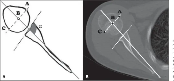
The glenoid version was assessed using the method de-scribed by Friedman et al.(6): the angle formed by the line drawn at the anterior and posterior aspects of the glenoid margins and the line between the medial margin of the scapula and the midpoint of the glenoid is measured, then 90° is subtracted from the resulting angle (Figure 1). Both lines were drawn in the same image slice. Positive values were interpreted as glenoid cavity anteversion, and negative values were interpreted as glenoid cavity retroversion.
method) at the midpoint of the humeral head, thus obtain-ing the ratio between the measurements (PHHA = AB/AC × 100) (Figure 1). Humeral head displacement was consid-ered insignificant when the PHHA values were between 40% and 50%; posterior subluxation was defined as a PHHA be-tween 0% and 35%; and the entire humeral head being pos-terior to the scapular line was classified as total luxation. The greatest axial diameter of the affected and contralateral hu-meral heads was also measured by the method shown in Figure 1 (AC distance).
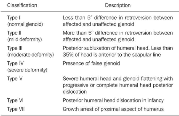
The axial slice level used for the measurements was defined separately for the affected and normal contralateral sides as the first image below the coracoid process that best showed the reference anatomical structures (glenoid margins, scapula margins, and humeral head). After the measurements, the degrees of glenohumeral dysplasia were classified accord-ing to the criteria established by Waters et al.(8), as shown in Figure 1.
Statistical analysis
Statistical analyses were performed with KyPlot 2.0™ software, to compare parameters assessed between BPBP-af-fected limbs and unafBPBP-af-fected, contralateral limbs. Statistical significance was defined as a p < 0.05. Unaffected, contralat-eral limbs were used as internal controls for this study.
RESULTS
Six males and four females, with a mean age of four years and nine months (range, 2–12 years), were included in this study. In six cases, the left shoulder was affected. CT and MRI were used in the evaluation of eight cases and four cases, respectively, both having been used in two cases.
The mean diameter of the affected humeral head was 1.93 cm, with a standard deviation of 0.52 cm. The con-tralateral humeral head measured an average of 2.33 cm, with a standard deviation of 0.71 cm. In all patients, there was marked asymmetry in the humeral head diameters and the difference was statistically significant (p < 0.01). This
indicates a developmental delay in the humeral head on the affected side (Figure 2).
Two cases showed no significant posterior displacement of the humeral head, five cases showed posterior subluxation of the humeral head, and the three remaining cases showed total luxation of the humeral head.
The mean PHHA for the affected shoulder was 24%, with a standard deviation of 18%, compared with 48% with stan-dard deviation of 6%, for the contralateral shoulder. As can be seen in Figure 3, the posterior translation of the humeral head was significant greater on the affected side (p < 0.05). The average glenoid version of the affected limbs (90–α) was –19.6° (95% CI: –11.1° to –28.0°) versus +1.6° (95% CI: 1.0° to 3.1°) in the normal contralateral limbs. In all patients, the affected side presented retroversion of the gle-noid cavity, varying between –9° and –39°, and was signifi-cantly different from the unaffected side (p < 0.05), as de-picted in Figures 4 and 5.
According to the classification system devised by Wa-ters et al.(8), detailed in Table 1, our sample presented two Figure 2. Sample distribution regarding the diameter of the humeral head (cm) on the affected side and unaffected (contralateral) side.
• One case was excluded from this analysis because the non-affected contra-lateral limb was not evaluated.
Figure 1.A: Schematic drawing of glenoid version (90–α) and hu-meral head translation measure-ment methods. The percentage of the humeral head anterior to the scapular line (PHHA) was mea-sured according to the formula
PHHA = AB/AC × 100. B: Axial computed tomography scan of the glenohumeral joint, corresponding to an illustrative case.
DISCUSSION
Glenohumeral dysplasia secondary to BPBP is an un-common cause of severe limb dysfunction in young patients. Here, we have retrospectively reviewed 10 cases of BPBP. The main bone and articular alterations identified in those patients were posterior translation/subluxation of the humeral head, retroversion of the glenoid cavity, and developmental delay in the humeral head. The bone and anatomic alterations were statistically significant when compared against the con-tralateral unaffected limb (internal control).
The prevalence of bone deformities in BPBP varies across studies depending on the methodology and the evalu-ation criteria used. In a populevalu-ation-based study conducted by Sjöberg et al. in Sweden(9), approximately 25% of births with brachial plexus palsy were found to present persistent abnormalities. In a prospective cohort assessing 94 children with brachial plexus palsy, the authors found abnormalities on cross-sectional imaging in 38% of the cases, and 26 (62%) of the 42 patients who underwent tomography for surgical planning presented evidence of posterior subluxation of the humeral head(10).
In the present study, approximately 80% of the patients presented posterior subluxation or total luxation of the hu-meral head (Waters types III, IV, and VI). Only two cases
• One case was excluded from this analysis because the non-affected contra-lateral limb was not evaluated.
Figure 3. Sample distribution regarding the percentage of the humeral head anterior to the scapular line. A reference range for normality would be 40–50%.
• One case was excluded from this analysis because the non-affected contra-lateral limb was not evaluated.
Figure 4. Glenoid version distribution on the affected side and unaffected (con-tralateral) side.
Table 1—Deformity of the glenohumeral joint according to the criteria estab-lished by Waters et al.(8).
Classification
Type I (normal glenoid) Type II (mild deformity) Type III
(moderate deformity) Type IV
(severe deformity) Type V
Type VI Type VII
Description
Less than 5° difference in retroversion between affected and unaffected glenoid
More than 5° difference in retroversion between affected and unaffected glenoid
Posterior subluxation of humeral head. Less than 35% of head is anterior to the scapular line Presence of false glenoid
Severe humeral head and glenoid flattening with progressive or complete humeral head posterior dislocation
Posterior humeral head dislocation in infancy Growth arrest of proximal aspect of humerus
cases with mild deformity (Waters type II), three cases with moderate deformity (Waters type III), two cases with severe deformity (Waters type IV), and three cases of total poste-rior displacement of the humeral head (Waters type VI). In our sample, there were no cases classified as Waters types I, V, or VII.
(20%) presented mild deformities (Waters type II). The high frequency of advanced cases in our sample, compared with what has been reported in the literature(9,10), may be related to increased referral of highly complex cases, underdiagnosis of mild cases, or late diagnosis with unfavorable development. Recent studies have emphasized the importance of im-aging methods (ultrasound, CT, and MRI) in the early evalu-ation and classificevalu-ation of patients with brachial obstetrical plexus palsy(11–16). Multiple methods for measurement and classification of the glenoid dysplasia and its associated find-ings have been described in the literature. The methods used in this study were previously described by Friedman et al.(6) and Waters et al.(8), which are notable for their accuracy and reproducibility(3,6,8).
There is as yet no consensus in the literature regarding the role of different imaging methods in the diagnosis and monitoring of BPBP. Imaging assessment in the first three months after birth is rare because most cases present spon-taneous clinical remission. In infants between three and eigh-teen months of age, ultrasound may be used as a screening test in those who did not present satisfactory progress or who present with clinical worsening(16).
The importance of MRI in patients over 18 months of age should be highlighted, because it allows for morphologi-cal assessment, location of the humeral head epiphyseal car-tilage, and aids in the mapping of muscular structures for surgical planning. CT can be used as a complementary, or even standalone, technique for detailed evaluation of bone structures (Figure 6).
Proper evaluation of sectional imaging parameters, as described in this study, is important for correct classifica-tion of dysplasia severity and has a direct effect on treatment choice and post-surgical prognosis for patients(17–22). Patients with minimal (Waters type I) glenohumeral dysplasiawill benefit from latissimus dorsi and teres major tendon
trans-ference to the rotator cuff. Patients with mild or moderate (Waters type II or III) glenohumeral dysplasia, with reduc-ible subluxation or luxation, will be better served by treat-ment with open or arthroscopic reduction of the gleno-humeral joint, together with latissimus dorsi and teres ma-jor tendon transference to the rotator cuff, as well as length-ening of the pectoralis major tendon, subscapularis tendon, or both. Finally, older patients with severe (Waters type IV or higher) glenohumeral deformity, irreducible gleno-humeral luxation, or established arthrosis are candidates for humeral osteotomy(17–22). Recently, a combination of ante-version glenoid osteotomy, so-called glenoplasty, and ten-don transfer has come to be used as a therapeutic option in younger patients with moderate to severe glenohumeral dys-plasia(23).
The main limitations of this study are the retrospective design and the small sample size. In addition, images were not acquired using a standardized protocol and evaluations were performed on a mix of CT and MRI images. Further-more, readings were performed in tandem, and it was there-fore not possible to evaluate interobserver agreement. De-spite these limiting factors, the findings presented in this study demonstrate a useful and easily adoptable method for the evaluation of imaging studies in cases of BPBP.
CONCLUSION
The main deformities found in the affected limbs evalu-ated in this study were BPBP-associevalu-ated retroversion of the glenoid cavity, delayed development of the humeral head, and posterior translation of the humeral head. It is essential that the radiologists become familiar with this condition and its presentation, as well as with the diagnostic methods avail-able for its detection and classification. Such assessments may have significant therapeutic and prognostic implications in patients affected by BPBP.
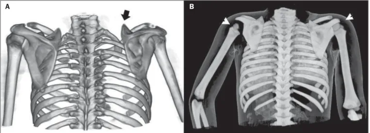
Figure 6. Examples of three-dimensional volume rendering of computed tomography images acquired from two different patients. A: Three-dimensional reconstruc-tion, posterior view. Female patient, 12 years old, history of high obstetric traumatic brachial plexus injury on the right. Note the elevation of the right scapula (arrow) and reduction of right scapula size. B: Posterior view. Volume rendering with skin referential of a 4-year-old male patient with obstetric brachial plexus injury on the left side. Note the elevation of the left scapula, reduced size of the humeral head (arrowheads), and positioning of the left limb (maintained in abduction and internal rotation).
Acknowledgments
This study received financial support from the Conselho Nacional de Desenvolvimento Científico e Tecnológico (CNPq) (Brazilian National Council for Scientific and Tech-nological Development).
REFERENCES
1. Ruchelsman DE, Grossman JA, Price AE. Glenohumeral deformity in children with brachial plexus birth injuries. Bull NYU Hosp Jt Dis. 2011;69:36–43.
2. Sibinski M, Wozniakowski B, Drobniewski M, et al. Secondary gleno-humeral joint dysplasia in children with persistent obstetric brachial plexus palsy. Int Orthop. 2010;34:863–7.
3. van der Sluijs JA, van Ouwerkerk WJ, de Gast A, et al. Deformities of the shoulder in infants younger than 12 months with an obstetric lesion of the brachial plexus. J Bone Joint Surg Br. 2001;83:551–5. 4. Hoenecke HR Jr, Hermida JC, Flores-Hernandez C, et al. Accuracy of CT-based measurements of glenoid version for total shoulder arthroplasty. J Shoulder Elbow Surg. 2010;19:166–71.
5. Churchill RS, Brems JJ, Kotschi H. Glenoid size, inclination, and version: an anatomic study. J Shoulder Elbow Surg. 2001;10:327– 32.
6. Friedman RJ, Hawthorne KB, Genez BM. The use of computerized tomography in the measurement of glenoid version. J Bone Joint Surg Am. 1992;74:1032–7.
7. Randelli M, Gambrioli PL. Glenohumeral osteometry by computed tomography in normal and unstable shoulders. Clin Orthop Relat Res. 1986;(208):151–6.
8. Waters PM, Smith GR, Jaramillo D. Glenohumeral deformity sec-ondary to brachial plexus birth palsy. J Bone Joint Surg Am. 1998; 80:668–77.
9. Sjöberg I, Erichs K, Bjerre I. Cause and effect of obstetric (neona-tal) brachial plexus palsy. Acta Paediatr Scand. 1988;77:357–64. 10. Beischer AD, Simmons TD, Torode IP. Glenoid version in children
with obstetric brachial plexus palsy. J Pediatr Orthop. 1999;19:359– 61.
11. Reading BD, Laor T, Salisbury SR, et al. Quantification of humeral head deformity following neonatal brachial plexus palsy. J Bone Joint Surg Am. 2012;94:e136(1-8).
12. Pearl ML, Woolwine S, van de Bunt F, et al. Geometry of the
proxi-mal humeral articular surface in young children: a study to define normal and analyze the dysplasia due to brachial plexus birth palsy. J Shoulder Elbow Surg. 2013;22:1274–84.
13. Lippert WC, Mehlman CT, Cornwall R, et al. The intrarater and interrater reliability of glenoid version and glenohumeral sublux-ation measurements in neonatal brachial plexus palsy. J Pediatr Orthop. 2012;32:378–84.
14. Clarke SE, Chafetz RS, Kozin SH. Ossification of the proximal humerus in children with residual brachial plexus birth palsy: a magnetic resonance imaging study. J Pediatr Orthop. 2010;30:60– 6.
15. Pöyhiä TH, Lamminen AE, Peltonen JI, et al. Brachial plexus birth injury: US screening for glenohumeral joint instability. Radiology. 2010;254:253–60.
16. Sanchez TR1, Chang J, Bauer A, et al. Dynamic sonographic evalu-ation of posterior shoulder dislocevalu-ation secondary to brachial plexus birth palsy injury. J Ultrasound Med. 2013;32:1531–4.
17. Waters PM, Bae DS. The effect of derotational humeral osteotomy on global shoulder function in brachial plexus birth palsy. J Bone Joint Surg Am. 2006;88:1035–42.
18. Waters PM, Bae DS. Effect of tendon transfers and extra-articular soft-tissue balancing on glenohumeral development in brachial plexus birth palsy. J Bone Joint Surg Am. 2005;87:320–5.
19. Pearl ML, Edgerton BW, Kazimiroff PA, et al. Arthroscopic release and latissimus dorsi transfer for shoulder internal rotation contractures and glenohumeral deformity secondary to brachial plexus birth palsy. J Bone Joint Surg Am. 2006;88:564–74. 20. Kozin SH, Chafetz RS, Barus D, et al. Magnetic resonance imaging
and clinical findings before and after tendon transfers about the shoulder in children with residual brachial plexus birth palsy. J Shoul-der Elbow Surg. 2006;15:554–61.
21. Waters PM, Bae DS. The early effects of tendon transfers and open capsulorrhaphy on glenohumeral deformity in brachial plexus birth palsy. J Bone Joint Surg Am. 2008;90:2171–9.
22. Crouch DL, Plate JF, Li Z, et al. Computational sensitivity analysis to identify muscles that can mechanically contribute to shoulder deformity following brachial plexus birth palsy. J Hand Surg Am. 2014;39:303–11.