Letters to the Editor
Radiol Bras. 2015 Nov/Dez;48(6):399–403
402
http://dx.doi.org/10.1590/0100-3984.2015.0074 tumors are constituted of plasmacytoid cells, presenting
malig-nant degeneration and producing a specific immunoglobulin mol-ecule(3–7).
The incidence of EMP is higher in men than in women, at a 3–4:1 ratio, most frequently occurring around the age of 50– 60(1,4,6,7). It is estimated that such tumor represents 2–4% of
plas-macytoid neoplasms whose most relevant representative is the multiple myeloma(1,3–7), the latter representing up to 1% of all
general malignancies(8).
Approximately 80–90% of EMP cases involve craniocervical structures (upper aerodigestive tract; larynx; nasopharynx; tonsilla; nasal and paranasal cavities)(1–8), but the number of cases does
not reach 1% of all neoplastic head and neck lesions(5). Other sites
such as gastrointestinal and urogenital tracts, central nervous system, thyroid, parathyroid glands, salivary glands, lymph nodes, skin, lungs, and breasts are uncommon(2,3,5,6). Lymph node
in-volvement in pulmonary hila is extremely rare, with rates as low as less than 2% of cases(2).
Generally, they present as masses with nonspecific soft parts density(3). Histologically, such tumors do not originate directly
from the bone marrow and cannot be distinguished from mul-tiple myelomas. Also the differentiation from plasmacytoid cell granulomas and other inflammatory reactions is difficult, essen-tially requiring immunophenotyping(1,4).
The diagnosis of EMP is made after rigorous investigation to rule out the presence of multiple myeloma, highlighting the histological confirmation by means of immunohistochemical analysis, biopsy/bone marrow puncture showing < 5% of plasma-cytoid atypia; to rule out the presence of osteolytic lesions, serum and urinary protein dosage and electrophoresis (to rule out the pres-ence of M and Bpres-ence-Jones proteins, respectively); and non-exist-ence of anemia(1–4,6,7).
EMP may be the initial manifestation of multiple myeloma, with progression in about 30% of cases(1,2,7).
Lenara Renó Arbex Coelho1, Gabriel Pinheiro Coelho1, Rodolfo Mendes Queiroz1, Marcus Vinicius Nascimento Valentin1
1. Documenta – Hospital São Francisco, Ribeirão Preto, SP, Brazil. Mailing Address: Dr. Rodolfo Mendes Queiroz. Documenta – Centro Avançado de Diagnóstico por Imagem. Rua Bernardino de Campos, 980, Centro. Ribeirão Preto, SP, Brazil, 14015-130. E-mail: rod_queiroz@ hotmail.com.
Treatments of choice include radiotherapy due the high ra-diosensitivity in 80–100% of cases, and surgery for localized le-sions(1,3–5,8). With such treatments, one observes recurrence and
dissemination rates between 20% and 40%(1,,2,5–7), and ten-year
survival in 70% of cases(1,5–7).
REFERENCES
1. Luh SP, Lai YS, Tsai CH, et al. Extramedullary plasmacytoma (EMP): report of a case manifested as a mediastinal mass and multiple pulmo-nary nodules and review of literature. World J Surg Oncol. 2007;5:123. 2. Nakayama K, Okada D, Koizumi K, et al. Excision of extramedullary plas-macytoma in a hilar lymph node. Japanese Journal of Lung Cancer. 2006; 46:723–6.
3. Ooi GC, Chim JC, Au WY, et al. Radiologic manifestations of primary solitary extramedullary and multiple solitary plasmacytomas. AJR Am J Roentgenol. 2006;186:821–7.
4. Bertolami A, Henriques AC, Penha FG, et al. Plasmocitoma extramedular. Arq Med ABC. 2005;30:58–60.
5. Ching ASC, Khoo JBK, Chong VFH. CT and MR imaging of solitary extramedullary plasmacytoma of the nasal tract. AJNR Am J Neuroradiol. 2002;23:1632–6.
6. Galieni P, Cavo M, Pulsoni A, et al. Clinical outcome of extramedullary plasmacytoma. Haematologica. 2000;85:47–51.
7. Lee SY, Kim JH, Shin JS, et al. A case of extramedullary plasmacytoma arising from the posterior mediastinum. Korean J Intern Med. 2005;20: 173–6.
8. Ferrari S, Tecchio C, Turri G, et al. Unusual case of solitary intraparen-chymal brain plasmacytoma. J Clin Oncol. 2012;30:e350–2.
PET/CT and brown fat in the evaluation of treatment response in Hodgkin lymphoma
PET/CT e gordura marrom na avaliação da resposta terapêutica no linfoma de Hodgkin
Dear Editor,
A female, 15-year-old patient presented with insidious onset of weight loss and low fever. Hodgkin’s lymphoma was diagnosed after biopsy of a palpable enlarged lymph node. 18
F-FDG PET/ CT was performed during the initial staging, demonstrating
hy-permetabolic mediastinal, axillary and cervical lymph node enlarge-ment (Figure 1). The findings were interpreted as lymphoma in activity in the mentioned sites. At basal PET/CT study, one could not observe metabolic activity in brown fat. Chemotherapy was initiated with adriblastine, bleomycine, vinblastine and dacar-bazine at days D1 and D15 for every 28-day cycles.
Six chemotherapy cycles were uneventfully performed. A new FDG PET/CT performed after about three months to evaluate the therapeutic response demonstrated complete regression of all the lesions interpreted as lymphoma in activity at the first study.
Letters to the Editor
Radiol Bras. 2015 Nov/Dez;48(6):399–403
403
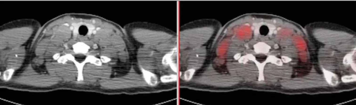
Also, the onset of activity was observed in fat tissue with typical brown fat distribution (at the neck base and shoulders – Figure 2).
Brown fat is an adipose tissue specialized in the generation of heat by means of glucose metabolization (differently from the white fat whose function is just storing energy under the form of lipids)(1,2).
As compared with white fat, brown fat has abundant vascu-larization and innervation by the sympathetic nervous system. Many times, the metabolic activity in the brown fat may obscure intermingled hypermetabolic lesions (metastatic lymph nodes, for example)(1,2).
The onset of activity in brown fat with disappearance of le-sions suspicious for active lymphoma after chemotherapy comple-tion in children and adolescents is described in the literature and is related to a complete therapeutic response in the lymphoma(3,4).
Such an inverse relationship between the absence of tumor and presence of brown adipose tissue has been observed in both fe-male and fe-male patients regardless their body mass index and tem-perature(3,4). The possible mechanisms for brown fat suppression
by the lymphoma still remain unknown. However, patients with malignant lymphomas present with high levels of tumor necrosis factor alpha, an important cytokine capable of inducing a great number of biological effects in multiple systems, including apoptotic degeneration of brown adipocytes(1–4).
PET/CT using FDG has been widely adopted as the main imaging modality in the evaluation of lymphomas(5,6). The
iden-tification of brown adipose tissue in humans by PET/CT has re-vived the interest in the function and relevance of those cells, since there was a concept that they were seen only in neonates, and currently they are identified by PET/CT also in children and young adults(7,8). The knowledge that the brown adipose tissue is a
pre-dictor of disease state contributes to a correct analysis of images
Laís Bastos Pessanha1, André Ribeiro Nogueira de Oliveira1, Luiz Felipe Alves Guerra1, Diego Lima Nava Martins1, Ronaldo Garcia Rondina1, Melissa Bozzi Nonato Mello1
1. Universidade Federal do Espírito Santo (UFES), Vitória, ES, Brazil. Mailing Address: Dra. Laís Bastos Pessanha. Departamento de Clínica Médica/CCS/UFES. Avenida Marechal Campos, 1468, Nazareth. Vitória, ES, Brazil, 29043-900. E-mail: [email protected].
from children and adolescents with lymphoma, being useful in the follow-up and clinical therapeutics of those patients.
REFERENCES
1. Cannon B, Nedergaard J. Brown adipose tissue: function and physiologi-cal significance. Physiol Rev. 2004;84:277–359.
2. Kleis M, Daldrup-Link H, Mathay K, et al. Diagnostic value of PET/CT for the staging and restaging of pediatric tumors. Eur J Nucl Med Mol Imaging. 2009;36:23–36.
3. Gilsanz V, Hu HH, Kajimura S. Relevance of brown adipose tissue in infancy and adolescence. Pediatr Res. 2013;73:3–9.
4. Gilsanz V, Hu HH, Smith ML, et al. The depiction of brown adipose tissue is related to disease status in pediatric patients with lymphoma. AJR Am J Roentgenol. 2012;198:909–13.
5. Bitencourt AGV, Lima ENP, Chojniak R, et al. Correlation between PET/ CT results and histological and immunohistochemical findings in breast carcinomas. Radiol Bras. 2014;47:67–73.
6. Soares Junior J, Fonseca RP, Cerci JJ, et al. Lista de recomendações do exame PET/CT com 18
F-FDG em oncologia. Consenso entre a Sociedade Brasileira de Cancerologia e a Sociedade Brasileira de Biologia, Medicina Nuclear e Imagem Molecular. Radiol Bras. 2010;43:255–9. 7. Oliveira CM, Sá LV, Alonso TC, et al. Suggestion of a national
diagnos-tic reference level for 18
F-FDG/PET scans in adult cancer patients in Brazil. Radiol Bras. 2013;46:284–9.
8. Curioni OA, Souza RP, Amar A, et al. Value of PET/CT in the approach to head and neck cancer. Radiol Bras. 2012;45:315–8.