Letters to the Editor
Radiol Bras. 2015 Nov/Dez;48(6):399–403
399
0100-3984 © Colégio Brasileiro de Radiologia e Diagnóstico por Imagem
Letters to the Editor
Plantar vein thrombosis: a rare differential diagnosis in patients with plantar pain
Tromboflebite plantar: um diagnóstico diferencial raro em pacientes com dor plantar
Dear Editor,
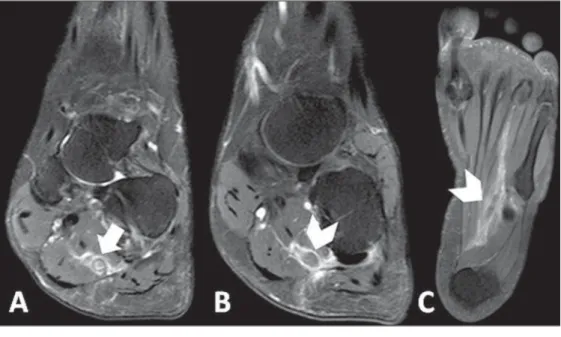
A female, 42-year-old, smoking patient with previous history of thrombotic thrombocytopenic purpura, undergoing corticos-teroid and plasmapheresis therapy was admitted to the hospital presenting with important pain in the plantar region of her right foot, with difficulty in ambulation starting about one week ago. The patient denied the occurrence of any trauma, previous sur-gery or recent travel. At physical examination, only hyperalgesia was observed at palpation. Magnetic resonance imaging (MRI) of the right foot demonstrated thickening and failure in filling in the whole extent of the lateral plantar vein associated with perivenular enhancement and edema of adjacent structures, sug-gesting the presence of plantar vein thrombosis (Figure 1).
Supplementary ultrasonography showed the presence of hypoechogenic material within de lateral plantar vein in associa-tion with ectasia and non-compressibility of the vessel, as well as absence of flow at Doppler study – findings which corroborated the initial hypothesis (Figure 2). The patient was discharged with recommendations for rest and treatment with nonsteroidal anti-inflammatory drugs (NSAIDs), and presented with symptoms improvement at clinical follow-up.
Plantar vein thrombosis (PVT) is a rare condition(1) charac-terized by development of an intraluminal thrombus in plantar veins, with less than 100 cases published in the literature(2). It preferentially affects women of mean age 58.2 years(3). Gener-ally, the lateral plantar vein is most affected (in 96% of cases), followed by the middle plantar vein (in 41%)(3). Several causal fac-tors are speculated, among them traumas(4,5), paraneoplastic syn-dromes(6,7), postoperative conditions(4,7), thrombophilias(2,6), use of contraceptive drugs(2,4), immobilization(5,6) and HIV infection(7). However, most cases are classified as being idiopathic(2).
Cardinal signs and symptoms of PVT include recent onset(7,8) of plantar pain(1–3) and local edema with a typically unilateral pre-sentation(1) in association with limited ambulation(2).
Considering the wide spectrum of differential diagnoses, the imaging diagnosis methods represent a useful tool for a correct characterization of the disease.
Ultrasonography is considered to be the main imaging method for the diagnosis of PVT(1,3), demonstrating hypoechoic venous content(2,6) associated with ectasia(2,5,6), loss of vascular compressibility(2,4) and absence of flow at Doppler study(2,6). How-ever, plantar veins are neglected at routine examinations(1,2,4), which may explain the low rate of diagnosis of the disease.
MRI has gained prominence as besides making the diagno-sis of PVT it can rule out other possible causes of plantar pain(2). Main findings include characterization of intraluminal filling de-fect in plantar veins(2,4,6) in association with edema(5,6) and perivenular enhancement(5). Such findings were observed in 100% of cases in one of the greatest studies approaching the theme(5). On the other hand, only in 2008 the first case of PVT diagnosed at MRI was described(9).
Figure 1. A: Proton density-weighted sequence with fat saturation demonstrat-ing parietal thickendemonstrat-ing of lateral plantar vein in association with perivascular edema (arrow). B,C: Contrast enhanced T1-weighted sequence showing intralu-minal filling defect and perivascular en-hancement (arrowheads).
Figure 2. Ultrasonography of plantar region showing hypoechoic material within the lateral plantar vein in association with ectasia and non-compressibility of the vessel, as well as absence of flow at Doppler study.
Letters to the Editor
Radiol Bras. 2015 Nov/Dez;48(6):399–403
400
http://dx.doi.org/10.1590/0100-3984.2015.0075
Maurício Fabro1, Sara Raquel Madalosso Fabro1, Rafael
Santiago Oliveira Sales1, Cesar Augusto Machado1, Gustavo
Lopes de Araújo1
1. Hospital Santa Catarina de Blumenau, Blumenau, SC, Brazil. Mailing Address: Dr. Maurício Fabro. Rua Tobias Barreto, 266, ap. 304, Vila Nova. Blumenau, SC, Brazil, 89035-070. E-mail: [email protected].
There is no defined treatment for PVT(1,3), and possible treat-ment strategies include use of anticoagulant drugs(1–4,6), NSAIDs(1,3,6), elastic socks(3,6) and rest(6). However, the different therapies have shown similar results.
The most important complications of PVT include throm-bosis extension into deep veins in the leg(7) and occurrence of
pul-monary embolism(1).
Amongst the differential diagnosis of PVT, plantar fasciitis(2,4,5), tendinous involvement(3,5), bursitis(5), Morton’s neuroma(4,5), stress fractures(2,4,5), sesamoiditis(5) and ganglion cysts(5). No descrip-tion of death associated with PVT is found in the literature.
REFERENCES
1. Barros M, Nascimento I, Barros T, et al. Plantar vein thrombosis and pulmonary embolism. Phlebology. 2015;30:66–9.
2. Bruetman JE, Andrews JA, Finn BC, et al. Plantar vein thrombosis as a cause of local pain. Medicina (B Aires). 2014;74:87–8.
3. Czihal M, Röling J, Rademacher A, et al. Clinical characteristics and course of plantar vein thrombosis: a series of 22 cases. Phlebology. 2014. [Epub ahead of print].
4. Karam L, Tabet G, Nakad J,et al. Spontaneous plantar vein thrombosis: state of the art. Phlebology. 2013;28:432–7.
5. Miranda FC, Carneiro RD, Longo CH, et al. Tromboflebite plantar: achados em ressonância magnética. Rev Bras Ortop. 2012;47:765–9. 6. Geiger C, Rademacher A, Chappell D, et al. Plantar vein thrombosis due to busy night duty on intensive care unit. Clin Appl Thromb Hemost. 2011;17:232–4.
7. Barros MV, Labropoulos N. Plantar vein thrombosis – evaluation by ul-trasound and clinical outcome. Angiology. 2010;61:82–5.
8. Bernathova M, Bein E, Bendix N, et al. Sonographic diagnosis of plantar vein thrombosis: report of 3 cases. J Ultrasound Med. 2005;24:101–3. 9. Siegal DS, Wu JS, Brennan DD, et al. Plantar vein thrombosis: a rare
cause of plantar foot pain. Skeletal Radiol. 2008;37:267–9.
Pulmonary neoplasia mimicking fungus ball
Neoplasia pulmonar simulando bola fúngica
Dear Editor,
We report the case of a 74-year-old man smoking 80 ciga-rette packages per year, with history of pulmonary tuberculosis for 50 years. Two years ago, the patient underwent chest computed tomography that demonstrated centrilobular and paraseptal em-physema, besides sparse bullae, the largest one located in the right lower lobe, with a small nodular mass inside, measuring about 0.8 cm in diameter (Figure 1A).
The patient didn’t return for follow-up and after two years pre-sented with progressive dyspnea whose onset had occurred two months ago, in association with cough, weight loss and pain in the lower third of the right hemithorax. A new chest computed tomography demonstrated a mass with spiculated margins, adja-cent to the posterior portion of the largest bulla, occupying the whole bulla where the nodular mass had been seen at the previ-ous computed tomography images (Figure 1B). Also, interstitial thickening suggestive of carcinomatous lymphangitis was ob-served, besides bilateral pleural effusion.
Pericardium biopsy and cytological analysis of pleural effu-sion revealed adenocarcinoma, raising the hypothesis of lung ad-enocarcinoma with metastasis to the pleura and pericardium. A chemotherapy protocol with gemcitabine and carboplatin was ini-tiated. The patient presented worsening of the respiratory condi-tion, progressing to death after two months.
Lung cancer frequently presents like a nodule or solitary lung mass(1,2). However, the disease presentation forms are quite vari-able and some typical findings may be observed. One of such find-ings is growth from a preexisting cystic mass, mimicking a fun-gus ball. Thus, a cystic image showing either focal or diffuse wall thickening progressing to a nodular mass should include lung tumor in the differential diagnosis(3), particularly in cases where the nodule is attached to the wall and does not move with change in decubitus.
Other conditions which may present the finding of fungus ball include Rasmussen aneurysms, hydatid cysts, abscesses and intra-cavitary hematomas, besides fungal diseases themselves (aspergillo-sis, nocardia(aspergillo-sis, actinomyco(aspergillo-sis, candidia(aspergillo-sis, coccidioidomycosis)(2,4). As the neoplasm develops in previous pulmonary lesions, it is found especially in fibroatelectatic or granulomatous areas
result-Figure 1. HRCT scan at the level of the lung bases (A) showing two bullae at right, with a small nodular mass measuring about 0.8 cm in diameter inside the small bulla (arrow). On B, scan acquired two years later, with a section of the same region, showing the presence of a mass with spiculated borders, adjacent to the posterior portion of the largest bulla, occupying the small bulla where the nodular mass had been seen at the previous CT images. Also, observe the presence of interstitial thickening suggestive of carcinomatous lymphangitis, besides bilateral pleural effusion.