www.bjorl.org
Brazilian
Journal
of
OTORHINOLARYNGOLOGY
ORIGINAL
ARTICLE
The
role
of
facial
canal
diameter
in
the
pathogenesis
and
grade
of
Bell’s
palsy:
a
study
by
high
resolution
computed
tomography
夽
Onur
Celik
a,
Gorkem
Eskiizmir
a,
Yuksel
Pabuscu
b,
Burak
Ulkumen
a,∗,
Gokce
Tanyeri
Toker
caCelalBayarUniversity,SchoolofMedicine,DepartmentofOtorhinolaryngology,Manisa,Turkey bCelalBayarUniversity,SchoolofMedicine,DepartmentofRadiology,Manisa,Turkey
cGeliboluStateHospital,DepartmentofOtorhinolaryngology,Gelibolu,Turkey
Received20February2016;accepted23March2016 Availableonline29April2016
KEYWORDS
Facialcanal; Facialnerve; Bell’spalsy; Idiopathicfacial paralysis; Computed tomography
Abstract
Introduction:TheexactetiologyofBell’spalsystillremainsobscure.Theonlyauthenticated findingisinflammationandedemaofthefacialnerveleadingtoentrapmentinsidethefacial canal.
Objective: ToidentifyifthereisanyrelationshipbetweenthegradeofBell’spalsyanddiameter ofthefacialcanal,andalsotostudyanypossibleanatomicpredispositionoffacialcanalfor Bell’spalsyincludingpartswhichhavenotbeenstudiedbefore.
Methods:Medicalrecordsandtemporalcomputedtomographyscansof34patientswithBell’s palsywereutilizedinthisretrospectiveclinicalstudy.Diametersofbothfacialcanals(affected andunaffected)ofeachpatientweremeasuredatlabyrinthinesegment,geniculateganglion, tympanic segment, second genu, mastoid segment and stylomastoid foramen. The House-Brackmann(HB)scaleofeachpatientatpresentationand3monthsafterthetreatmentwas evaluatedfromtheirmedicalrecords.Thepairedsamplest-testandWilcoxonsigned-ranktest wereusedforcomparisonofwidthbetweentheaffectedsideandunaffectedside.TheWilcoxon signed-ranktestwasalsousedforevaluationofrelationshipbetweenthediameteroffacial canalandthegradeoftheBell’spalsy.Significantdifferenceswereestablishedatalevelof
p=0.05(IBMSPSSStatisticsforWindows,Version21.0.;Armonk,NY,IBMCorp).
Results:Thirty-four patients --- 16 females, 18 males; mean age±Standard Deviation, 40.3±21.3-withBell’spalsy wereincludedinthestudy.Accordingtothe HBfacialnerve gradingsystem;8patientsweregradeV,6weregradeIV,11weregradeIII,8weregradeIIand 1patientwasgradeI.Themeanwidthatthelabyrinthinesegmentofthefacialcanalinthe
夽
Pleasecitethisarticleas:CelikO,EskiizmirG,PabuscuY,UlkumenB,TokerGT.Theroleoffacialcanaldiameterinthepathogenesis andgradeofBell’spalsy:astudybyhighresolutioncomputedtomography.BrazJOtorhinolaryngol.2017;83:261---8.
∗Correspondingauthor.
E-mail:[email protected](B.Ulkumen).
PeerReviewundertheresponsibilityofAssociac¸ãoBrasileiradeOtorrinolaringologiaeCirurgiaCérvico-Facial.
http://dx.doi.org/10.1016/j.bjorl.2016.03.016
affectedtemporalbonewassignificantlysmallerthantheequivalentintheunaffected tempo-ralbone(p=0.00).Therewasnosignificantdifferencebetweentheaffectedandunaffected temporalbonesatthegeniculateganglion(p=0.87),tympanicsegment(p=0.66),secondgenu (p=0.62),mastoidsegment(p=0.67)andstylomastoidforamen(p=0.16).Wedidnotfindany relationshipbetweentheHBgradeandthefacialcanaldiameterattheleveloflabyrinthine segment(p=0.41),tympanicsegment(p=0.12),mastoidsegment(p=0.14),geniculate gan-glion(p=0.13)andstylomastoidforamen(p=0.44),whilewefoundsignificantrelationshipat thelevelofsecondgenu(p=0.02).
Conclusion:Wefoundthediameteroflabyrinthinesegmentoffacialcanalasananatomicrisk factor for Bell’spalsy.We also found significantrelationship between theHB grade andFC diameteratthelevelofsecondgenu.Futurestudies(MRI-CTcombinedor3Dmodeling)are neededtopromotethispossiblerelevanceespeciallyatsecondgenu.Thus,inthefutureitmay bepossibletoselectivelydecompressparticularsegmentsinhighgradeBPpatients.
© 2016 Associac¸˜ao Brasileira de Otorrinolaringologia e Cirurgia C´ervico-Facial. Published by Elsevier Editora Ltda. This is an open access article under the CC BY license (http:// creativecommons.org/licenses/by/4.0/).
PALAVRAS-CHAVE
Canalfacial; Nervofacial; ParalisiadeBell; Paralisiafacial idiopática; Tomografia computadorizada
OpapeldodiâmetrodocanalfacialnapatogeniaegraudeparalisiadeBell:estudo portomografiacomputadorizadadealtaresoluc¸ão
Resumo
Introduc¸ão:AetiologiaexatadaparalisiadeBellaindapermaneceobscura.Osúnicosachados confirmadossãoainflamac¸ãoeedemadonervofacial(NF)quelevamaoaprisionamentono canalfacial.
Objetivo:Identificar seháalgumarelac¸ãoentreograudeparalisiadeBelleodiâmetrodo canalfacialetambémestudarqualquerpossívelpredisposic¸ãoanatômicadocanalfacialpara aparalisiadeBellincluindoaspartesqueaindanãoforamestudadas.
Método: Osprontuáriosmédicoseexamesdetomografiacomputadorizadade34pacientescom paralisiadeBellforamavaliadosnesteestudoclínicoretrospectivo.Osdiâmetrosdeambosos canaisfaciais(acometidosenãoacometidos)decadapaciente forammedidos nosegmento labiríntico, gângliogeniculado, segmento timpânico, segundo joelho,segmentomastoide e forameestilomastoideo.AsescalasdeHouse-Brackmann(HB)decadapacientenaapresentac¸ão inicialetrêsmesesapósotratamentoforamavaliadas apartirdeseusprontuários.Oteste
t de amostraspareadas eo testedos postos sinalizados deWilcoxon foramutilizados para comparac¸ãode largura entreolado acometidoe olado não acometido. Otestede postos sinalizadosdeWilcoxontambémfoiusadoparaavaliac¸ãodarelac¸ãoentreodiâmetrodocanal facialeograudeparalisiadeBell.Diferenc¸assignificativasforamestabelecidasemumnível dep=0,05(IBMSPSSStatisticsforWindows,versão21.0;Armonk,NY,IBMCorp).
Resultados: Foramincluídos 34 pacientes -16 mulheres,18 homens; idade média±desvio padrão(DP),40,3±21,3comparalisiadeBell.Deacordocomosistemadeclassificac¸ãodo nervofacialdeHB,oitopacienteseramdegrauV,seisdegrauIV,11degrauIII,oitodegrau IIeumdegrauI.A larguramédianosegmentolabiríntico docanalfacialnoossotemporal acometidofoisignificativamentemenordoqueoequivalentenoossotemporalnãoacometido (p=0,00).Nãohouvediferenc¸asignificativaentreosossostemporaisacometidosenão acometi-dosnogângliogeniculado(p=0,87),segmentotimpânico(p=0,66),segundojoelho(p=0,62), segmentomastoide(p=0,67)eforameestilomastoideo(p=0,16).Nãohouverelac¸ãoentreo graudeHBeodiâmetrodocanalfacialnoníveldosegmentolabiríntico(p=0,41),segmento timpânico (p=0,12), segmento mastoideo(p=0,14), gângliogeniculado (p=0,13) eforame estilomastoideo (p=0,44), mas houve uma relac¸ão significativano nível do segundo joelho (p=0,02).
Conclusão:Odiâmetrodosegmentolabiríntico docanal facialfoi fatorderisco anatômico paraaparalisiadeBell.Tambémhouverelac¸ãosignificativaentreograudeHBeodiâmetro doCFnoníveldosegundojoelho.Estudosfuturos(RM-TCcombinadasoumodelagem3D)são necessáriosparapromoveressapossívelrelevânciaespecialmentenosegundojoelho.Assim, nofuturo,podeserpossíveldescomprimirsegmentosespecíficosempacientescomaltograu dePB.
Introduction
Bell’s palsy (BP) is a lower motor neuron disease that is characterized by sudden unset of unilateral facial pare-sis/paralyzes of varying intensities. It is a diagnosis of exclusioninwhichathoroughsearchfailstoidentifyother knowncausesofacuteunilateralperipheralfacialparalysis. TheincidenceofBell’spalsyis20---30patientsper100,000 population per year though it is higher in patients older than65years(59of100,000).Manandwomenareequally affected but it is slightly more common among pregnant women.1,2
Despite theplenitude ofresearchregarding the patho-physiologyof Bell’s palsy, the exact etiology stillremains obscure.Mainassertedcauses of Bell’spalsy includeviral infection,ischemicneuropathy,microcirculatory failureof thevasanervorum,geneticpredispositionandautoimmune reactions.Ofthese,thereactivationoflatentherpes sim-plexvirustypeIandherpeszostervirushasbeenthemost widelyacceptedcause.3,4
Whatever the etiology is, the only authenticated find-ing is inflammation and edema of the facial nerve (FN) leading to entrapment inside the facial canal (FC) which triggers‘‘ischemicneuropathy’’.5---9Inthelightofthis phe-nomenon;wecanreadilypropose‘‘thewidthoftheFC’’as amajorriskfactorasrevealedbypreviousresearchers.2,10,11 Eventhoughthereisconsensusonthe‘‘entrapmenttheory’’ regardingtheBell’spalsy,controversyexistsamong differ-entresearchersaboutwhichpartoftheFCisinvolved.11,12 The width of the facial canal in Bell’s palsypatients has been studied at the level of labyrinthine, tympanic and mastoidsegments,butithasnotbeenstudiedatthelevel ofgeniculateganglion,secondgenuandstylomastoid fora-men.Further,tothebestofourknowledgetherelationship betweenthewidthofthefacialcanalandgradeoftheBell’s palsyalsohasnotbeenstudiedbefore.Inthisstudy, com-putedtomographyimageswereusedtomeasurethewidthof theFCatparticularlevels(labyrinthinesegment,geniculate ganglion,tympanicsegment,secondgenu,mastoidsegment andstylomastoid foramen) todetermine if anysignificant differenceexistsbetweentheaffectedandunaffectedsides of Bell’s palsy cases. By this means, we aim to reveal a possible anatomic predisposition for Bell’s palsy. We also investigate ifthereisany relationship betweenthe grade ofthefacialparalyzesandthediameterofthefacialcanal.
Methods
The medical records of thirty-four patients with unilat-eralBell’spalsywereutilizedinthisretrospectiveclinical study.ThestudywasapprovedbytheEthicalCommitteeof the institution (approval protocol number 20478486-371). The inclusion criterion wasacute onsetof idiopathic uni-lateral facialnerve paralyzes while the exclusion criteria were any evidence of vascular, traumatic, oncologic, or other infectious etiologies. All patients in the study had received corticosteroid therapy and the intensity of par-alyzes had been recorded at presentation and 3 months after the treatment, according to House-Brackmann (HB) facialnervegrading system.13 Grading ofeach BP patient was carried out by the senior author to preclude any
inter-observervariability.Inaddition,thefacialdigitalvideo recordingofeachBPpatienthasbeenroutinelystoredinour clinic.
Radiologic analysis was done on temporal tomography scanswhichhadbeenobtainedbyaToshiba(Aquilion)128 multi-slicecomputedtomography (CT)withparametersof 120mKV and180mA, rotationtime 1s, 512×512 matrix, and180mmfieldofviewusingthebonealgorithm.Axialand sagittal1mmthickCTcontiguoussectionsoftemporalbone wereevaluatedbymeasuringthediameterofFCatdifferent portions.Measurementsat themiddlepartoflabyrinthine segment,tympanicsegmentandgeniculate ganglionwere done at the axialplane while measurements at the mid-dlepartofmastoidsegmentandstylomastoidforamenwere doneatthesagittalplane.Bothbonycanals(affectedand unaffected)ofeachpatientweremeasuredforcomparison. Allmeasurementswere evaluatedby oneradiologist, who wasblindedastothesideoftheparalysis.
Thedataarepresentedasmean±SD.TheShapiro---Wilk testwasemployedfortheevaluationofdistribution(testof normality).ThepairedsamplesttestandWilcoxon signed-ranktest wereusedforcomparisonof widthbetweenthe affectedsideandunaffectedside.TheWilcoxonsigned-rank testwasalsousedforevaluation ofanypossible relation-shipbetweenthediameteroffacialcanalandthegradeof theBell’s palsy. Linearregression wasdone toclarify the effectofcanaldiameteronHBgradeatthe2ndgenu. Sig-nificantdifferenceswereestablished atalevel ofp=0.05 (IBMSPSSStatisticsforWindows,Version21.0.;Armonk,NY, IBMCorp.).
Results
Thirty-four patients (16 females, 18 males); mean age±Standard Deviation (SD) 40.3±21.3 --- with Bell’s palsy were included in the study. According to the HB facial nerve grading system; 8 patients were grade V, 6 were grade IV, 11 were grade III, 8 were grade II and 1 patientwasgradeI.Completerecoverywasachievedin20 patients after 3 months of treatment by corticosteroids. Regardingthe remainder;4 weregradeI,6 weregrade II and4 were grade III. None of the patients had recurrent disease.Resultsof themeasurementsat thelabyrinthine, tympanic and mastoid segment are presented in Table 1 while theresults at the geniculateganglion, second genu andstylomastoidforamenarepresented inTable2forthe affectedandunaffectedside.
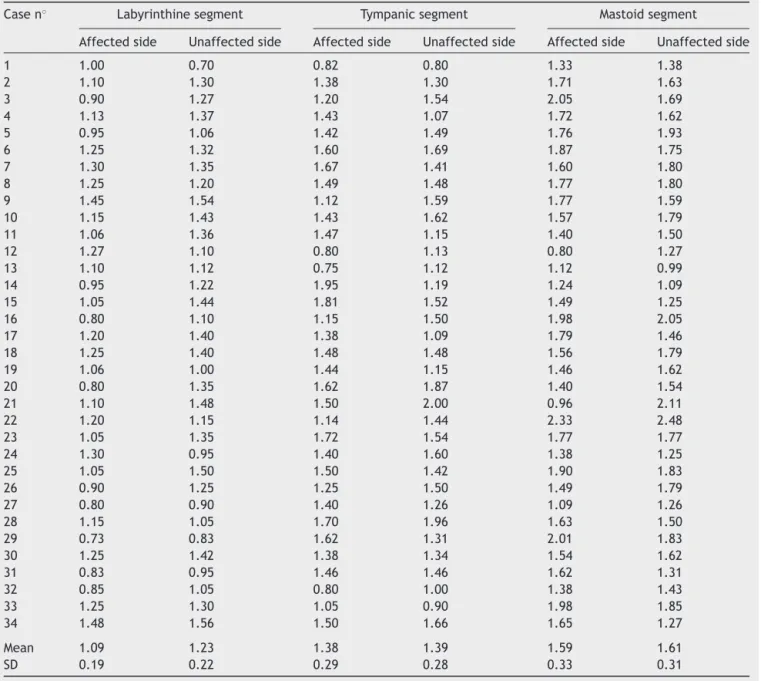
Table1 Diametersofthefacialcanalatthelabyrinthine,horizontalandmastoidsegmentsinpatientswithBell’spalsy.All valuesreportedasmm.
Casen◦ Labyrinthinesegment Tympanicsegment Mastoidsegment
Affectedside Unaffectedside Affectedside Unaffectedside Affectedside Unaffectedside
1 1.00 0.70 0.82 0.80 1.33 1.38
2 1.10 1.30 1.38 1.30 1.71 1.63
3 0.90 1.27 1.20 1.54 2.05 1.69
4 1.13 1.37 1.43 1.07 1.72 1.62
5 0.95 1.06 1.42 1.49 1.76 1.93
6 1.25 1.32 1.60 1.69 1.87 1.75
7 1.30 1.35 1.67 1.41 1.60 1.80
8 1.25 1.20 1.49 1.48 1.77 1.80
9 1.45 1.54 1.12 1.59 1.77 1.59
10 1.15 1.43 1.43 1.62 1.57 1.79
11 1.06 1.36 1.47 1.15 1.40 1.50
12 1.27 1.10 0.80 1.13 0.80 1.27
13 1.10 1.12 0.75 1.12 1.12 0.99
14 0.95 1.22 1.95 1.19 1.24 1.09
15 1.05 1.44 1.81 1.52 1.49 1.25
16 0.80 1.10 1.15 1.50 1.98 2.05
17 1.20 1.40 1.38 1.09 1.79 1.46
18 1.25 1.40 1.48 1.48 1.56 1.79
19 1.06 1.00 1.44 1.15 1.46 1.62
20 0.80 1.35 1.62 1.87 1.40 1.54
21 1.10 1.48 1.50 2.00 0.96 2.11
22 1.20 1.15 1.14 1.44 2.33 2.48
23 1.05 1.35 1.72 1.54 1.77 1.77
24 1.30 0.95 1.40 1.60 1.38 1.25
25 1.05 1.50 1.50 1.42 1.90 1.83
26 0.90 1.25 1.25 1.50 1.49 1.79
27 0.80 0.90 1.40 1.26 1.09 1.26
28 1.15 1.05 1.70 1.96 1.63 1.50
29 0.73 0.83 1.62 1.31 2.01 1.83
30 1.25 1.42 1.38 1.34 1.54 1.62
31 0.83 0.95 1.46 1.46 1.62 1.31
32 0.85 1.05 0.80 1.00 1.38 1.43
33 1.25 1.30 1.05 0.90 1.98 1.85
34 1.48 1.56 1.50 1.66 1.65 1.27
Mean 1.09 1.23 1.38 1.39 1.59 1.61
SD 0.19 0.22 0.29 0.28 0.33 0.31
Test ofnormalityvalues;for every segmentwas calcu-latedbytheShapiro-Wilktest.Exceptforthemeasurements of geniculate ganglion of unaffected group and stylomas-toid foramen of the affected group, the distribution was normal(p>0.05). So we use ‘‘paired samples t test’’ for comparisonofaffectedandunaffectedsidesoflabyrinthine
segment,tympanicsegment,secondgenuandmastoid
seg-ment.Themeanwidthat thelabyrinthinesegmentofthe
facialcanalintheaffectedtemporalbonewassignificantly smallerthantheequivalentintheunaffectedtemporalbone (p=0.00).Therewasnosignificantdifferencebetweenthe
affected and unaffected temporalbones at the tympanic
segment(p=0.66),secondgenu(p=0.62),andmastoid
seg-ment (p=0.67). We apply Wilcoxon signed-rank test for
valuesofgeniculateganglionandstylomastoidforamendue toabnormaldistributionof data.Therewasnosignificant
differencebetweentheaffected andunaffectedtemporal
bonesatthelevelofgeniculateganglion(p=0.87)and sty-lomastoidforamen(p=0.16).
WeuseSpearmentestfortheevaluationofthe relation-shipbetweenBell’s palsygradeandfacialcanaldiameter.
We did not find any relationship between the HB grade
and thefacialcanal diameterat the levelof labyrinthine
segment (p=0.41), tympanic segment (p=0.12),
mas-toid segment(p=0.14), geniculateganglion (p=0.13) and stylomastoidforamen(p=0.44);converselywefounda sig-nificantrelationshipat thelevelofsecondgenu(p=0.02). Thus,weapplylinearregression(p=0.015)toevaluatethe affect of canal diameter on HB grade specifically at the
level of 2nd genu. The mean HB grade and mean canal
diameter of 2nd genu (affected side) were; 3.35±1.18
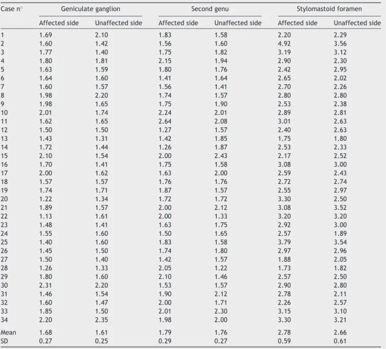
Table2 Diametersofthefacialcanalatthegeniculateganglion,secondgenuandstylomastoidforameninpatientswithBell’s palsy.Allvaluesreportedasmm.
Casen◦ Geniculateganglion Secondgenu Stylomastoidforamen
Affectedside Unaffectedside Affectedside Unaffectedside Affectedside Unaffectedside
1 1.69 2.10 1.83 1.58 2.20 2.29
2 1.60 1.42 1.56 1.60 4.92 3.56
3 1.77 1.40 1.75 1.82 3.19 3.12
4 1.80 1.81 2.15 1.94 2.90 2.30
5 1.63 1.59 1.80 1.76 2.42 2.95
6 1.64 1.60 1.41 1.64 2.65 2.02
7 1.60 1.57 1.56 1.41 2.70 2.26
8 1.98 2.20 1.74 1.57 2.80 2.80
9 1.98 1.65 1.75 1.90 2.53 2.38
10 2.01 1.74 2.24 2.01 2.89 2.81
11 1.62 1.65 2.64 2.08 3.01 2.63
12 1.50 1.50 1.27 1.57 2.40 2.63
13 1.43 1.31 1.42 1.85 1.75 1.80
14 1.72 1.44 1.26 1.87 2.53 2.33
15 2.10 1.54 2.00 2.43 2.17 2.52
16 1.70 1.41 1.75 1.58 3.08 3.00
17 2.00 1.62 1.63 2.00 2.59 2.43
18 1.57 1.57 1.76 1.76 2.72 2.74
19 1.74 1.71 1.87 1.57 2.55 2.97
20 1.22 1.34 1.72 1.72 3.30 2.50
21 1.89 1.57 2.00 2.12 3.08 3.52
22 1.13 1.61 2.00 1.33 3.20 3.20
23 1.48 1.41 1.63 1.75 2.92 3.00
24 1.55 1.60 1.50 1.65 2.57 1.89
25 1.40 1.60 1.83 1.58 3.79 3.54
26 1.45 1.50 1.74 1.80 2.97 2.96
27 1.50 1.40 1.42 1.57 1.88 2.05
28 1.26 1.33 2.05 1.22 1.73 1.82
29 1.80 1.60 2.10 1.46 2.57 2.50
30 2.31 2.20 1.53 1.57 2.90 2.80
31 1.46 1.54 1.90 2.12 2.78 2.11
32 1.60 1.47 2.00 1.71 2.26 2.57
33 1.85 1.50 2.01 2.30 3.15 3.10
34 2.20 2.35 1.98 2.00 3.30 3.21
Mean 1.68 1.61 1.79 1.76 2.78 2.66
SD 0.27 0.25 0.29 0.27 0.59 0.61
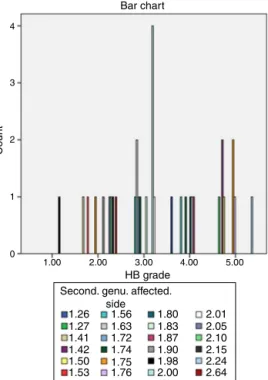
canaldiameter(R2=0.017).Noautocorrelationwasfoundin
theDurbin-Watsontest(2.059).ThedistributionofHBgrade accordingtoFCdiameteratthelevelof2ndgenuisshown inFig.2.
Discussion
Edema of the facial nerve leading to entrapment within the osseous fallopian canal is the main underlying mech-anism in Bell’s palsy.3---9,14 Conversely, reactivationof HSV typeIhasbeenchargedwithbeingthebestknown trigger-ingfactor.Soleantiviraltherapyhasnoprovedtobeofno benefitover placebo. Ithas alsobeen proved that antivi-ral plus corticosteroid therapy,when compared with sole corticosteroidtherapy,leadstonosignificantimprovement inprognosis.15 Theonlytreatmentthathasbeenprovedto
beeffectiveis corticosteroids: thatalsosubstantiatesthe theoryofedemainducedentrapmentneuropathy.7,8 Facial nervedecompressionisanothertreatmentmodalitythatis indicatedinalimitednumberofhighgradecases unrespon-sivetomedicaltherapy.16,17Thus,toknowwhichpartofthe facialnerveismorepronetoentrapmentbecomesmoreof anissue,especiallywhiledeterminingonthetypeofsurgical approach.
Figure1 CTimagesofthelefttemporalboneofa12 year-oldgirlwithleft Bell’spalsy.Thearrowindicatesthefacialnerve canal.(A)Labyrinthinesegment,(B)tympanicsegment,(C)mastoidsegment.
InBell’spalsypatientsthewidthofthefallopiancanalhas beenfoundtobenarroweratthemiddlepartofmeatal fora-menandlabyrinthinesegment.2 Ourlabyrinthine segment measurementscorroboratethe aforementionedfinding. In anotherstudywithBell’spalsypatients,themean diame-tersoffacialcanalandfacialnervewerefoundtobesmaller thanthecontrolgroupatthelevelthetympanicandmastoid segments.Inthesamestudyitwasfoundthatthenarrowest partsofthefacialcanalwerethelabyrinthineandtympanic segmentsrespectively.12 Likewise,inourstudyconsidering
4
3
1.26 1.27 1.41 1.42 1.50 1.53
1.56 1.63 1.72 1.74 1.75 1.76
1.80 1.83 1.87 1.90 1.98 2.00
2.01 2.05 2.10 2.15 2.24 2.64
2
1
0
1.00 2.00 3.00
HB grade Bar chart
Count
4.00 5.00
Second. genu. affected. side
Figure2 DistributionofcasesaccordingtoHBgradewith rel-evantFCdiametersatthelevelofsecondgenuofaffectedside. Colorvaluesreportedinmm.
themeanfacialcanaldiameters(Tables 1and2);the nar-rowestpartswerethelabyrinthineandtympanicsegments respectivelybothfortheaffectedandunaffectedsites.
Althoughthelabyrinthinesegmentis knownasthe nar-rowestpartoftheFC,controversyexistsaboutwhichpart ismorepronetoneuralentrapment.11,12Similarly,itisalso controversialwhichmeasurementofthediameterofa par-ticularsegmentorthemeancross-sectionalarea---istobe takenintoconsideration.Mayetal.havereportedthe nar-rowestpartoftheFCastheentryoftheinternalacoustic meatuswitha meandiameter of0.68mm.20 In ourstudy, inpatients1,8,12,19,22,24and28thediameterofthe affected sidewasfound tobe greater at thelabyrinthine segmentleading toan unignorableSD (Table1). Although itseemstocontradicttheentrapmenttheoryat first con-sideration, itmay berelatedtorelatively thicker(dueto edema)facialnerveinthesepatients,becauseFN/FC pro-portion is more reliable than the sole FC diameter when it comestoentrapment. As a matter offact, despite the wholepicturerevealinganarrowerbonyFCinaffectedside theremaybesomesporadiccaseshavingalargerbonycanal whencomparedtounaffectedside.Onemorereasonforthis contradictionmaybethecomplexcourseofthefacialcanal. Itisnotalwayspossibletomeasurethediameterfully per-pendicular.Inrelationtothat(consideringthecross-sections ofFC)itwasfoundthattheshapeofthecanalcanbe cir-cular,ellipticorkidneyshaped.2Accordingtothesefindings it was assertedthat measurement of cross-sectional area wouldbemoreaccurateratherthanmeasuringtheFC diam-eter.Forexampleinanotherstudy,themeancross-sectional areaoflabyrinthinesegmentwasevaluatedandtheaffected sitewasfoundtobenarrower.11
at the level of tympanic and mastoid segment of Bell’s palsypatients. The supremacy of this techniquewas that itwarranted thecalculation of FNtoFCratioin termsof diameteratpreferredsegments.Asamatteroffact, calcu-latingthisproportionwouldbemorereasonablebecauseof beingthemainindicatoroffreespacebetweentheFCand FN.It would not be inaccurate tohypothesize that when there is less space around the FN, thereis more risk for the entrapment tooccur. Thus,the sole measurement of FCdiameterwithoutmeasuringthenerveitselfmaybe con-founding.Thisproportionmayalsobecalculatedbycoupling thedataacquiredfrommagneticresonanceimagingofthe FNandcomputedtomographyimagingofFC,whichhasnot yetbeendone.
In this study, we use computed tomography images to measure the diameter of the FC at particular levels (labyrinthinesegment,geniculate ganglion,tympanic seg-ment, second genu, mastoid segment and stylomastoid foramen) of both the affected and unaffected sides of Bell’spalsypatients.RegardingtheetiologyofBell’spalsy, labyrinthine,tympanicandmastoidsegmentswerestudied before,butasfarasweknowgeniculateganglion,second genu andstylomastoid foramen have not been studied to date.Wealsorevealedasignificantlyimportantnarrowing atthelabyrinthinesegmentofaffectedsidewhencompared with the same level of the unaffected side. This finding has been shown before by the majority of the previous studies.2,5,7,8ButinthestudyofVienaetal.,theresponsible segmentswerefoundtobethetympanicandmastoid.Their argumentassertingtheproportionoftheFC/FNdiameteras themain indicatorregarding theentrapment theorylooks moresuperiorandreasonablethanthemeremeasurement of FC.Only tomeasurethe bonycanalmay be confound-ing (which is the case in majority of the relatedstudies) due tovariable ratioof FC/FN diameter. Forthis reason, studiesaimingtofindthisratiowillbemoreprecise.This canbe doneeither by 3Dreconstructionfrom histopatho-logical slides asdefined by Viena et al., or by utilizing a combinationofMRIandCT.ByutilizingMRIandCTimages we can measure the cross-sectional area for FC (CT) and FN(MRI)separatelyandthen calculate theFN/FC propor-tion.AsaresultwethinkthatisolatedmeasurementofFC maybe erroneous,whichis the case inmost of the stud-ies.
Our investigation provided at least baselinevalues for the level of geniculate ganglion, second genu and stylo-mastoidforamen.Wefoundmeandiametersoftheseparts narrowerinaffectedsidesofBell’spalsypatientsbutitwas statisticallyinsignificant.Inthelightofourresultswe can say thatthese partsdonotplay a rolein the pathogene-sisregardingtheneuronalentrapmenttheory.Butthereare somereportssuggestingaroleforgeniculateganglion with-outconsideringitswidth.Kimetal.assertedthelengthof greatersuperficialpetrosalnerve(whichisanindicatorof distancebetweenthegeniculateganglionand sphenopala-tineganglion) asa riskfactor for Bell’s palsy. Theyfound thelengthofthegreatersuperficialpetrosalnerveshorter intheaffectedsideofBell’s palsypatients.10 Buttomake suchadeductionmorestudiesshouldbecarriedoutinwhich controlgroupsareutilized.
The other circumstance that we investigate was the possiblerelationship betweenthe HBgradeandthe mean
diameterof FC.Wefoundstatisticallysignificant relation-ship only at 2nd genu of facialnerve (p=0.02). Although wefoundnosignificantdifferencebetweenmeandiameters ofthe affected and unaffectedside at 2nd genu,we can saythat it may play some rolein the pathogenesis of BP inadifferentway.Thisrelationshipmayalsobeacluefor theaforementioned FN/FCproportion. Although we were notable tocalculate thisproportion,future MRI-CT com-binedstudiesmayclarifythisrelevanceespeciallyfor2nd genu.Besides, accordingto ourlinearregression analyses wefoundanintermediatenegativeaffectofcanaldiameter onHBgradeat2ndgenu;and83%ofthiseffectwasfound tobecausedbydifferentfactors(R2=0.017).Thesefactors maybe the orientation or angle of the 2nd genu. Future studiesconcerningtheseparameterswouldbepromising.
Conclusion
We found the diameter of labyrinthine segment of facial canal asan anatomicrisk factor for Bell’s palsy. We also foundasignificantrelationshipbetweentheHBgradeandFC diameteratthelevelofsecondgenu.Futurestudies(MRI-CT combinedor3Dmodeling)areneededtoconfirmthis possi-blerelevanceespeciallyatsecondgenu.Thus,inthefuture itmaybepossibletoisolateanddecompressparticular seg-mentsinhighgradeBPpatients.
Conflicts
of
interest
Theauthorsdeclarenoconflictsofinterest.
References
1.MattoxDE.Clinicaldisordersofthefacialnerve.In:Richardson MA,editor.Cummingsotolaryngology---headandnecksurgery. 5thed.Philadelphia,PA:Mosby/Elsevier;2010.p.2391---403. 2.KefalidisG,RigaM,ArgyropoulouP,KatotomichelakisM,
Gou-verisC,PrassopoulosP,etal.Isthewidthofthelabyrinthine portionof thefallopian tube implicated inthe pathophysio-logyofBell’spalsy:aprospectiveclinicalstudyusingcomputed tomography.Laryngoscope.2010;120:1203---7.
3.BibasT,JiangD,GleesonJ.Disordersofthefacialnerve.In: GleesonM,editor.Scott-Brown’sotorhinolaryngologyheadand neck surgery. 7th ed.London, UK: EdwardArnold; 2008. p. 3883---6.
4.MurakamiS,MizobuchiM,NakashiroY, DoiT,HatoN, Yanag-iharaN.Bellpalsyandherpessimplexvirus:identificationof viral DNA in endoneurial fluidand muscle. Ann Intern Med. 1996;124:27---30.
5.YetiserS,KazkayasM,AltinokD,KaradenizY.Magnetic reso-nanceimagingoftheintratemporalfacialnerveinidiopathic peripheralfacialpalsy.ClinImaging.2003;27:77---81.
6.Fisch U. Surgery for Bell’s palsy. Arch Otolaryngol. 1981;107:1---11.
7.SchwaberMK,LarsonTCIII,ZealearDL,CreasyJ. Gadolinium-enhancedmagneticresonanceimaginginBell’spalsy. Laryngo-scope.1990;100:1264---9.
9.TienR,DillonWP,JacklerRK.Contrast-enhancedMR imaging ofthefacialnervein11patientswithBell’spalsy.AJRAmJ Roentgenol.1990;155:573---9.
10.KimJ,JungGH,ParkSY,KoSH,LeeWS.Anatomical consider-ation ofthetemporalboneasapathogenesis ofBell’spalsy. MedHypotheses.2011;77:705---7.
11.MuraiA,KariyaS,TamuraK,DoiA,KozakuraK,OkanoM,etal. ThefacialnervecanalinpatientswithBell’spalsy:an inves-tigation by high-resolution computed tomography with mul-tiplanarreconstruction.EurArchOtorhinolaryngol.2013;270: 2035---8.
12.ViannaM,AdamsM,SchachernP,Lazarini PR,PaparellaMM, Cureoglu S. Differences in thediameter offacial nerve and facial canal in Bell’s palsy----a 3-dimensional temporal bone study.OtolNeurotol.2014;35:514---8.
13.House JW, Brackmann DE. Facial nerve grading system. OtolaryngolHeadNeckSurg.1985;93:146---7.
14.Engström M, Berg T, Stjernquist-Desatnik A, Axelsson S, PitkärantaA,HultcrantzM,etal.Prednisoloneandvalaciclovir
inBell’spalsy:arandomised,double-blind,placebo-controlled, multicentretrial.LancetNeurol.2008;7:993---1000.
15.GagyorI,MadhokVB,DalyF,SomasundaraD,SullivanM, Gam-mieF,etal.AntiviraltreatmentforBell’spalsy(idiopathicfacial paralysis).CochraneDatabaseSystRev.2015;9:11.
16.KimSH,JungJ,LeeJH,ByunJY,ParkMS,YeoSG.Delayedfacial nervedecompressionforBell’spalsy.EurArchOtorhinolaryngol. 2015;7:30.
17.CannonRB,GurgelRK,WarrenFM,SheltonC.Facialnerve out-comesaftermiddlefossadecompressionforBell’spalsy.Otol Neurotol.2015;36:513---8.
18.LeeHY,ParkMS,ByunJY,ChungJH,NaSY,YeoSG.Agreement betweentheFacialNerveGradingSystem2.0andthe House-BrackmannGradingSysteminPatientswithBellPalsy.ClinExp Otorhinolaryngol.2013;6:135---9.
19.BrennerMJ,NeelyJG.Approachestogradingfacialnerve func-tion.SeminPlastSurg.2004;18:13---22.