Universidade de Lisboa
Faculdade de Farmácia
S-Adenosylhomocysteine Promotes
Endothelial Dysfunction and Activation: a
Role for Hypomethylation in Vascular Disease
Maria Madalena Henriques Serras Vicente Barroso
Orientadores: - Prof. Doutora Maria Rita Mouzinho de
Albuquerque de Azevedo e Castro
- Prof. Doutora Diane Elizabeth Handy
Tese especialmente elaborada para obtenção do grau de
Doutor em Farmácia
(Especialidade: Biologia Celular e Molecular)
Universidade de Lisboa
Faculdade de Farmácia
S-Adenosylhomocysteine Promotes
Endothelial Dysfunction and Activation: a
Role for Hypomethylation in Vascular Disease
Maria Madalena Henriques Serras Vicente Barroso
Orientadores:
- Prof. Doutora Maria Rita Mouzinho de Albuquerque de
Azevedo e Castro
- Prof. Doutora Diane Elizabeth Handy
Tese especialmente elaborada para obtenção do grau de
Doutor em Farmácia
(Especialidade: Biologia Celular e Molecular)
Júri:
Presidente: Prof. Doutora
Cecília Maria Pereira Rodrigues
Vogais:
- Prof. Doutor
Joseph Loscalzo
- Prof. Doutora
Diane E. Handy
- Prof. Doutor João António Nave Laranjinha
- Prof. Doutora
Paula Alexandra Quintela Videira
- Prof. Doutor
Luís Afonso Brás Simões do Rosário
- Prof. Doutora Maria Isabel Ginestal Tavares de Almeida
- Prof. Doutora Maria Rita Mouzinho de Albuquerque de Azevedo e Castro
2015
Universidade de Lisboa
Faculdade de Farmácia
S-Adenosylhomocysteine Promotes
Endothelial Dysfunction and Activation: a
Role for Hypomethylation in Vascular Disease
S-Adenosilhomocisteína Promove Disfunção e
Ativação Endotelial: um Papel para a
Hipometilação na Doença Vascular
Tese especialmente elaborada para obtenção do grau de
Doutor em Farmácia
(Especialidade: Biologia Celular e Molecular)
The studies presented in this thesis were performed at the Metabolism and Genetics Group, The Research Institute for Medicines (iMed.ULisboa), and at the Department of Biochemistry and Human Biology, Faculdade de Farmácia da Universidade de Lisboa, Portugal, under the supervision of Professor Rita Azevedo e Castro, and at the Cardiovascular Division, Department of Medicine, Brigham and Women’s Hospital and Harvard Medical School, Harvard University, Boston, USA, under the supervision of Professor Diane E. Handy. This work was financially supported by Fundação para a Ciência e a Tecnologia (F.C.T.), Lisboa, Portugal (SFRH/BD/73021/2010).
vii
TABLE OF CONTENTS
Abbreviations ix Summary xiii Sumário xv Chapter 1 General Introduction 1 Chapter 2Aims and Outline 27
Chapter 3
Cellular Hypomethylation is Associated with Impaired Nitric Oxide Production by Cultured Human Endothelial Cells
31
Chapter 4
4.1. Inhibition of Cellular Methyltransferases Promotes Endothelial Cell Activation by Suppressing Glutathione Peroxidase-1 Expression
47
4.2. S-Adenosylhomocysteine Alters Methylation of Cellular RNA 75 Chapter 5
The Role of DNA Hypomethylation in S-Adenosylhomocysteine-Induced Endothelial Activation
95
Chapter 6
S-Adenosylhomocysteine Induces Inflammation Through NFkB: a Possible Role for EZH2 in Endothelial Cell Activation
107
Chapter 7
General Discussion and Perspectives 129
List of Publications 147
Acknowledgements Agradecimentos
Abbreviations ix
ABBREVIATIONS
5-aza-dC 5-aza-2’-deoxycytidine 5-mC 5-methylcytosine 5-MTHF 5-methyltetrahydrofolate 5,10-meTHF 5,10-methylenetetrahydrofolate ADA adenosine-2’,3’-dialdehyde AdCtrl control adenoviral vectorAdGPx-1 adenoviral construct with the human glutathione peroxidase-1 cDNA AdIkBDN adenovirus carrying the dominant negative IkB
ADMA asymmetric dimethylarginine
Ad-βGal adenoviral construct with a CMV-LacZ cassette ALKBH8 alkylation repair homolog 8
Am 2′-O-methyladenosine ATP adenosine triphosphate
BHMT betaine-homocysteine methyltransferase
BNIP3 BCL2/adenovirus E1B 19kDa interacting protein 3 CBS cystathionine β-synthase
CDKN1A cyclin-dependent kinase inhibitor 1A CDKN2A cyclin-dependent kinase inhibitor 2A CGL cystathionine γ-lyase Cm 2′-O-methylcytidine cm5U 5-carboxymethyluridine Ctrl control CVD cardiovascular disease DAPI 4',6-diamidino-2-phenylindole DNMT DNA-methyltransferase DTNB 5,5’-dithio-bis(2-nitrobenzoic acid) DTT dithiothreitol
eNOS endothelial nitric oxide synthase EZH2 enhancer of zeste homolog 2 FoxO1 forkhead box O 1
Gm 2′-O-methylguanosine GPx1 glutathione peroxidase-1 GRP glucose-regulated protein
Abbreviations
x
H2O2 hydrogen peroxide
H3K9me3 histone H3 lysine 9 trimethylation H3K27me3 histone H3 lysine 27 trimethylation HCAEC human coronary artery endothelial cells Hcy homocysteine
HDM histone demethylase HHcy hyperhomocysteinemia
HKMT histone lysine methyltransferase HUVEC human umbilical vein endothelial cells i6A isopentenyladenosine
ICAM-1 intercellular adhesion molecule-1 IkB inhibitor of kappa B
IkBDN inhibitor of kappa B dominant negative mutant IKK IκB kinase complex
IL interleukin
JMJD3 jumonji domain containing 3 demethylase
LC-MS/MS liquid chromatography-tandem mass spectrometry LDH lactate dehydrogenase
LDL low-density lipoprotein
LDLR low-density lipoprotein receptor L-NNA L-NG-nitroarginine m1A 1-methyladenosine m1G 1-methylguanosine m26A N6,N6-dimethyladenosine m2G N2-methylguanosine m5C 5-methylcytidine m5U 5-methyluridine m6A N6-methyladenosine m7G 7-methylguanosine
MAT methionine adenosyltransferase mcm5U 5-methoxycarbonylmethyluridine
mcm5Um 5-methoxycarbonylmethyl-2′-O-methyluridine MFI mean fluorescence intensity
MS methionine synthase
MsrB1 methionine sulfoxide reductase B1
Abbreviations
xi MTT 3-(4,5-dimethylthiazol-2-yl)-2,5-diphenyltetrazolium bromide
NAC N-acetylcysteine NFκB nuclear factor kappa B NIK NFkB-interacting kinase NO nitric oxide
NOX NADPH oxidase NT no treatment
PBS phosphate-buffered saline PDGF platelet-derived growth factor
PECAM-1 platelet endothelial cell adhesion molecule-1 PKC protein kinase C
PRC polycomb repressive complex PRMT1 protein arginine methyltransferase-1
qRT-PCR quantitative reverse transcription polymerase chain reaction ROS reactive oxygen species
SAH S-adenosylhomocysteine
SAHH S-adenosylhomocysteine hydrolase SAM S-adenosylmethionine
Sec selenocysteine
SELE E-selectin coding gene
SelH selenoprotein H SelM selenoprotein M
Shc Src homology 2 domain containing SHMT serine hydroxymethyltransferase siRNA small interference RNA
SOD superoxide dismutase
SREBF1 sterol regulatory element binding transcription factor-1 TET ten-eleven translocation enzymes
THF tetrahydrofolate TNF tumor necrosis factor
TRM112 tRNA methyltransferase 112 homolog tRNA[Ser]Sec selenocysteine-carrying tRNA (Sec-tRNA) TrxR thioredoxin reductase
Um 2′-O-methyluridine
Abbreviations
xii
UTX ubiquitously transcribed tetratricopeptide repeat on X chromosome demethylase
VCAM-1 vascular adhesion molecule-1 WBC white blood cells
Summary
xiii
SUMMARY
Homocysteine has been established as a risk factor for cardiovascular disease (CVD) by mechanisms incompletely defined. S-Adenosylhomocysteine (SAH) is the metabolic precursor of homocysteine that accumulates in the setting of hyperhomocysteinemia and is a negative regulator of most cell methyltransferases.
This thesis project investigated whether methylation imbalance, caused by excess SAH, disrupts endothelium homeostasis and favors the establishment of a pro-atherogenic phenotype.
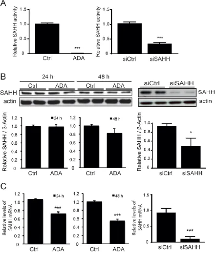
To experimentally address this possibility, studies were conducted in human endothelial cells, in which SAH accumulation was induced using either a pharmacologic or a siRNA approach. As the major regulator of vascular homeostasis, the endothelium exerts a number of vasoprotective effects that are largely mediated by nitric oxide (NO), the most potent endogenous vasodilator. Decreased NO bioavailability is a principal manifestation of underlying endothelial dysfunction, an early marker of atherosclerosis and CVD. To determine whether excess SAH alters NO bioavailability, the expression and activity of endothelial nitric oxide synthase (eNOS), and NO production were monitored in cells. These experiments showed that excess SAH increased the levels of eNOS mRNA but caused a decrease in eNOS protein and activity, to decrease cellular production of NO.
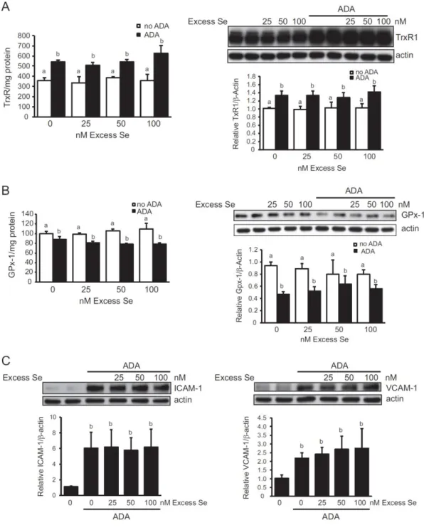
Another important feature of endothelial dysfunction is oxidative stress. Studies in endothelial cells revealed that a hypomethylating environment, induced by excess SAH, impairs, not only NO production, but also the cellular redox state. Glutathione peroxidase-1 (GPx-1) is a selenoprotein and a major cellular antioxidant. A link between homocysteine-associated suppression of GPx-1 and endothelial dysfunction had been reported previously; however, the causal molecular mechanisms remained unresolved. The experiments presented here demonstrate a specific mechanism by which SAH-mediated hypomethylation suppresses GPx-1 expression and leads to inflammatory activation of endothelial cells. The expression of a subset of selenoproteins (including GPx-1) is dependent on a specific methylation of the selenocysteine-tRNA (Sec-tRNA). Thus, SAH accumulation was found to inhibit the formation of this methylated isoform of Sec-tRNA resulting in decreased GPx-1 expression, as well as alterations in the expression of other selenoproteins, to promote oxidative stress and a pro-inflammatory activation state in endothelial cells.
The observation that Sec-tRNA methylation is decreased by excess SAH, suggests that other RNA species may also be targets for SAH-mediated hypomethylation. Therefore, the effect of SAH on methylation modifications was determined in total and size-fractionated RNA samples from our cell model. Additionally, to confirm these observations, RNA methylation was analyzed in tissue samples from a hyperhomocysteinemic mouse model, where SAH accumulation results from a genetic disorder affecting homocysteine metabolism. Conditions of excess SAH altered the content of some
Summary
xiv
RNA methylation modifications, suggesting that specific RNA methyltransferases may be more susceptible to inhibition by SAH.
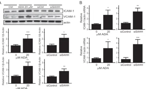
The activation of endothelial cells that occurs during atherogenesis is characterized by the up-regulation of adhesion molecules, which by recruiting circulating leukocytes favor their transendothelial migration. In a series of studies, the physiological relevance of SAH-induced endothelial cell activation was demonstrated by determining that these SAH-activated cells promoted leukocyte adhesion and migration. Further, the role of DNA hypomethylation on the SAH-induced up-regulation of adhesion molecules was examined. ICAM-1 (intercellular adhesion molecule 1) was found to be up-regulated by SAH accumulation as well as by a DNA methyltransferase inhibitor, suggesting that its expression may be regulated by DNA methylation. Analysis of its promoter methylation; however, showed that it was demethylated in untreated cells, suggesting that it may be regulated by factors other than DNA promoter methylation in response to excess SAH.
To understand better the factors involved in the pro-inflammatory activation of endothelial cells, the role of NFkB (nuclear factor kappa B) in SAH-induced responses was examined. These studies establish a role for NFkB in the endothelial cell response to SAH and further link these responses to a suppression of the epigenetic regulator EZH2 (enhancer of zeste homolog 2). EZH2 is a methyltransferase that regulates gene expression by mediating a repressive histone methylation. These results identify EZH2 as a new target of SAH regulation important in inflammatory responses, demonstrating that EZH2 suppression and NFkB activation mediated by excess SAH accumulation may contribute to its adverse effects in the vasculature.
Overall, these studies implicate SAH as a key modulator of epigenetic mechanisms by compromising RNA, DNA, and histone methylation. More importantly, our results clearly present SAH as a key player in the disruption of endothelial homeostasis, supporting a role for SAH as an important mediator of homocysteine-associated vascular disease.
Key words: S-Adenosylhomocysteine, hyperhomocysteinemia, hypomethylation stress, endothelial dysfunction, pro-inflammatory phenotype.
Sumário
xv
SUMÁRIO
A doença cardiovascular é a principal causa de morte nos países desenvolvidos, facto que reflete a insuficiência das atuais medidas terapêuticas e de prevenção para esta patologia. O aumento do conhecimento sobre os mecanismos moleculares na origem da doença cardiovascular poderá assim contribuir para a resolução deste importante problema de saúde pública. A elevação dos níveis circulantes de homocisteína é um fator de risco para a ocorrência de doença cardiovascular; no entanto, os mecanismos fisiopatológicos subjacentes à relação entre a hiperhomocisteinémia e a doença cardiovascular, embora intensamente estudados, permanecem ainda por elucidar.
A homocisteína é um aminoácido sulfurado, em cujo metabolismo as vitaminas do grupo B atuam como importantes cofatores ou substratos. Em estudos clínicos, nos quais a concentração de homocisteína plasmática foi significativamente reduzida através da administração destas vitaminas, não foram observados os efeitos cardio-protetores esperados. Uma teoria alternativa propõe a S-adenosilhomocisteína (SAH), o precursor metabólico da homocisteína que se acumula em situações em que esta se encontra elevada, como um marcador mais fidedigno do risco de doença cardiovascular associado a situações de hiperhomocisteinémia. Sendo a SAH um regulador negativo da atividade enzimática da maioria das metiltransferases celulares, que atuam sobre diversos compostos (incluindo moléculas como DNA, RNA e proteínas), a sua acumulação poderá originar, teoricamente, um ambiente celular de hipometilação.
A presente tese investigou a possibilidade de o desequilíbrio das reações de metilação celulares, provocado pela elevação dos níveis intracelulares de SAH, perturbar a homeostase endotelial, favorecendo o estabelecimento de um fenótipo pró-aterogénico e a ocorrência de doença cardiovascular. Para tal, desenvolveu-se um modelo de estudo com base em células endoteliais humanas, nas quais a acumulação de SAH foi induzida através de abordagens genéticas (RNA de interferência) ou farmacológicas (inibição da atividade da enzima responsável pela síntese de SAH, a SAH hidrolase).
O endotélio é o principal regulador da homeostasia vascular, exercendo uma série de efeitos vasoprotetores que são largamente mediados pelo óxido nítrico (NO), um potente vasodilatador endógeno. A diminuição da biodisponibilidade do NO é uma manifestação importante da disfunção endotelial e um marcador precoce de aterosclerose e doença cardiovascular. O trabalho experimental desenvolvido revelou que o excesso de SAH diminui os níveis endoteliais de NO, os quais estão diretamente relacionados com a diminuição, quer dos níveis de proteína eNOS (sintase do óxido nítrico endotelial), quer da sua atividade enzimática.
Outra característica importante da disfunção endotelial é um aumento de stress oxidativo. Várias selenoproteínas, incluindo a glutationa peroxidase-1 (GPx-1), são componentes enzimáticos
Sumário
xvi
dos principais sistemas antioxidantes celulares. Estudos anteriores relataram a existência de uma associação entre a supressão de GPx-1, induzida por um excesso de homocisteína, e a ocorrência de disfunção endotelial, sem no entanto conseguir esclarecer os mecanismos moleculares subjacentes. Adicionalmente, encontra-se descrito que a expressão de um subconjunto de selenoproteínas, entre as quais a GPx-1, depende da metilação específica do RNA de transferência necessário à incorporação do aminoácido selenocisteína (Sec-tRNA). Interessantemente, os resultados das nossas experiências revelaram que um ambiente de hipometilação, induzido por um excesso de SAH, altera não só a produção de NO, mas também o estado redox celular. Efetivamente, as experiências aqui apresentadas demonstraram a existência de um mecanismo específico através do qual a hipometilação suprime a expressão de GPx-1 e leva à ativação inflamatória das células endoteliais. Os resultados revelaram que a acumulação de SAH inibe a formação da isoforma metilada do Sec-tRNA, o que resulta na diminuição da expressão da GPx-1, bem como em alterações na expressão de outras selenoproteínas, promovendo, desta forma, um estado de stress oxidativo e de ativação pró-inflamatória das células endoteliais.
A observação de que a metilação do Sec-tRNA é reduzida por um excesso de SAH sugere a possibilidade de que outras espécies de RNA possam ser igualmente alvos da hipometilação mediada pela acumulação intracelular de SAH. Para averiguar esta possibilidade, foram quantificadas modificações de metilação, quer em RNA total, quer em diferentes espécies de RNA obtidas por fracionamento de acordo com o seu tamanho molecular, extraídas de amostras do nosso modelo celular. Adicionalmente, estas observações foram avaliadas em amostras de tecido obtidas a partir de um modelo animal de hiperhomocisteinémia, no qual a acumulação de SAH resulta de uma deficiência da enzima CBS (cistationina β-sintetase) envolvida no catabolismo da homocisteína. Em ambas as condições estudadas, os resultados revelaram alterações nos níveis de modificações por metilação do RNA, sugerindo que as metiltransferases específicas de RNA são suscetíveis à inibição da sua atividade enzimática pela acumulação de SAH.
O estado de ativação de células endoteliais que ocorre durante a aterogénese caracteriza-se por um aumento da expressão de citocinas e moléculas de adesão, as quais recrutam leucócitos circulantes, favorecendo a sua migração transendotelial e a formação das placas de ateroma. A relevância fisiológica da ativação endotelial induzida pela SAH foi demonstrada numa série de estudos, nos quais se observou um aumento da expressão de moléculas de adesão e um acréscimo na capacidade das células endoteliais promoverem a adesão de leucócitos e a sua subsequente transmigração.
A metilação do DNA é um mecanismo epigenético que pode regular a expressão genética, estando a hipometilação das regiões promotoras tipicamente associada com a ativação da expressão dos genes correspondentes. Por esta razão, após a confirmação de que SAH induz uma hipometilação global do DNA no modelo celular utilizado, foi examinado o papel desta hipometilação
Sumário
xvii na sobre-expressão de moléculas de adesão. Os resultados revelaram que a expressão endotelial de ICAM-1 (intercellular adhesion molecule-1), uma importante molécula de adesão, era regulada positivamente não só por uma acumulação de SAH induzida no nosso modelo celular, mas também pela utilização de um inibidor específico das metiltransferases do DNA. No entanto, a quantificação dos níveis de metilação no promotor do gene ICAM1 revelaram um estado de hipometilação nas células controlo, ou seja, com níveis normais de SAH. Estes dados demonstram que a indução da expressão de ICAM-1 por SAH é mediada por fatores independentes da hipometilação do seu promotor.
Como referido, o excesso de SAH induz um fenótipo pró-inflamatório nas células endoteliais. Assim, e com o intuito de compreender melhor os mecanismos moleculares subjacentes, foram examinadas as vias de ativação do NFkB (nuclear factor kappa B), um conhecido promotor da resposta inflamatória. Os resultados estabelecem um papel da ativação do NFkB nesta resposta inflamatória, sugerindo que a supressão do regulador epigenético EZH2 (enhancer of zeste homolog 2) possa contribuir para esta resposta. O EZH2 é uma histona metiltransferase que regula negativamente a expressão genética através da metilação específica das histonas (nomeadamente ao estabelecer a trimetilação da lisina 27 na histona H3, ou seja a marca epigenética H3K27me3). A acumulação de SAH em células endoteliais resultou numa diminuição da expressão e atividade de EZH2, que subsequentemente determinou um aumento de expressão de citocinas pro-inflamatórias e moléculas de adesão celular. Estes resultados revelaram assim um novo alvo para a SAH na regulação da resposta inflamatória endotelial, demonstrando que a supressão de EZH2 e a ativação de NFkB, mediadas pelo excesso de SAH, contribuem para os seus efeitos adversos no endotélio.
Considerados em conjunto, estes estudos implicam a SAH como um metabolito chave na modulação de mecanismos epigenéticos, ao comprometer a metilação do DNA, do RNA e das histonas, e mostram claramente que a acumulação de SAH desempenha um papel determinante na perturbação da homeostasia do endotélio. Como referido anteriormente, a SAH é o precursor metabólico da homocisteína. Os resultados aqui apresentados demonstram um papel importante da SAH como mediador da doença vascular associada à hiperhomocisteinémia, suportando a hipótese da SAH ser um possível marcador mais fidedigno do que a própria homocisteína.
Palavras-chave: S-adenosilhomocisteína, hiperhomocisteinémia, stress de hipometilação, disfunção endotelial, fenótipo pro-inflamatório.
CHAPTER
1
General Introduction
1. Homocysteine Metabolism 2. Hyperhomocysteinemia
3. Hyperhomocysteinemia and Cardiovascular Disease
3.1. Homocysteine and Endothelial Dysfunction and Activation 3.2. S-Adenosylhomocysteine and Cardiovascular Disease
4. S-Adenosylhomocysteine and Cell Hypomethylation
4.1. Targets of S-Adenosylhomocysteine-Mediated Methylation Inhibition
4.1.1. DNA 4.1.2. RNA 4.1.3. Protein
Chapter 1
3
1. Homocysteine Metabolism
Homocysteine was identified by the Nobel laureate Vincent DuVigneaud in 1932 during his pioneering studies on sulfur amino-acid chemistry. In fact, he first discovered the disulfide homocystine, and only later identified its reduced form, an intermediate of methionine metabolism, which he named homocysteine (1).
Homocysteine metabolism is shown in Figure 1. Homocysteine is formed by the demethylation of the essential amino acid, methionine, via the formation of two intermediate compounds, S-adenosylmethionine (SAM) and S-adenosylhomocysteine (SAH) (2). Methionine is first converted to SAM through the catalytic action of methionine adenosyltransferase enzymes (MATs). MAT transfers the adenosyl moiety of ATP to the methionine molecule, forming SAM, a highly energetic compound. SAM is the major methyl-donor compound for cellular methylation reactions, which are catalyzed by specific methyltransferases that target important biomolecules, such as DNA, RNA, proteins, and lipids (2). Nevertheless, in mammals, most methyl groups transferred from SAM are used in creatine formation, in phosphatidylcholine synthesis, and in the generation of sarcosine from glycine (3). After the transfer of a methyl group, the methylated product is formed and SAM is converted to SAH. Importantly, because SAH has nearly the same, or even higher affinity for the catalytic site of most SAM-dependent methyltransferases than SAM, SAH is a potent inhibitor of these methyltransferases and the SAM/SAH ratio is often used as an indicator for intracellular methylation capacity (4).
SAH is further converted into homocysteine and adenosine by SAH hydrolase (SAHH), which is ubiquitously expressed in mammalian tissues. The formation of homocysteine from methionine via the transmethylation pathway is the only pathway for homocysteine biosynthesis in humans (2).
Importantly, the SAH hydrolase reaction is reversible and presents a thermodynamic equilibrium that strongly favors SAH synthesis rather than its hydrolysis (5). However, under normal physiological conditions the reaction is directed towards SAH hydrolysis due to the rapid metabolism or export of homocysteine. Homocysteine is located at a metabolic branch point and can be either conserved by remethylation back to methionine or irreversibly degraded to cysteine via the transsulfuration pathway (5).
Homocysteine can be remethylated back to methionine by two alternative metabolic pathways, the folate-dependent or independent remethylation pathways (Figure 1). 5-Methyltetrahydrofolate (5-MTHF) is the active folate derivative and the main circulating form of folate in plasma. In the folate dependent-pathway, 5-MTHF supplies the methyl group used by methionine synthase (MS) to remethylate homocysteine and produce methionine and tetrahydrofolate (THF). THF is then converted to 5,10-methylenetetrahydrofolate (5,10-MeTHF) in the presence of serine and vitamin B6 by the enzyme serine hydroxymethyltransferase (SHMT). After reduction by 5,10-methylenetetrahydrofolate reductase (MTHFR), 5,10-MeTHF is converted into 5-MTHF, which will be available for the remethylation of a second molecule of homocysteine. MTHFR uses flavin
General Introduction
4
adenine dinucleotide (FAD; the active form of vitamin B2) as a cofactor. The folate-dependent
remethylation pathway is present in nearly all cells (3, 6). Additionally, in liver and kidney, remethylation also occurs by the folate independent pathway in which methyl groups are donated by betaine (also known as trimethylglycine, a derivative of choline oxidation), in a reaction catalyzed by the enzyme betaine-homocysteine methyltransferase (BHMT) (3, 6).
Figure 1 - Schematic representation of homocysteine metabolism. MAT (methionine
adenosyltransferase), SAHH (S-adenosylhomocysteine hydrolase), CBS (cystathionine
-synthase), CGL (cystathionine γ-lyase), MS (methionine synthase), SHMT (serine hydroxymethyltransferase), MTHFR (5,10-methylenetetrahydrofolate reductase), BHMT (betaine-homocysteine methyltransferase).
Homocysteine can be irreversibly metabolized through the transsulfuration pathway, which is mainly confined to the liver, kidney, small intestine and pancreas (7). In the first step of this pathway, cystathionine β-synthase (CBS) catalyzes the condensation of homocysteine and serine to form cystathionine using pyridoxal phosphate (PLP or vitamin B6) as a co-factor (5). Cystathionine is
further metabolized to produce cysteine by another PLP-requiring enzyme, cystathionine γ-lyase (CGL) (5, 8). In addition to protein synthesis, cysteine is used in the synthesis of glutathione, an important cellular antioxidant (5, 9). The sulfur end product of cysteine metabolism is sulfate, which can be excreted by the kidneys (10).
In addition to the homocysteine metabolic pathways, and to maintain the optimal intracellular levels of homocysteine, cells may also export homocysteine. The mechanisms that regulate homocysteine export into the extracellular compartment are not completed defined. Nevertheless, two types of transport of homocysteine across the cell membranes were suggested: one involving the
Chapter 1
5 removal of the reduced form of homocysteine (with a free thiol group) to the extracellular compartment, and the other the transport of the oxidized, disulfide forms of homocysteine, that are most abundant in plasma, into cells (11). In plasma, the majority (98-99%) of homocysteine is rapidly oxidized, reacting with free thiol-containing molecules (including small thiol molecules such as homocysteine or cysteine, and proteins with free cysteines, such as albumin) to form disulfides. The remaining 1-2% of plasma homocysteine remains in its reduced form (5, 11). Total plasma homocysteine includes the sum of the circulating homocysteine molecules either in its reduced or oxidized forms.
Homocysteine can be cleared from the organism by urinary excretion, but 98-99% of plasma homocysteine is reabsorbed in the kidney (12). The remethylation and transsulfuration pathways play major roles in maintaining circulating homocysteine levels. Nevertheless, when renal function is compromised, homocysteine levels often become elevated (5, 12).
2. Hyperhomocysteinemia
The intracellular concentration of homocysteine is under tight control. Once formed in the cell, homocysteine is either metabolized to cysteine or remethylated to methionine or exported from the cell. Hence, cellular export of homocysteine reflects the balance between homocysteine production and its catabolism. Persistent elevation of homocysteine in the blood defines the condition called hyperhomocysteinemia. In healthy adults, fasting homocysteine concentrations in plasma are usually within a 5-15 µmol/L range (13–15). Hyperhomocysteinemia is classified according to the levels of homocysteine accumulated, as mild (15-30 µmol/L), moderate (31-100 µmol/L), or severe (>100 µmol/L) (14, 16, 17).
Several factors can contribute to the circulating levels of homocysteine. In fact, plasma homocysteine levels are influenced by various non-genetic as well as genetic determinants.
Non-genetic determinants of plasma homocysteine include inadequate concentrations of B vitamins that play a vital role in the metabolism of homocysteine. As mentioned in the previous section, folate is a primary substrate for homocysteine remethylation and other B vitamins, such as vitamin B12, vitamin B6, and vitamin B2, are co-factors for the major homocysteine regulating enzymes
(Figure 1). As such, plasma concentrations of homocysteine are inversely related to plasma concentrations of folate, vitamin B12 and vitamin B6, as well as to the intake of these vitamins (18). The
most consistent association has been found with lower folate intake or with lower plasma concentrations of folate (17). A modest inverse relationship has been reported between plasma concentrations of vitamin B2 and homocysteine; however, this association is restricted to subjects
carrying the MTHFR 677TT genotype (see below). In addition, normal kidney function maintains optimal plasma levels of homocysteine, and impaired renal function is often associated with mild to moderate hyperhomocysteinemia (5, 12). Plasma homocysteine concentrations increase with age. In
General Introduction
6
fact, homocysteine levels approximately double from childhood to old age (2, 5). Gender also significantly affects concentration of plasma homocysteine, with males having higher levels than females (5, 17). A rise in plasma homocysteine concentrations with menopause has been also described (17). Lifestyle factors can also modulate the circulating levels of homocysteine. Tobacco smoking, high coffee consumption, alcoholism, and lack of exercise are examples of lifestyle factors associated with mild hyperhomocysteinemia (19). Several pharmacological agents can also disturb homocysteine metabolism, such as certain anti-carcinogenic agents (e.g. methotrexate or sulfasalazine) and anticonvulsants (e.g. carbamazepine or phenytoin) (22). Lastly, pathologic conditions, such as hypothyroidism, psoriasis, lymphoblastic leukemia and other malignancies, were also associated with hyperhomocysteinemia (22).
Genetic determinants cause mild to severe hyperhomocysteinemia. Severe hyperhomocysteinemia is caused by rare genetic defects in either homocysteine transsulfuration or remethylation pathways. CBS deficiency or classical homocystinuria, an autosomal recessive disorder that affects the transsulfuration pathway, is the most common inborn error of homocysteine metabolism (20). In addition to severe hyperhomocysteinemia and homocystinuria, the biochemical phenotype of CBS deficiency also includes hypocysteinemia, and hypermethioninemia, due to increased homocysteine remethylation (20). Phenotypic consequences of CBS deficiency include thromboembolism and vascular occlusion, skeletal abnormalities, dislocation of the optic lenses, marfanoid features, and varying degrees of neurological impairment. In these patients, early homocysteine-lowering treatment significantly reduces the risk of life-threatening vascular events despite imperfect biochemical control of homocysteine levels (20).
Other genetic causes of hyperhomocysteinemia include disorders that cause cobalamin (vitamin B12) deficiency. Inborn errors of cobalamin metabolism can affect its absorption, transport, as
well as its intracellular metabolism, namely adenosylcobalamin synthesis, methionine synthase function, or both (21).
Several pieces of evidence lead to the conclusion that the more common mild forms of hyperhomocysteinemia are, at least partially, genetically based (22). For this reason, almost every gene involved in homocysteine metabolism has been analyzed for functional polymorphisms that could affect the circulating concentrations of homocysteine, and many genetic variants have been identified. However, one MTHFR polymorphism has been the most consistently associated with plasma homocysteine variability. In fact, the major known genetic determinant of plasma homocysteine levels in the general population is the 677C>T variation in the MTHFR gene (23). MTHFR is a FAD-dependent enzyme involved in the folate-dependent remethylation of homocysteine. The common MTHFR C677T polymorphism determines the synthesis of a thermolabile enzyme with reduced activity (24). Homozygosity for the thermolabile MTHFR variant increases the plasma concentrations of homocysteine by approximately 25% in individuals with low folate status (25).
Chapter 1
7
3. Hyperhomocysteinemia and Cardiovascular Disease
Cardiovascular diseases (CVD) are the number one cause of death in the Western industrialized world. Meta-analyses have shown that, in the general population, mild hyperhomocysteinemia is an independent risk factor for CVD, with a prevalence around 5% (19, 26, 27).
The hypothesis that elevated homocysteine may contribute to vascular disease was suggested by McCully (1969) on the basis of postmortem observations of widespread arterial disease in young patients with markedly elevated homocysteine concentrations due to different genetic causes (1, 6, 28). These observations formed the basis for the so-called Homocysteine Theory that suggests the importance of hyperhomocysteinemia in the development of atherosclerosis. McCully raised the important question of whether mild to moderate elevations of homocysteine, common in the general population, would increase the risk of vascular disease (1, 29).
In 1976, Wilcken and Wilcken provided the first evidence for an association between impaired homocysteine metabolism and CVD in the general population (30). Since these observations, data from a large number of clinical and epidemiological studies have implied an important role for mild hyperhomocysteinemia as an independent risk factor for CVD and related-mortality (26). Large meta-analysis studies concluded that every increase of 5 μmol/L in plasma concentration of homocysteine increases the risk of CVD by approximately 20%, independently of traditional risk factors, such as diabetes, hypertension, smoking and hypercholesterolemia (31).
3.1. Homocysteine and Endothelial Dysfunction and Activation
Increased levels of plasma homocysteine are associated with various forms of vascular disease, including arterial and venous thrombosis, in which a common feature is endothelial dysfunction, an early step in atherogenesis. The term 'endothelial dysfunction' refers to the impairment of the normal homeostatic properties of the vascular endothelium, which include endothelium-dependent regulation of vascular tone, hemostasis, and inflammation. A decrease of nitric oxide bioavailability and an impairment of cell redox balance are major features of endothelial dysfunction (Figure 2). Endothelial dysfunction often leads to a pro-inflammatory state (endothelial activation) that precedes the formation of atherosclerotic plaques (32). The detrimental effect of homocysteine on the endothelium may explain the increased risk for CVD associated with hyperhomocysteinemia (33).
General Introduction
8
Figure 2 - Schematic representation of major steps of the atherosclerosis process.
The mechanisms by which homocysteine promotes endothelial dysfunction are not fully understood, although several effects known to disturb endothelial homeostasis have been associated with hyperhomocysteinemia (summarized in Figure 3).
Figure 3 - The harmful effects of homocysteine in the endothelium (red dots indicate that these
Chapter 1
9 Different studies have reported increased oxidative stress and impaired vasodilation, due to decreased nitric oxide (NO) bioavailability, in the context of hyperhomocysteinemia. Cellular oxidative stress is characterized by an increase in oxidants over antioxidants, which leads to an imbalance of the redox state. Increased levels of reactive oxygen species (ROS), such as hydrogen peroxide, superoxide anions and hydroxyl radicals, contribute to a more oxidant environment (34). There are several means by which homocysteine can promote ROS accumulation, these include: homocysteine’s auto-oxidation, endothelial nitric oxide synthase (eNOS) uncoupling, and the inhibition of the activity of important antioxidant enzymes, such as glutathione peroxidase-1 (GPx-1) or superoxide dismutase (SOD) (35–37). The reaction of superoxide with NO produces the strong oxidant peroxynitrite (ONOO-) and, simultaneously, decreases NO bioavailability (33). NO is considered a major endogenous anti-atherosclerotic molecule, as it is a potent vasodilator, and it inhibits monocyte and platelet adhesion, smooth muscle cell proliferation, and low density lipoprotein (LDL) oxidation (38, 39). Increased plasma levels of homocysteine have been associated with impaired vascular tone, due to the decrease in NO bioavailability and increase of endothelin-1 (ET-1), a potent vasoconstrictor (35). Vascular dysfunction was observed in an animal model of diet-induced mild hyperhomocysteinemia (40). Moreover, oxidative stress was shown to contribute to this association in various in vivo studies (41, 42). In addition, decreased levels of NO were observed in several vascular beds obtained from mice with mild hyperhomocysteinemia (35). Importantly, elevated homocysteine levels have also been associated with an increase of asymmetric dimethylarginine (ADMA) in endothelial cells (43). ADMA is a by-product of the hydrolysis of methylated proteins and an endogenous inhibitor of NO synthases (43). Therefore, ADMA has been suggested as a mediator of reduced NO availability during hyperhomocysteinemia (44).
Moreover, homocysteine induces endothelial inflammation and activation of the coagulation cascade, further contributing to the progression of atherosclerotic lesions. In studies using cell and animal models, homocysteine triggered the activation of NFkB, a transcription factor known to stimulate the production of cytokines, chemokines, leukocyte adhesion molecules, and hematopoietic growth factors (45). In other studies using vascular endothelial cells, homocysteine increased the expression of important adhesion molecules, such as E-selectin and vascular cell adhesion protein-1 (VCAM-1) (46, 47). These adhesion molecules and cytokines enhance the binding of leukocytes to the endothelium and promote their transmigration to the vessel wall. More recently, it was shown that in humans, plasma levels of homocysteine correlate with those of interleukin 6 (IL-6) and interleukin-1 (IL-1) receptor antagonist (39, 48). Cell culture studies also showed that homocysteine induces activation of the coagulation cascade, favoring a pro-coagulant state and platelet adhesion. Endothelial cells exposed to homocysteine had increased levels of tissue factor (TF) and enhanced activation of factor V to Va (2). Plasma of patients with coronary artery disease showed a correlation between homocysteine and fibrinogen, an acute-phase protein involved in vascular inflammation and
General Introduction
10
a marker of endothelial dysfunction. Von Willebrand factor, another marker of adverse changes to the endothelium, was also found increased in plasma from hyperhomocysteinemic patients with premature arterial disease (2).
Endoplasmic reticulum (ER) stress is another deleterious effect associated with high concentrations of homocysteine. ER stress is characterized by an accumulation of misfolded proteins in the ER lumen, which triggers the unfolded protein response (UPR) and increases the expression of stress response genes, such as GRP78 and GRP94. ER stress can induce inflammation and apoptosis, which can favor the progression of atherosclerotic lesions (39).
Several pathways contribute to homocysteine’s harmful effects on the endothelium to promote vascular disease progression. For example, both ROS release and ER stress can activate apoptosis (45). Oxidative stress contributes to LDL oxidation and the formation of foam cells, which are determinants of atherosclerotic plaque growth. Together with enhanced activation of coagulation pathways, decreased NO levels lead to increased platelet aggregation. Furthermore, as has been shown in vascular cells from animal models of hyperhomocysteinemia, homocysteine-induced peroxynitrite can induce apoptosis and reduce cyclin A levels, promoting cell cycle arrest and preventing replacement of damaged endothelial cells (36, 39). Thus, effects of excess homocysteine on the endothelium promote a cascade of events that will affect surrounding cells and tissue, contributing to CVD.
3.2. S-Adenosylhomocysteine and Cardiovascular Disease
As discussed above, several studies concluded that elevations of plasma homocysteine in the mild to moderate range were an independent risk factor for CVD in general population. Subsequently, to unequivocally establish the causal effect of increased plasma homocysteine in the promotion of CVD, several randomized controlled studies were conducted to examine whether homocysteine-lowering B-vitamin therapy would decrease the occurrence of adverse cardiovascular events. Surprisingly, despite a substantial, quick and long lasting effect on lowering the concentrations of plasma homocysteine, the majority of these intervention trials and the subsequent follow-up meta-analysis studies have shown no clear clinical benefit of these vitamin treatments on vascular disease risk and mortality (49–53). An alternative theory proposes that SAH, rather than homocysteine, may be a more accurate measure of CVD risk (54–57). SAH is the homocysteine precursor that accumulates in the setting of hyperhomocysteinemia and growing evidence shows that B-vitamin treatments that reduce plasma homocysteine fail to lower plasma (58) and intracelular SAH (59).
To date, association studies between SAH and CVD are still scarce, possibly due, in part, to the sample preparation methodologies necessary to preserve SAH that can be difficult in large-scale studies. Nevertheless, experiments in mice have established an association between excess SAH
Chapter 1
11 and CVD (60), suggesting SAH as a more sensitive biomarker for atherosclerosis than homocysteine (56). A prospective cohort study in coronary angiographic patients showed that increased plasma SAH levels are significantly correlated with an increase in CVD events (57). Moreover, a cross-sectional cohort study of 402 individuals with low CVD risk, found an association between SAH and subclinical atherosclerosis, implicating SAH as a marker of early atherosclerotic disease (61). Taken together, these observations support the concept that SAH, rather than homocysteine, may be the culprit in the CVD risk that has been associated with hyperhomocysteinemia. Nevertheless, the underlying molecular mechanisms are still elusive.
4. S-Adenosylhomocysteine and Cell Hypomethylation
SAH is the homocysteine precursor that accumulates in the setting of hyperhomocysteinemia due to the reversibility of SAH hydrolase reaction. As discussed above, the SAH hydrolase reaction is reversible and presents a thermodynamic equilibrium that strongly favors SAH synthesis rather than its hydrolysis. Therefore, increased homocysteine levels can lead to SAH accumulation as well. In fact, several studies report SAH accumulation under conditions of hyperhomocysteinemia in humans and in animal models (62–65).
Homocysteine metabolism is biochemically linked to the methylation status of several biomolecules. In fact, cell methyltransferases use SAM as a methyl donor to methylate various targets, including DNA, RNA, and proteins (66). A common feature of these reactions is the production of SAH. SAH is, simultaneously, a by-product and a potent inhibitor of the activity of SAM-dependent methyltransferases. In fact, SAH has nearly the same (or greater) affinity for the catalytic site of most methyltransferases than SAM (4). Hence, homocysteine-induced SAH accumulation may inhibit the activity of cell methyltransferases, thereby disrupting methylation homeostasis and promoting a hypomethylating environment. Targets of SAH-mediated methylation inhibition include key determinants of gene expression: DNA, RNA, and proteins. Therefore, DNA, RNA, or protein hypomethylation are potential mechanistic links between hyperhomocysteinemia and CVD and may contribute to the molecular basis of homocysteine-induced vascular toxicity.
4.1. Targets of S-Adenosylhomocysteine-Mediated Methylation Inhibition
4.1.1. DNA
DNA methylation plays an important role in the epigenetic regulation of gene expression. Epigenetics refers to heritable traits that are not a consequence of the DNA sequence (67, 68). Epigenetic marks, such as DNA methylation, regulate gene expression during development and adulthood, and they can determine cell and tissue specific gene expression.
General Introduction
12
Methylation of the carbon 5 position of the cytosine ring (Figure 4) is the most common modification on the double helix, and this cytosine methylation can modulate the transcriptional potential of genomic DNA (69).
Figure 4 – Chemical structure of 5-methylcytosine.
In differentiated mammalian cells, cytosine methylation can occur in any nucleotide context; however, more than 98% occurs in cytosines (C) that are followed by guanines (G), in a CpG dinucleotide context (70).
CpG islands are sequences present in the genome that have a CG-rich base composition and high density of CpG dinucleotides (71). The majority of the annotated gene promoters, including promoters of housekeeping genes, tissue-specific genes and developmental regulator genes, contain CpG islands (71).
DNA methylation is generally associated with a repressed chromatin state and inhibition of transcriptional initiation. Repression by DNA methylation has been demonstrated for promoters that contain CpG islands and also for promoters with low CpG density (72).
Cytosine methylation in DNA is accomplished by the action of three DNA methyltransferases (DNMTs): DNMT1, DNMT3A, and DNMT3B. DNMT1 is responsible for maintenance of the DNA methylation patterns during replication, while the DNMT3A and DNMT3B function as de novo methyltransferases (69). Importantly, SAH has been shown to inhibit the in vitro methyltransferase activity of each of the DNMT enzymes (73). Accordingly, excess SAH levels have been strongly associated with altered gene expression in several studies (74–76). Therefore, SAH-induced damage to the endothelium can be partially due to impaired activity of DNMTs.
SAH has been shown to disturb promoter methylation of different genes, including, the PDGF (platelet-derived growth factor) gene (74), the stress response-related p66Shc (76), genes involved in the regulation of cholesterol biosynthesis, such as SREBF1 (sterol regulatory element binding transcription factor-1) and LDLR (LDL receptor) (75), and those involved in cell stress and cell cycle regulation, such as the BNIP3 (BCL2/adenovirus E1B 19kDa interacting protein 3) gene (77). Additionally, the inflammatory genes IL1B (interleukin 1 beta), IL6, IL8 (interleukin 8), and ICAM1 (intercellular adhesion molecule-1), that were shown to be regulated by DNA methylation in cancer cells (78, 79), are possible targets of SAH-mediated regulation.
Several lines of evidence support the notion that an elevation of homocysteine can lead to DNA hypomethylation, secondary to inhibition of DNMTs by SAH, and that DNA hypomethylation may contribute to atherosclerotic disease mechanisms (62, 64, 68, 80, 81). For instance, plasma
Chapter 1
13 homocysteine concentration correlates positively with SAH levels and negatively with lymphocyte DNA methylation status in healthy individuals and in patients with CVD (64, 65). In another example, a significant reduction in the genomic methyl cytosine content was detected in advanced human atherosclerotic lesions (82) and in vascular lesions of mice lacking apolipoprotein E (ApoE), a well-established animal model of atherosclerosis (83). Similarly, another recent study showed that DNA hypomethylation is present in human atherosclerotic carotid plaques (84). Although several studies report global hypomethylation in atherosclerotic plaques (67, 84), some report global hypermethylation (85). The basis for these differences is unclear, but may relate to the stage of the plaque (86), or whether or not the plaque is symptomatic (i.e., had a rupture) (80). Nonetheless, many studies have identified specific genes that are hypomethylated in atherosclerotic plaques and can be determinants for disease (81). Additional analysis is necessary to determine whether SAH or homocysteine can potentiate the hypomethylation of specific genes that contribute to atherosclerosis.
4.1.2. RNA
Although much less studied than other SAM-methylation targets, RNA is highly methylated. There are almost one hundred different methylation modifications in RNA described so far (87). However, due, in part, to the highly demanding techniques necessary for studying RNA modifications, the function of most RNA modifications is unknown. Different RNA species, such as tRNA, rRNA, mRNA, snRNA, and miRNA, are differently methylated. Interestingly, some RNA methyltransferases target different RNA species, while others are specific for a single RNA species (88).
The importance of RNA methylation is illustrated by the 5’ methylation cap in mRNA, which is critical for mRNA stability and efficient gene expression (89). Several mechanisms have been proposed for tRNA and rRNA methylation-mediated regulation of translation and codon decoding (88). RNA methylation has been shown to affect basic cellular processes, such as RNA decoding and synthesis, maturation, transport, and splicing. The role of methylation in RNA function depends on the RNA molecule that is being methylated, as well as the location within that molecule, and within the nucleotide.
Even though RNA methylation can occur on nitrogen, oxygen, and carbon, nitrogen is the most modified atom within the pyrimidine or purine base rings (88). Nucleotides are usually methylated on the nucleotide base or ribose, and 2′-O-methylation is the most common type of ribose methylation (90). Recently, the fat mass and obesity-associated protein (FTO) and the alkylation repair homolog-5 (ALKBH5) were reported to mediate m6A (N6-methyladenosine)* demethylation (89, 91, 92). The discovery that RNA methylation can be a highly dynamic modification raises the question
*
General Introduction
14
of whether these modifications can regulate gene expression. RNA methylation is per se an addition of information to the RNA sequence and its flexibility further increases RNA complexity and diversity.
The lack of information on the function of specific RNA methylations, as well as the fact that several RNA methyltransferases still lack characterization, makes the study of these modifications in normal and disease conditions challenging. There are no studies on the effects of hyperhomocysteinemia on RNA methylation, although cell culture studies, as well as studies using purified enzymes, have shown that specific RNA methylation modifications are sensitive to SAH accumulation (93–96). One study conducted in rabbits experiencing myocardial infarction reported altered patterns of liver tRNA methylation in these stressed rabbits compared to controls (97). In particular, m1A (1-methyladenosine)* content was decreased in rabbits experiencing myocardial
infarction, whereas m7G (7-methylguanosine)* and m1G (1-methylguanosine)* content was increased (97); however, the specific function of each of these modifications remains unclear as is the molecular basis for these changes (97). Several studies have also reported hypo- and hyper-methylation of specific tRNA modifications in different types of cancers, such as, hepatoma, adenocarcinoma, breast and lymphoid cancers (90, 98–103). Likewise, impaired rRNA methylation was found in leukemic blast cells, where rRNA maturation was compromised (104, 105). Additionally, genome-association studies have also implicated different gene variants of the mRNA demethylase FTO gene in cancer, neuronal development, and renal and cardiovascular diseases (92, 106).
tRNA is the most heavily modified RNA species and some of these modification may have important roles in mediating cellular stress responses (107). In 2010, the activity of ALKBH8, a mammalian methyltransferase that is able to methylate a number of different tRNA residues, was reported (107). Its activity includes modifications that are essential for the correct translation of a subset group of selenoproteins (proteins that contain selenocysteine in their polypeptide chain) (107, 108). Selenocysteine (Sec) carrying tRNA (Figure 5) recognizes a UGA codon, which is a common signal for translation termination, as a target for Sec incorporation (107, 109, 110). Thus, Sec-tRNA is essential for efficient translation of selenoproteins. There are two major Sec-tRNA isoforms, mcm5U (5-methoxycarbonylmethyluridine) and mcm5Um (5-methoxycarbonyl-methyl-2'-O-methyluridine), which differ by a single methyl group at the ribose of the U at position 34 (Figure 5) (107, 109). ALKBH8 is responsible for the conversion of cm5U (5-carboxymethyluridine) to mcm5U, and the 2’-O-ribose methylation to form mcm5Um doesn’t occur without the prior synthesis of the mcm5U (107, 108). Several selenoproteins rely on the presence of mcm5Um to be efficiently translated, including GPx-1. GPx-1 is a major antioxidant in human cells and its impaired translation is associated with high homocysteine levels (37). Songe-Møller et al. found that mice lacking ALKBH8 showed decreased mcm5U and mcm5Um content in tRNA with a consequent decrease in GPx-1 expression and increased susceptibility for oxidative stress (107). Subsequent studies have also shown that a deficiency of ALKBH8 suppresses the expression of GPx-1 and other selenoproteins to
Chapter 1
15 cause oxidative stress in mouse embryonic fibroblasts (111). Importantly, in addition to GPx-1, many other selenoproteins are involved in cell detoxification and redox regulation. Furthermore, selenium deficiency has also been associated with oxidative stress and cancer (109, 112). Therefore, impaired Sec-tRNA methylation may contribute to endothelial dysfunction due to its key role in the expression of the selenoproteome.
Figure 5 - Human selenocysteine (Sec)-tRNA.
The sequence and structure of the Sec-tRNA is shown at the top and the chemical modifications of the wobble uridine at position 34 of the anticodon (U34) are shown at the bottom. mcm5U, 5-methoxycarbonyl-methyluridine; mcm5Um, 5-methoxycarbonyl-methyl-2′-O-methyluridine.
4.1.3. Protein
Protein methylation is a widespread post-translational modification that modulates protein function and increases the structural diversity of the proteome. Furthermore, protein methylation can be reversible, similar to phosphorylation or acetylation, adding another means to regulate protein-protein interactions, protein stability, protein localization, and/or enzymatic activity (113). Protein methylation has been reported for many different amino acids, such as histidine, cysteine, aspartic acid, glutamic acid, serine, and threonine, although methylation has been most commonly observed on lysine and arginine residues (114). Currently, 50 different protein lysine methyltransferases (PKMTs) and 11 protein arginine methyltransferases (PRMTs) are known (115). PKMTs can catalyze the transfer of one, two, or three methyl groups from SAM to lysine residues in a protein polypeptide chain, producing mono-, di-, or tri-methyl-lysine residues. Likewise, PRMTs can also mono-, di-, or tri-methylate arginine residues. Additionally, PRMTs-mediated di-methylation can
General Introduction
16
be symmetric, generating symmetric dimethylarginine (SDMA) residues, or asymmetric, producing ADMA residues (116, 117).
PKMTs and PRMTs methylate a wide variety of proteins involved in diverse cell processes, such as transcriptional regulation, protein localization, signal transduction, RNA metabolism, and DNA repair (117–119).
The methylation of histones is a well-known epigenetic mechanism that regulates gene expression. Histones are nuclear proteins that package the DNA into units referred to as nucleosomes, which constitute the fundamental units of chromatin (67). Histones are susceptible to different types of modifications, including lysine and arginine methylation. Methylation of histone can influence gene expression by modulating chromatin structure (67, 120). The effects of histone methylation on chromatin activation depend on the specific residues that are methylated (120). Methylation of lysines 4, 36 and 79 in histone H3 are mainly associated with active transcription, while methylation at lysines 9 and 27 are associated with gene repression and heterochromatin formation. Furthermore, the methylation status (mono-, di-, or trimethylation) of a single lysine residue can be determinant for gene expression. For example, trimethylated histone H3 lysine 4 (H3K4) is a mark of active promoters, while di- and trimethylated histone H3 lysine 9 (H3K9) residues are strongly associated with transcriptional repression (120). Methylation of the histone H3 arginines 17 and 26 is associated with active chromatin, whereas the methylation of histone H3 arginine 2 and 8, and methylation of histone H4 arginine 3, are associated with gene repression and heterochromatin formation (121, 122).
Histone methylation is also highly dynamic. The removal of methyl moieties at arginine and lysine residues is carried out by histone demethylases. JMJD6 is the only known histone arginine demethylase in humans and it is responsible for the removal of the H3R2me2, H4R3me1, and H4R3me2 marks (123). Histone lysine methylation is also reversible, and several demethylases have been identified. Histone lysine demethylases (KDM) were classified into different families (KDM 1-7) based on their substrate specificities and protein domain organization (117).
The establishment and maintenance of epigenetic gene silencing is fundamental to cell homeostasis (124). The Polycomb group (PcG) of proteins are negative epigenetic regulators of transcription and represent evolutionarily conserved multiprotein complexes: the Polycomb repressive complexes (PRCs) (125). There are two main PRCs: PRC1 and PRC2. PRC2 catalyzes the di-methylation and tri-methylation of histone H3 at lysine 27 (H3K27me2/3), which is a central feature of PcG-silenced chromatin; while PRC1 catalyzes lysine 119 mono-ubiquitylation of histone H2A (H2AUb1) (126). PRC2 complex is involved in the initiation of gene silencing, whereas PRC1 is responsible for stabilizing and maintaining gene repression. Enhancer of zeste homolog 2 (EZH2) is a SAM-dependent histone lysine methyltransferase (HKMT) that is the catalytic component of PCR2 and epigenetically silences gene expression by mediating the H3K27me3 methylation (124, 127).
Chapter 1
17 UTX (ubiquitously transcribed tetratricopeptide repeat on X chromosome demethylase) and JMJD3 (jumonji domain containing 3 demethylase) are H3K27-specific histone demethylases, which are capable of removing di- and tri-methylation of H3K27 (117).
G9a/GLP is another major epigenetic silencing machinery in eukaryotes. G9a and G9a-Like Protein (GLP) are two H3K9 HKMTs (128). Several different demethylases were reported to remove H3K9 methylation, including members of the KDM families 1, 3, 4, 5 and 7 (117). Very recently, functional crosstalk between PCR2 and G9a/GLP was reported (128). Notably, G9a enzymatic activity in stem cells was shown to control EZH2 recruitment and H3K27me3 content at a subset PRC2-target genes genome wide. In a more targeted study in differentiated cells, the activities of G9a and EZH2 were coordinately linked with the suppression of cyclooxygenase 2 (129). Experimental evidence suggests that G9a may contribute to the monomethylation of H3K27 to facilitate the docking of EZH2 and promote the establishment of the repressive H3K27me3 mark on PCR2 target genes (128). Interestingly, similar to EZH2, G9a also relies on SAM as the methyl-donor compound for its HKMT activity (129, 130).
Recent studies revealed that EZH2 epigenetically regulates cell proliferation, spreading, and angiogenesis in endothelial cells (131). Additionally, EZH2 target genes encode several inflammation mediators, including pro-inflammatory cytokines that can induce endothelial cell expression of
adhesion molecules (131). As SAM-dependent HKMTs, EZH2 and G9a may be targets for SAH-dependent inhibition of their methyltransferases activities. As such, these observations raise the
possibility that deregulation of epigenetic control mediated by EZH2/G9a, via SAH accumulation, may contribute to endothelial dysfunction and CVD. In support of this idea, excess SAH was found to cause a loss of the repressive histone marks, H3K27me3 and H3K9me3 (132, 133). Moreover, a reduction in global histone H3K27me3 in atherosclerotic plaques was just reported (134).
Nonetheless, the methylation of non-histone proteins has also been associated with disease. Perna et al. suggested, for the first time, the involvement of protein hypomethylation in hyperhomocysteinemia, reporting that SAH induced a decrease of methyl esterification of erythrocyte membrane proteins of patients with renal function impairment (135). Garcia and colleagues also observed that a decrease in the SAM/SAH ratio in rats resulted in impaired arginine methylation of proliferator-activated receptor-γ co-activator-1 (PGC-1α), a transcriptional co-activator that regulates genes involved in energy metabolism (136).
Our group reported the SAH-mediated effects on global protein arginine methylation in vitro and in vivo (137, 116, 138). In these studies, we observed that protein arginine methylation is a more susceptible target for SAH-mediated inhibition than DNA methylation (116). Furthermore, we found hypomethylation of proteins, including histones, in two independent animal models of hyperhomocysteinemia (137, 138). These results provide evidence that protein methylation is a target for SAH-mediated inhibition during hyperhomocysteinemia and lay the groundwork for further studies
General Introduction
18
of specific protein methylation targets that may contribute to vascular disease. In support of these findings, an alteration of histone (and DNA) methylation in human atherosclerotic carotid plaques has been recently reported (84).
Chapter 1
19
References
1. McCully, K. S. (2001) The biomedical significance of homocysteine. J. Sci. Explor. 15, 5–20
2. Durand, P., Prost, M., Loreau, N., Lussier-Cacan, S., and Blache, D. (2001) Impaired Homocysteine Metabolism and Atherothrombotic Disease. Lab. Investig. 81, 645–672
3. Schalinske, K. L., and Smazal, A. L. (2012) Homocysteine imbalance: a pathological metabolic marker.
Adv. Nutr. 3, 755–62
4. Ueland, P. M. (1982) Pharmacological and biochemical aspects of adenosylhomocysteine and S-adenosylhomocysteine hydrolase. Pharmacol. Rev. 34, 223–53
5. Castro, R., Rivera, I., Blom, H. J., Jakobs, C., and Tavares de Almeida, I. (2006) Homocysteine metabolism, hyperhomocysteinaemia and vascular disease: an overview. J. Inherit. Metab. Dis. 29, 3–20 6. Eldibany, M. M., and Caprini, J. A. (2007) Hyperhomocysteinemia and thrombosis: an overview. Arch.
Pathol. Lab. Med. 131, 872–84
7. Finkelstein, J. D. (1998) The metabolism of homocysteine: pathways and regulation. Eur. J. Pediatr. 157
Suppl , S40–4
8. Locasale, J. W. (2013) Serine, glycine and one-carbon units: cancer metabolism in full circle. Nat. Rev.
Cancer. 13, 572–83
9. Taysi, S., Keles, M. S., Gumustekin, K., Akyuz, M., Boyuk, A., Cikman, O., and Bakan, N. (2015) Plasma homocysteine and liver tissue S-adenosylmethionine, S-adenosylhomocysteine status in vitamin B6-deficient rats. Eur. Rev. Med. Pharmacol. Sci. 19, 154–60
10. Stipanuk, M. H., and Ueki, I. (2011) Dealing with methionine/homocysteine sulfur: cysteine metabolism to taurine and inorganic sulfur. J. Inherit. Metab. Dis. 34, 17–32
11. Blom, H. J. (2000) Consequences of homocysteine export and oxidation in the vascular system. Semin.
Thromb. Hemost. 26, 227–32
12. Van Guldener, C. (2006) Why is homocysteine elevated in renal failure and what can be expected from homocysteine-lowering? Nephrol. Dial. Transplant. 21, 1161–6
13. Clarke, R., Halsey, J., Bennett, D., and Lewington, S. (2011) Homocysteine and vascular disease: review of published results of the homocysteine-lowering trials. J. Inherit. Metab. Dis. 34, 83–91
14. Cacciapuoti, F. (2011) Hyper-homocysteinemia: a novel risk factor or a powerful marker for cardiovascular diseases? Pathogenetic and therapeutical uncertainties. J. Thromb. Thrombolysis. 32, 82–8
15. Cheng, X. (2013) Updating the relationship between hyperhomocysteinemia lowering therapy and cardiovascular events. Cardiovasc. Ther. 31, e19–26
16. Abraham, J. M., and Cho, L. (2010) The homocysteine hypothesis: still relevant to the prevention and treatment of cardiovascular disease? Cleve. Clin. J. Med. 77, 911–8
17. Maron, B. A., and Loscalzo, J. (2009) The treatment of hyperhomocysteinemia. Annu. Rev. Med. 60, 39– 54
18. Graham, I. M., and O’Callaghan, P. (2002) Vitamins, homocysteine and cardiovascular risk. Cardiovasc.
Drugs Ther. 16, 383–9
19. Kuo, H.-K., Sorond, F. A., Chen, J.-H., Hashmi, A., Milberg, W. P., and Lipsitz, L. A. (2005) The role of homocysteine in multisystem age-related problems: a systematic review. J. Gerontol. A. Biol. Sci. Med.
Sci. 60, 1190–201
20. Mudd, S. H., Skovby, F., Levy, H. L., Pettigrew, K. D., Wilcken, B., Pyeritz, R. E., Andria, G., Boers, G. H., Bromberg, I. L., and Cerone, R. (1985) The natural history of homocystinuria due to cystathionine beta-synthase deficiency. Am. J. Hum. Genet. 37, 1–31
21. Whitehead, V. M. (2006) Acquired and inherited disorders of cobalamin and folate in children. Br. J.