Universidade de Lisboa
Faculdade de Medicina de Lisboa
Thrombotic recurrent events after
Cerebral Vein Thrombosis
Bruno Miranda
Mestrado em Neurociências
2010
A impressão desta dissertação foi aprovada pela Comissão
Coordenadora do Conselho Científico da Faculdade de
Medicina de Lisboa em reunião de 23 de Novembro de 2010.
Universidade de Lisboa
Faculdade de Medicina de Lisboa
Thrombotic recurrent events after
Cerebral Vein Thrombosis
Bruno Miranda
Mestrado em Neurociências
Dissertação orientada pelo
Prof. Doutor José Ferro
Todas as afirmações efectuadas no presente documento são
da exclusiva responsabilidade do seu autor, não cabendo
qualquer responsabilidade à Faculdade de Medicina de
Lisboa pelos conteúdos nele apresentados.
TABLE OF CONTENTS
Abstract and key words (in Portuguese) 5
Abstract and key words (in English) 7
Introduction 9
Venous thromboembolic recurrences 10
Objective 11
Materials and Methods 12
Study population 12 Methods 12 Statistical analysis 13 Results 16 Discussion 25 References 32
RESUMO
Introdução: Após uma trombose dos seios venosos cerebrais (TSV), o
risco de recorrência trombótica está aumentado. O tempo até um segundo evento trombótico e os factores de risco associados à recorrência não foram adequadamente avaliados em estudos prospectivos.
Métodos: Foram utilizados dados do International Study on Cerebral Vein
and Dural Sinus Thrombosis (ISCVT) que incluiu 624 doentes com TSV seguidos durante um período de tempo com mediana de 13,9 meses. Os parâmetros de medida incluíram todos os eventos trombóticos venosos ou arteriais recorrentes sintomáticos, incluindo recorrência de TSV. Os potenciais preditores de recorrência, que incluíram características demográficas, achados imageológicos, anomalias trombofílicas, outros factores de risco para TSV e anticoagulação, foram avaliados por análise de sobrevivência usando o método de Cox.
Resultados: Dos 624 doentes incluídos, 43 (6,9%) teve pelo menos uma
recorrência trombótica. A taxa de recorrência foi de 5,1 por 100 pessoa-anos para qualquer tipo de recorrência após a TSV inicial, 4,1 por 100 anos para eventos tromboembólicos venosos, 1,5 por 100 pessoa-anos para recorrência de TSV e 0,8 por 100 pessoa-pessoa-anos para eventos trombóticos arteriais. De todos os eventos trombóticos, 65,2% (n=30) ocorreram no primeiro ano. Vinte e quatro (63,2%) eventos tromboembólicos venosos e 9 (64,3%) recorrências de TSV aconteceram no primeiro ano. O sexo masculino (HRs= 2,6; 95% CI, 1,4–5,1; p=0,004) e a presença de policitémia/trombocitémia (HRs= 4,4; 95% CI, 1,6–12,7;
p=0,005) foram os únicos factores associados a um aumento significativo de eventos tromboembólicos venosos na análise multivariada.
Conclusão: O risco de recorrência é baixo para uma segunda TSV, mas é
moderado quando se consideram os outros eventos tromboembólicos venosos. Os doentes do sexo masculino e com policitémia/trombocitémia têm mais frequentemente eventos tromboembólicos venosos após TSV.
Palavras-chave: Trombose dos seios venosos cerebrais; recorrência
ABSTRACT
Introduction: After cerebral vein and dural sinus thrombosis (CVT) there is
an increased risk of further thromboembolic events. Time to a second cerebral or systemic thrombotic event and risk factors for recurrence have not been investigated in large prospective studies.
Methods: Data was collected from the International Study on Cerebral Vein
and Dural Sinus Thrombosis (ISCVT), which included 624 CVT patients followed up for a median of 13.9 months. Outcome measures included all symptomatic venous or arterial thrombotic events, including CVT recurrence. Potential predictors of recurrence including demographic characteristics, imaging features, thrombophilic abnormalities, other risk factors for CVT and anticoagulation were analyzed by Cox survival analysis.
Results: Of the 624 included patients, 43 (6.9%) had at least one recurrent
thrombotic event. The rate of recurrence was 5.1 per 100 person-years for any thrombotic event after the initial CVT, 4.1 per 100 person-years for venous thromboembolic events (VTEs), 1.5 per 100 person-years for CVT recurrence and 0.8 per 100 person-years for arterial thrombotic events. Of all thrombotic events, 65.2% (n=30) occurred within the first year. Twenty-four (63.2%) VTEs and 9 (64.3%) CVT recurrences occurred within the first year. Male gender (HRs= 2.6; 95% CI, 1.4–5.1; p=0.004) and polycythemia/thrombocythemia (HRs= 4.4; 95% CI, 1.6–12.7; p=0.005) were the only factors associated with a significant higher risk of VTEs in multivariate survival analysis.
Conclusion: The risk of recurrence of CVT is low, but is moderate for other
VTEs. Recurrence of venous thrombosis after CVT is more frequent among men and in patients with polycythemia/thrombocythemia.
Key words: cerebral vein and dural sinus thrombosis; thrombotic
INTRODUCTION
Cerebral vein and dural sinus thrombosis (CVT) is a cerebrovascular disease characterized by the presence of a thrombus within a cerebral vein or dural sinus, causing a partial or total occlusion and preventing normal venous blood flow.1,2,3 It is very often seen as a challenging neurological condition due to the variable clinical presentation and outcome. This heterogeneity may be explained by the complex pathophysiological mechanisms involved in CVT.2 The obstruction of blood flow within the cerebral vein causes focal damage as result of the localized edema, parenchymal infarction or hemorrhagic lesions. On the other hand, the blockage of cerebrospinal fluid by occlusion of a dural venous sinus explains the development of intracranial hypertension, usually associated with this illness.
The estimated yearly incidence of CVT is 3 to 5 cases per million among adults2 and around 7 cases per million in children or neonates4. It is clinically classified as a rare type of stroke and contributes to less than 1% of all cerebrovascular events.3 In opposition to arterial stroke, it most often affects children as well as young to middle-aged adults. It is also more common in women, particularly during their fertile years.
In approximately 85% of the CVT cases, the cause or a predisposing factor can be identified.5,6 In fact, CVT should be regarded as a multifactorial disease as more than one cause or risk factor is found in almost half of the patients.5 Nevertheless, it is important to know that after extensive investigation, 15-20% of the cases are considered cryptogenic CVT because no cause or risk factor was found.5,6 When present, the etiological factors associated with CVT are often separated into inherited or acquired as well as infective or non-infective.7 Despite important, this division is not very helpful when evaluating future management. The risk of a new thrombotic event and the duration of treatment depend on whether the risk factor is transient or permanent. Inherited or acquired prothrombotic disorders, cancer and certain hematological conditions are examples of permanent risk factors. On the other hand, infections, mechanical
precipitants, pregnancy/puerperium and drugs, such as oral contraceptives, are transient or precipitating factors.7
The diagnosis of CVT requires a high degree of suspicion and is very often delayed.8 Although not specific, the most common symptoms and signs at presentation are headache, focal neurological deficits, papilledema, seizures and altered conscious state.9,10 Because of the clinical difficulties, neuroimaging studies are determinant establishing the definitive diagnosis. Nowadays, magnetic resonance imaging (MRI) is the modality of choice but computed axial tomography (CT) or in selected cases conventional cerebral angiography can also be used.2,3,11
The current treatment of acute CVT includes anticoagulation because it may reduce the risk of fatal outcome, severe disability and thrombotic recurrence without promoting a significant increase in hemorrhagic lesions.2,3,12,13
The increasing awareness of the condition along with the recent diagnostic and treatment advances have modified the clinical course of the disease. The prognosis of CVT is now more favorable than previously reported with an average rate of death or dependency of 15%5.
Venous thromboembolic recurrences
A potential long-term complication of CVT is the occurrence of new venous thromboembolic events (VTEs), including recurrent CVT.
After an episode of CVT, the risk of a second cerebral or systemic venous thromboembolic event and the risk factors for recurrence are poorly known. Accurate data are needed to identify patients with a high risk of recurrence and to guide antithrombotic treatment after the acute phase of CVT.
Current guidelines suggest oral anticoagulation therapy for three to twelve months after a first episode of CVT, depending on event-related
features and thrombophilic characteristics.14 These recommendations are extrapolated from studies on extracerebral venous thrombosis, which may be inaccurate, since the risk of thrombotic events after CVT appears to be lower.15
Previous studies report an overall recurrence rate after CVT of 2 to 3% for another CVT and around 5% for any venous thromboembolic event.5,15,16,17,18 In a retrospective study, the presence of thrombophilic risk factors did not predict recurrent venous thrombosis after a CVT, and the risk of thrombotic recurrence was also not influenced by anticoagulant therapy.18 However, in a recent pediatric cohort, G20210A prothrombin mutation and non-administration of anticoagulation before relapse were associated with recurrent venous thrombosis.19
OBJECTIVE
Using longitudinal data from the cohort of CVT patients of the International Study on Cerebral Vein and Dural Sinus Thrombosis (ISCVT)5, the main objective of this work was to investigate recurrent thrombotic events after a first episode of CVT.
The association between thrombotic recurrences with clinical imaging information, risk factors for CVT as well as with the duration of anticoagulant therapy was also studied.
Part of the results of this study has been published in a peer-reviewed journal.20
MATERIALS AND METHODS
Study population
This study includes all participants of the ISCVT cohort, a prospective multinational, multicentre, observational study that included 624 consecutive patients (aged >15 years) with symptomatic CVT, from 89 hospitals in 21 countries.5
Methods
The diagnosis of CVT was confirmed by conventional angiography, CT venography, MRI combined with MR venography, surgery or autopsy, according to established diagnostic criteria.21 Demographic and clinical data, date of confirmation of the diagnosis by imaging (considered as day 0), the location and number of occluded sinus and veins were recorded. The etiologic workup for CVT was done by the local investigators, with the help of an attachment to the inclusion form. A systematic search for thrombophilia was performed in 75% of the participating centers. Thrombophilia screening included protein S, protein C, antithrombin III, G20210A mutation, factor V Leiden, methylenetetrahydrofolate reductase (MTHFR) mutation, lupus anticoagulant and anticardiolipin antibodies.
Patients were categorized into four groups according to the identified risk factors for CVT: 1) those who only had transient risk factors (pregnancy or puerperium, infection, mechanical precipitants, oral contraceptives and other drugs with prothrombotic effect, recent surgery or dehydration); 2) those with only permanent risk factors (genetic or acquired thrombophilia, all
malignancies, polycythemia/thrombocythemia, severe anemia, vasculitis or other inflammatory prothrombotic systemic disorder); 3) patients with both transient and permanent risk factors; 4) patients without identifiable risk factors (cryptogenic CVT).
Patients were followed up at six months, twelve months and yearly thereafter, the majority by direct interview and observation by the local investigators. Information about recurrent symptomatic CVT (new neurological symptoms with new cerebral venous occlusion on repeated CT venography or MRI) and other symptomatic venous or arterial thrombotic events (confirmed by appropriate studies decided by the individual clinician) was registered. For this study all new thrombotic events were analyzed after day 0, which occurred either during hospitalization for the initial CVT or during follow-up after discharge.
The choice and duration of acute and post-discharge treatments was left to the decision of the treating physician, but all treatments and start-stop dates were recorded. Anticoagulation included intravenous or subcutaneous heparin in the acute phase and oral warfarin therapy (target international normalized ratio [INR] between 2.0 to 3.0).
Statistical analysis
The outcomes analyzed were: 1) any thrombotic event (arterial or venous); 2) any VTEs (recurrent CVT, lower limb deep vein thrombosis, pulmonary embolism, upper limb vein thrombosis, abdominal or pelvic venous thrombosis); 3) CVT recurrence; and 4) arterial thrombotic event
(ischemic stroke, transient ischemic attack, myocardial infarction or acute limb ischemia).
The Kaplan-Meier method22 was used to calculate the cumulative
incidence of the outcomes. In patients with more than one outcome event during the follow-up period, only the first event after the initial CVT was included for the survival curve analysis. The recurrence rate for each outcome measure was calculated as the number of events over the total number of person-years. The period of observation was calculated from day 0 (diagnosis of initial CVT) until the outcome of interest, death or end of follow-up, whichever came first.
As potential predictors of thrombotic recurrence, the following variables were selected a priori for the analysis: 1) demographic factors: age (analyzed as continuous variable and also dichotomized according to the median age of the cohort), elderly patient (≥ 65 years) and gender; 2) imaging features: location and number of occluded sinuses or veins (dichotomized as ≤ 2 and > 2 sinuses or veins involved); 3) thrombophilic risk factors: genetic or acquired thrombophilia, presence of specific genetic abnormalities (protein S, protein C, antithrombin III deficiencies, G20210A mutation, factor V Leiden and MTHFR mutation), the presence of more than one genetic risk factor, severe thrombophilia (presence of protein S, protein C, antithrombin III deficiencies, more than one genetic abnormality or antiphospholipid syndrome), or acquired trombophilia (antiphospholipid syndrome, nephrotic syndrome, hyperhomocysteinemia); 4) the four etiological groups described in the methods section, women ≤ 50 years of age on oral contraceptives, women with CVT during pregnancy or
post-partum period and some etiologies known to be associated with increased
risk of venous thromboembolism such as malignancy and
polycythemia/thrombocythemia.
Univariate Cox proportional-hazards models were used to calculate hazard ratios (HRs) and 95% CI’s for risk factors and to evaluate the impact of anticoagulation therapy on recurrence. Multivariate survival analysis was applied to identify independent risk factors associated with the outcome thrombotic recurrences of any type and VTEs. There were too few recurrences of CVT and new arterial thrombotic events to allow multivariate survival analysis. In the final statistical model it was included variables which contributed significantly (p≤0.05) to the outcome in the univariate analysis as well as variables that, even if not significant in the univariate analysis, were considered important for thrombotic recurrence on the basis of published data: age ≥ 65 years, male gender, malignancy, cryptogenic CVT, presence of thrombophilia and duration of oral anticoagulant therapy (days). Cox regression was done by a backward stepwise selection of variables and a p value of 0.05 was adopted as the limit for inclusion of a covariant. The analysis was not corrected for multiple comparisons. It was used SPSS Statistics 17.0 for Windows (SPSS Inc, Chicago, Ill) for all analyses.
RESULTS
Six hundred and twenty-four patients were included in the present analysis. Information at the end of the study follow-up was available for 98.7% of the patients. The median period of observation was 13.9 months (range 1 - 43.1 months). A total of 43 (6.9%) of the 624 patients had at least one thrombotic recurrence. A detailed distribution of the thrombotic recurrences after initial CVT is presented in Table 1.
Table 1: Distribution of all thrombotic events after the initial cerebral vein
and dural sinus thrombosis during the follow-up period. Months after initial CVT 0 to 3 months n > 3 to 6 months n > 6 to 12 months n > 12 months n Total n VTEs 11 5 8 14 38 CVT recurrence 1 4 4* 5 14 Lower limb DVT 6* 1 3† 6 16 Pulmonary embolism 1* 0 1* 2 4 Upper limb DVT 2 0 0 1 3 Mesenteric VT 1 0 0 0 1
Arterial thrombotic events 3 2 1 2 8
Ischemic stroke 1 1 0 0 2
TIA 0 0 1 1 2
Acute limb ischemia 2 1 0 1† 4
All thrombotic recurrences 14 7 9 16 46
*Two patients had two different venous thromboembolic events diagnosed during the same acute hospital admission. †One patient had two different thrombotic recurrences at different periods of time.
Abbreviations: VTEs, Venous thromboembolic events; CVT, cerebral vein and dural sinus thrombosis; DVT,
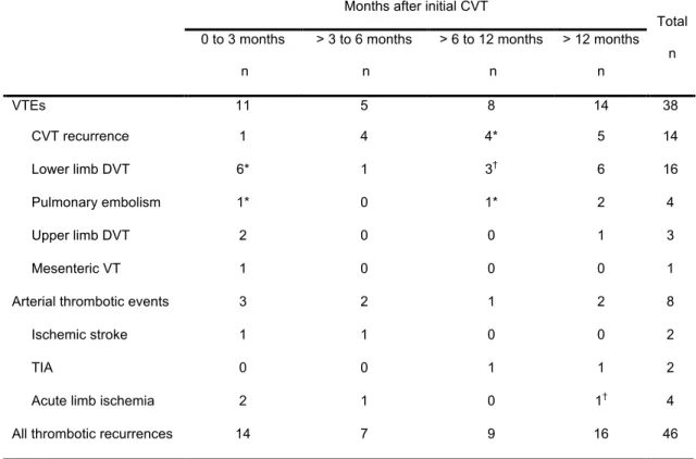
Twenty-one (45.6%) thrombotic recurrences occurred in the first six months and 30 (65.2%) within the first year. Two patients had two recurrences diagnosed during the same acute hospital admission (one had recurrent CVT and pulmonary embolism; the other had lower limb deep vein thrombosis and pulmonary embolism). One patient with acute limb ischemia later had deep leg vein thrombosis. Two patients suffered fatal thrombotic events (one recurrent CVT and one acute limb ischemia). The cumulative incidence of thrombotic recurrence after three and six months was 2.2% and 3.4% respectively. Cumulative incidence gradually increased to 5.0% after one year, 7.9% after two years and 14.3% after three years (Figure 1). The rate per 100 person-years for any thrombotic event was 5.1.
Figure 1: Kaplan-Meier estimation of all thrombotic events after the initial
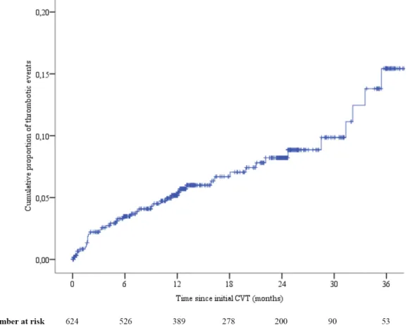
A total of 36 (5.8%) of the 624 patients had at least one venous thromboembolic event. Sixteen (42%) VTEs occurred in the first six months and 24 (63.2%) within the first year. The cumulative probability of venous thrombotic recurrence was 1.7% at three months, 2.6% at six months, 4.0% at twelve months, 6.5% at two years and 12.8% at three years (Figure 2). The venous thromboembolic recurrence rate was 4.1 per 100 person-years.
Figure 2: Kaplan-Meier estimation of venous thrombotic events after the
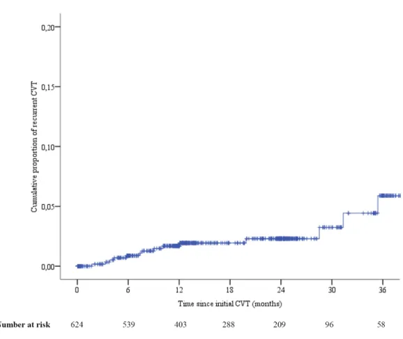
Fourteen (2.2%) patients had an episode of recurrent CVT. One patient suffered a fatal recurrent CVT. The cumulative incidence of a recurrent CVT event after three, six and twelve months was 0.2%, 0.9% and 1.7%, respectively. The two-year cumulative incidence was 2.3% and increased to 5.7% at three years (Figure 3). Five (35.7%) of the total CVT recurrences occurred in the first six months and 9 (64.3%) within the first year. The CVT recurrence rate was 1.5 per 100 person-years.
Figure 3: Kaplan-Meier estimation of recurrent cerebral vein and dural sinus
Eight patients (1.3%) had a new arterial thrombotic event. The cumulative probability of an arterial thrombotic event was 0.5% at three months, 0.9% at six months, 1.1% at twelve months, 1.7% at one and two years (Figure 4). The rate of arterial thrombosis was 0.8 per 100 person-years.
Figure 4: Kaplan-Meier estimation of arterial thrombotic events after the
initial cerebral vein and dural sinus thrombosis.
The results of the univariate analysis of the effects of the demographic, imaging features, thrombophilic risk factors and other risk factors on the incidence of a recurrence are shown in Table 2.
Thrombotic recurrences Overall group (n=624) Recurrent CVT (Patients=14) All venous (Patients=36) All arterial (Patients=8) All events (Patients=43) n (%) n (%) HR (95% CI) † n (%) HR (95% CI) ‡ n (%) HR (95% CI) ¥ n (%) HR (95% CI) ∂
Demographics
Age > 37 312 (50.0) 4 (1.3) 0.4 (0.1–1.3) 19 (6.1) 1.2 (0.6–2.2) 7 (2.2) 7.3 (0.9–59.5) 25 (8) 1.4 (0.8–2.7)
Age ≥ 65 51 (8.2) 0 (0) - 4 (7.8) 1.6 (0.6–4.5) 4 (7.8) 12.6 (3.2–50.8) § 8 (15.7) 3.0 (1.4–6.6) §
Male gender 159 (25.5) 9 (5.7) 6.2 (2.1–18.6) § 16 (10.1) 2.7 (1.4–5.3) § 3 (1.9) 1.9 (0.5–7.9) 18 (11.3) 2.4 (1.3–4.5) §
Imaging features
Superior sagittal sinus 387 (62.1) 11 (2.8) 2.3 (0.6–8.3) 26 (6.7) 1.7 (0.8–3.4) 4 (1.0) 0.6 (0.2–2.5) 29 (7.5) 1.3 (0.7–2.5) Left lateral sinus 279 (44.8) 8 (2.9) 1.7 (0.6–4.8) 17 (6.1) 1.1 (0.6–2.1) 4 (1.4) 1.2 (0.3–4.8) 21 (7.5) 1.2 (0.6–2.1) Right lateral sinus 257 (41.3) 8 (3.1) 1.8 (0.6–5.3) 15 (5.8) 1.0 (0.5–1.9) 4 (1.6) 1.4 (0.4–5.7) 18 (7.0) 1.0 (0.5–1.8)
Straight sinus 112 (18.0) 0 (0) - 4 (3.6) 0.6 (0.2–1.7) 1 (0.9) 0.7 (0.1–5.6) 5 (4.5) 0.6 (0.2–1.6)
Cortical veins 107 (17.2) 1 (0.9) 0.4 (0.1–3.1) 6 (5.6) 1.1 (0.4–2.5) 0 (0) - 6 (5.6) 0.9 (0.4–2.0)
Jugular veins 74 (11.9) 0 (0) - 1 (1.4) 0.2 (0–1.8) 1 (1.4) 1.1 (0.1–9.1) 2 (2.7) 0.4 (0.1–1.7)
Deep venous system 68 (10.9) 1 (1.5) 0.7 (0.1–5.4) 2 (2.9) 0.5 (0.1–2.3) 1 (1.5) 1.4 (0.2–11.2) 3 (4.4) 0.7 (0.2–2.2) > 2 sinuses or veins 179 (28.7) 4 (2.2) 1.0 (0.3–3.2) 9 (5.0) 0.8 (0.4–1.8) 2 (1.1) 0.9 (0.2–4.3) 11 (6.1) 0.9 (0.4–1.7) Thrombophilic RF Any thrombophilia 214 (34.3) 3 (1.4) 0.4 (0.1–1.6) 15 (7.0) 1.2 (0.6–2.3) 4 (1.9) 1.7 (0.4–6.9) 18 (8.4) 1.2 (0.6–2.2) Genetic thrombophilia 140 (22.4) 2 (1.4) 0.5 (0.1–2.3) 11 (7.9) 1.4 (0.7–2.9) 4 (2.9) 3.2 (0.8–12.9) 14 (10.0) 1.6 (0.8–3.0) Protein S deficiency 33 (7.5) 1 (3.0) 1.3 (0.2–10.2) 2 (6.1) 0.8 (0.2–3.2) 1 (3.0) 2.2 (0.3–19.2) 3 (9.1) 1.0 (0.3–3.4) Protein C deficiency 21 (4.8) 0 (0) - 1 (4.8) 0.7 (0.1–5.4) 0 (0) - 1 (4.8) 0.6 (0.1–4.5) Antithrombin deficiency 4 (0.9) 0 (0) - 0 (0) - 0 (0) - 0 (0) - Factor V Leiden 33 (7.5) 1 (3.0) 1.8 (0.2–14.4) 2 (6.1) 1.1 (0.3–4.5) 1 (3.0) 2.5 (0.3–21.6) 3 (9.1) 1.4 (0.4–4.5) Prothrombin G20210A 37 (8.4) 0 (0) - 4 (10.8) 1.9 (0.7–5.5) 2 (5.4) 5.7 (1.0–31.2) § 5 (13.5) 2.0 (0.8–5.2) MTHFR mutation 43 (9.9) 0 (0) - 2 (4.7) 0.7 (0.2–2.8) 1 (2.3) 1.6 (0.2–13.8) 2 (4.7) 0.5 (0.1–2.3) > 1 genetic RF 36 (7.7) 1 (2.8) 1.3 (0.2–10.1) 2 (5.6) 0.8 (0.2–3.5) 2 (5.6) 5.8 (1.1–32.0) § 3 (8.3) 1.1 (0.3–3.6) Severe thrombophilia 102 (16.3) 1 (1.0) 0.3 (0.1–2.3) 4 (3.9) 0.5 (0.2–1.4) 3 (2.9) 2.8 (0.7–11.6) 6 (5.9) 0.7 (0.3–1.6) Acquired thrombophilia 98 (15.7) 1 (1.0) 0.3 (0–2.5) 6 (6.1) 0.9 (0.4–2.1) 1 (1.0) 0.7 (0.1–5.5) 7 (7.1) 0.8 (0.4–1.9) Etiological group Transient RF only 216 (34.6) 5 (2.3) 1.1 (0.4–3.4) 9 (4.2) 0.7 (0.3–1.4) 0 (0) - 9 (4.2) 0.5 (0.3–1.1) Pregnancy/Puerperium* 77 (20.2) 0 (0) - 4 (5.2) 1.4 (0.4–4.2) 0 (0) - 4 (5.2) 1.3 (0.4–3.8) Oral contraceptives* 207 (54.3) 3 (1.4) 2.4 (0.2–22.7) 7 (3.4) 0.6 (0.2–1.6) 0 (0) - 7 (3.4) 0.5 (0.2–1.4) Permanent RF only 138 (22.1) 3 (2.2) 1.0 (0.3–3.4) 12 (8.7) 1.8 (0.9–3.5) 3 (2.2) 2.1 (0.5–8.7) 15 (10.9) 1.9 (1.0–3.6) § Malignancy 46 (7.4) 0 (0) - 0 (0) - 1 (2.2) 2.2 (0.3–18.2) 1 (2.2) 0.4 (0–2.6) Poly/thrombocythemia 18 (2.9) 1 (5.6) 3.1 (0.4–24.1) 4 (22.2) 4.9 (1.7–14.0) § 0 (0) - 4 (22.2) 3.9 (1.4–11.1) § Transient+Permanent RF 192 (30.8) 3 (1.6) 0.5 (0.1–1.9) 10 (5.2) 0.8 (0.4–1.6) 3 (1.6) 1.3 (0.3–5.5) 12 (6.3) 0.8 (0.4–1.5) Cryptogenic CVT 78 (12.5) 3 (3.8) 2.1 (0.6–7.5) 5 (6.4) 1.2 (0.5–3.2) 2 (2.6) 2.6 (0.5–12.9) 7 (9.0) 1.5 (0.7–3.4) *Analyses among 381 women ≤ 50 years of age. †Hazard ratios (HRs) represent the comparison between patients with and without CVT recurrence. ‡Hazard ratios (HRs) represent the comparison between patients with and without VTEs. ¥ Hazard ratios (HRs) represent the comparison between
patients with and without arterial trombotic events. ∂Hazard ratios (HRs) represent the comparison between patients with and without any thrombotic
recurrence. §p-value ≤ 0.05. The percentages for each specific genetic abnormality were calculated using the number of performed tests as denominator.
Abbreviations: CVT, cerebral vein and dural sinus thrombosis; VTEs, venous thromboembolic events (recurrent CVT, lower limb deep vein thrombosis, pulmonary embolism, upper limb vein thrombosis, abdominal or pelvic venous thrombosis); RF, risk factor.
Table 2: Univariate Cox proportional hazards analysis of predictors for
When age was analyzed as a continuous variable, no association was found for CVT recurrence (HRs= 0.9; 95% CI, 0.9–1.0; p=0.071) or VTEs (HRs= 1.0; 95% CI, 0.9–1.0; p=0.950). Male gender (HRs= 6.2; 95% CI, 2.1–18.6; p=0.001) was an important risk factor for CVT recurrence.
Male gender (HRs= 2.7; 95% CI, 1.4–5.3; p=0.003) and
polycythemia/thrombocythemia (HRs= 4.9; 95% CI, 1.7–14.0; p=0.003) were associated with an increased risk of venous thrombotic recurrence. In a recent meta-analysis, the presence of MTHFR mutation was not significantly associated with a higher risk of CVT.23 Therefore an additional analysis was performed on the role of genetic thrombophilia excluding the MTHFR mutation cases. However, no significant association was found between this genetic thrombophilia excluding MTHFR mutation and recurrent CVT (HRs= 0.6; 95% CI, 0.1–3.1; p=0.572) or VTEs (HRs= 1.3; 95% CI, 0.6–2.8; p=0.533).
The risk of thrombotic recurrence of any type was higher among elderly patients (HR= 3.0; 95% CI, 1.4–6.6; p=0.005), men (HR= 2.4; 95% CI, 1.3–4.5; p=0.004) and patients who had one permanent risk factor only
(HR= 1.9; 95%CI, 1.0–3.6; p=0.043). The presence of
polycythemia/thrombocythemia was also associated with an increased risk of all thrombotic recurrences (HR= 4.0; 95% CI, 1.4–11.1; p=0.009).
In the acute phase, 520 (83.3%) patients received anticoagulation therapy in therapeutic dosages with intravenous heparin or subcutaneous low-molecular-weight heparin. Oral anticoagulation after acute heparin therapy was prescribed to 476 (76.3%) patients. Information about the end date of oral anticoagulant therapy was not available for five patients. In
those who continued anticoagulation, the median duration of treatment was 7.5 months. Details about anticoagulation treatment in patients with and without recurrence are presented in Table 3.
Thrombotic recurrences
No recurrence* Recurrent CVT VTEs Arterial events Any event
n (%) n (%) n (%) n (%) n (%)
Anticoagulation continued (n=476) 446 (93.7) 11 (2.3)† 30 (6.3)‡ 5 (1.0)§ 34 (7.1)
3 months or less (n=36) 29 (80.6) 0 (0) 7 (19.4) 3 (8.3) 10 (27.8)
Between 3 and 6 months (n=94) 89 (94.7) 2 (2.1) 5 (5.3) 1 (1.1) 6 (6.4)
Between 6 and 12 months (n=205) 195 (95.1) 5 (2.4) 10 (4.9) 0 (0) 10 (4.9)
12 months or more (n=136) 128 (94.1) 4 (2.9) 8 (5.9) 1 (0.7) 8 (5.9)
* For 5 of the 442 patients continuing anticoagulation the end date of therapy was not available. †Two of these patients had the
CVT recurrence after finishing the anticoagulation regimen. ‡Nine of these patients had the CVT recurrence after finishing the
anticoagulation regimen. §Three of these patients had the arterial event after finishing the anticoagulation regimen.
Abbreviations: CVT, cerebral vein and dural sinus thrombosis; VTEs, venous thromboembolic events (recurrent CVT, lower limb deep vein thrombosis, pulmonary embolism, upper limb vein thrombosis, abdominal or pelvic venous thrombosis).
Table 3: Anticoagulation after the acute phase of cerebral vein and dural
sinus thrombosis in included patients with and without venous thromboembolic events.
At the time of recurrence, 23 (53.5%) patients with any thrombotic event, 21 (58.3%) with VTEs, 9 (64.3%) with a CVT recurrence and 2 (25%) with an arterial thrombotic event were on anticoagulant therapy. Oral anticoagulation after the acute phase was not associated with a significantly lower risk of thrombotic recurrence of any type (HRs= 1.0; 95% CI, 0.5–2.1; p=0.946), VTEs (HRs= 1.4; 95% CI, 0.6–3.4; p=0.459), CVT recurrence (HRs= 1.0; 95% CI, 0.3–3.7; p=0.981) or arterial thrombotic recurrence (HRs= 0.4; 95% CI, 0.1–1.8; p=0.244). In patients on oral anticoagulation, the duration of anticoagulant therapy was also not significantly associated with thrombotic events of any type (HRs= 1.0; 95% CI, 0.99–1.00; p=0.579)
or venous thromboembolic recurrences (HRs= 1.0; 95% CI, 0.99–1.00; p=0.963).
In the final Cox regression analysis model, male gender (HRs= 2.0; 95% CI, 1.1–3.9; p=0.030) and polycythemia/thrombocythemia (HRs= 3.2; 95% CI, 1.1–9.2; p=0.027) were associated with a higher risk of thrombotic recurrences of any type. For venous thrombotic recurrences, male gender (HRs= 2.6; 95% CI, 1.4–5.1; p=0.004) and polycythemia/thrombocythemia (HRs= 4.4; 95% CI, 1.6–12.7; p=0.005) were also the only significant independent risk factors.
DISCUSSION
The main new findings of this study are the description of the risk of a recurrent cerebral or a new systemic thrombotic event after CVT and the identification of male gender and polycythemia/thrombocythemia as independent predictors of venous thromboembolism after CVT. The large number of included subjects from a variety of countries and centers, the prospective design and the good quality of follow-up data are the main strengths of this study. Limitations include the fact that local investigators decided the investigations for symptomatic extracerebral thrombotic recurrences and thrombophilia screening was not performed in 25% of the centers. The median follow up in the ISCVT study was 13.9 months. Considering that most patients suffering a CVT are relatively young and have a life expectancy of several decades, a study with a longer follow-up might detect more recurrent events and additional predictors. Furthermore, thrombotic recurrences were not centrally adjudicated. These two factors could, therefore, underestimate the number of outcome events.
Patients with a first episode of a VTE of any kind are at an increased risk of further thrombotic episodes. This risk of venous thromboembolic recurrence not only varies with time after the initial event, but also depends on certain risk factors for recurrence. These factors are in some way different from the risk factors for the initial event and can be divided according to three main characteristics: a) patient-related features: male gender and certain molecular thrombophilias (antiphospholipid syndrome, antithrombin, protein C or protein S deficiency, homozygous factor V Leiden or prothrombin G20210 mutation and combined abnormalities); b)
associated features such as the presence of a cancer, a known temporary risk factor for VTE or history of a previous cryptogenic VTE; c) event-related features such as the initial location (proximal DVT and PE have a higher risk of thromboembolic recurrence than other locations).24 Information about risk factors for thrombotic recurrence after CVT is limited. One previous retrospective study did not identify any clinical predictor for recurrence18, but a recent small prospective study found a higher risk of venous thrombotic recurrence in males and in those with severe thrombophilia.25 When we
compare the results of the present study with studies on extracerebral venous thrombosis, male gender has also been associated with a higher risk of venous thrombotic recurrence26, whereas the influence of aging is more controversial.24 It is important to note that a significant proportion of women had the initial CVT while on oral contraceptives and during pregnancy or post-partum period. These factors are established transient risk factors for venous thrombosis27,28 and could explain the lower rate of further thrombotic events among women. However, a significant difference in thrombotic recurrence could not be found between women with and without these gender-specific risk factors, which favors the existence of further variables involved. The association between polycythemia or thrombocythemia and venous thrombotic events is well established29 and it is now confirmed for CVT.
Current European guidelines on the treatment of CVT after the acute phase propose different durations of oral anticoagulation therapy in the presence of transient risk factors, genetic thrombophilia or idiopathic CVT.14 In the ISCVT cohort, the different etiological groups and the presence of
genetic or acquired thrombophilia was not associated with a higher risk of thrombotic recurrence. Nonetheless, it is difficult to interpret these findings because the modest number of patients with specific genetic prothrombotic conditions and the low event rate make this study underpowered to investigate the role of specific prothombotic conditions in the recurrence of venous events. In a pediatric CVT cohort, prothrombin G20210A mutation predicted recurrent venous thromboembolism.19 The same study also reported persistent venous occlusion as an independent predictor of venous thrombotic recurrence. Martinelli and colleagues25, in a recent prospective study found a significantly higher risk of venous thromboembolic recurrence in patients with severe thrombophilia (antithrombin, protein C or protein S deficiency, more than one thrombophilic abnormality and positive antiphospholipid antibodies) when compared to those without or with mild thrombophilia. These results were obtained from a study performed in a tertiary thrombosis centre and include a much higher rate of patients with thrombophilic conditions, which could explain the differences between both studies. It is, therefore, important to further investigate this particular finding and one possibility for a future project could be a meta-analysis evaluating genetic abnormalities in CVT patients and the risk of recurrent VTEs.
Studies in adult CVT patients found that venous recanalization occurs mostly within four months after CVT irrespective of anticoagulation.30
Furthermore, no significant differences in relapses or outcome were found in those with incomplete or no recanalization.31,32 In this study, baseline imaging features such as location and thrombus load were not associated with thrombotic recurrence.
The cumulative incidence of recurrent thrombosis after CVT at six, twelve and twenty-four months was 3.4%, 5.0% and 7.9%, respectively. The overall rate of all thrombotic recurrences in the ISCVT cohort was 5.1 per 100 person-years. These recurrences had a fatal outcome in two patients (4.7%). Six months after a CVT, the likelihood of VTEs was 2.6% and the risk increased to 6.5% after two years. In the ISCVT cohort, it was found an overall rate of VTEs after CVT of 4.1 per 100 person-years. Regarding CVT recurrence, the cumulative probability increased in a similar way from 0.9% at six months to 2.3% at two years. The rate per 100 person-years of CVT recurrence found was 1.5. In a systematic review, Dentali and colleagues15 calculated an overall rate of 2.8% for CVT recurrence and 3.7% for extracerebral venous thrombotic events but the results and duration of follow-up varied widely between studies. A retrospective study18 reported a
CVT recurrence rate of 2.2 per 100 person-years and a rate of 5.0 per 100 person-years for recurrent venous thromboembolism. Moreover, the majority of the recurrences occurred within the first year after CVT. More recently, a prospective study with a median follow-up of 72 months reported an overall incidence of 1.58% year for VTEs after CVT and 0.53% patients-year for recurrent CVT, despite a prevalence of thrombotic recurrences at 16 months similar to the ISCVT cohort (1.3% for CVT and 3.0% for other manifestations of venous thromboembolism).25 In the same study, almost
half of the recurrences occurred within the first year after discontinuation of anticoagulant therapy. In the ISCVT cohort, it was also found that 63.2% of VTEs and 64.3% of CVT recurrences occurred within the first year after CVT. In addition, there was a steady increase in the risk of outcome events
and the Kaplan Meier curves showed a rather linear relation to time. The relatively short period of observation and the smaller number of patients in follow-up at later time points might explain the high event rates found within the first year. The venous thrombotic recurrence rate in this study was similar to the rate found in smaller prospective studies17,33,34 but CVT recurrence was less frequent than two retrospective analyses16,35 (Table 4). Venous thrombosis after CVT seems to be less frequent than the recurrence rate reported after deep vein thrombosis or pulmonary embolism.36,37 One
possible reason is the absence in CVT of the hydrostatic factor that can promote thrombosis in patients with predisposing conditions.
Study
Nº of patients
with follow-up Length of follow-up
CVT n (%) All VTEs n (%) Prospective ISCVT20 624 Median= 16 m; Mean= 18.6±11 m 14 (2.2) 38 (6.1) Martinelli et al25 145 Median= 6 y 5 (3.4) 15 (10.3) Kenet et al19* 384 Median= 36 m 13 (3.4) 22 (5.7) VENOPORT17** 84 Median= 12 m ; Mean= 12±7 m 0 5 (6) Breteau et al. 34 48 Median= 36 m; (range 12- 60 m) 0 3 (5.5)
Baumgartner et al.30 33 12 m 0 0
Rondepierre et al.33 18 31 m 0 1 (5.5)
Retrospective
Gosk-Bierska et al.18 154
Mean= 36±47 m 10 (6.5) 23 (14.9) Preter et al.16 77 Median= 63 m; Mean= 77.8 m 9 (11.7) 20 (26)
Mehraein et al.28 82 Mean= 10 y 5 (6.1) Not reported
Maqueda et al.35 54 Median= 2.4 y; Mean= 3.5 y 1 (1.9) 8 (14.8)
*Study in patients aged < 18 years. **Prospective part of the study. Abbreviations: m, months; y, years; CVT, Cerebral vein thrombosis; VTEs, Venous thromboembolic events.
Table 4: Studies investigating the frequency of recurrent thrombotic events
Recent evidence suggests an association between venous thrombosis and subsequent arterial thrombotic events.38 In the current report, the risk of an arterial thrombotic event after CVT was 0.8 per 100 person-years, considerably lower than that of venous thrombotic recurrence. This study could not find an association between anticoagulation or duration of anticoagulation and prevention of thrombotic recurrence. A significant number of patients (58.3%) had the thrombotic recurrence while on anticoagulation and the proportion was even higher (64.3%) among patients with recurrent CVT. These findings could be explained by differences in sample size and prognostic characteristics between individuals who continued anticoagulation and those who did not continue anticoagulant therapy. Furthermore, the level of anticoagulation with INR monitoring was not evaluated and could influence the results. Gosk-Bierska and colleagues8, also reported no benefit in survival or recurrence for those receiving oral anticoagulants, but a higher risk of recurrent events was found in children not taking anticoagulation.9 As previously mentioned, recent
results also found a very high rate of recurrence within the first year after discontinuation of anticoagulant therapy.24 We need to be cautious when interpreting these results as all these observational studies did not have the appropriate design for the assessment of the efficacy and appropriate duration of anticoagulant therapy after the acute phase of cerebral venous thrombosis.
In conclusion, the findings of this study suggest that CVT may be a different clinical entity when compared with deep vein thrombosis in respect to thrombotic recurrence. These results may have implications for current
clinical practice and future research. Knowing the risk of a thrombotic recurrence is important for patients, and also to guide secondary prevention. A systematic review including all new CVT studies and focusing on the risk factors for venous thrombotic recurrence after CVT, particularly genetic abnormalities, will provide important evidence for treatment guidance. Nevertheless, the efficacy and safety of short or extended anticoagulation after CVT should be adequately assessed by a randomized controlled trial.
REFERENCES
1 Ribes MF. Des recherches faites sur la phlébite. Revue Médicale
Française et Etrangère et Journal de Clinique de l’Hôtel-Dieu et de la Charité de Paris 1825; 3: 5-41.
2 Stam J. Thrombosis of the cerebral veins and sinuses. N Engl J Med
2005; 352: 1791–8.
3 Bousser MG, Ferro JM. Cerebral venous thrombosis: an update.
Lancet Neurol 2007; 6: 162–70.
4 De Veber G, Andrew M, Adams C, et al. Cerebral sinovenous
thrombosis in children. N Engl J Med 2001; 345: 417-423.
5 Ferro JM, Canhao P, Stam J, Bousser MG, Barinagarrementeria F.
Prognosis of cerebral vein and dural sinus thrombosis: results of the International Study on Cerebral Vein and Dural Sinus Thrombosis (ISCVT). Stroke 2004; 35: 664–70.
6 Agnelli G, Verso M. Epidemiology of Cerebral Vein and Sinus
Thrombosis. Front Neurol Neurosci 2008; 23: 16-22.
7 de Freitas GR, Bogousslavsky J. Risk factors of Cerebral Vein and
Sinus Thrombosis. Front Neurol Neurosci 2008; 23: 23-54.
8 Ferro J, Canhao P, Stam J, Bousser MG, Barinagarrementeria F, et
al. Delay in the Diagnosis of Cerebral Vein and Dural Sinus Thrombosis: influence on outcome. Stroke 2009; 40:3133-8.
9 Ferro JM. Cerebral venous thrombosis. J Neuroradiol 2002; 29:
10 Paciaroni M, Palmerini F, Bogousslavsky. Clinical Presentations of
Cerebral Vein and Sinus Thrombosis. Front Neurol Neurosci 2008; 23: 77-88.
11 Selim M, Caplan L. Radiological Diagnosis of Cerebral Venous
Thrombosis. Front Neurol Neurosci 2008; 23: 96-111.
12 Einhäupl KM, Villringer A, Meister W, Mehraein S, Garner C, Pellkofer
M, et al. Heparin treatment in sinus venous thrombosis. Lancet 1991; 338: 597-600.
13 de Bruijn SF, Stam J, for the Cerebral Venous Sinus Thrombosis
Study Group. Randomized, placebo-controlled trial of anticoagulant treatment with low-molecular-weight heparin for cerebral sinus thrombosis. Stroke 1999; 30: 484-488.
14 Einhäupl K, Stam J, Bousser MG, de Bruijn SF, Ferro JM, Martinelli I,
Masuhr F. EFNS guideline on the treatment of cerebral venous and sinus thrombosis in adult patients. Eur J Neurol 2010; 17: 1229–35.
15 Dentali F, Gianni M, Crowther MA, Ageno W. Natural history of
cerebral vein thrombosis: a systematic review. Blood 2006; 108: 1129–34.
16 Preter M, Tzourio C, Ameri A, Bousser MG. Long-term prognosis in
cerebral venous thrombosis. Follow-up of 77 patients. Stroke 1996; 27: 243–6.
17 Ferro JM, Lopes MG, Rosas MJ, Ferro MA, Fontes J. Long-term
prognosis of cerebral vein and dural sinus thrombosis. results of the VENOPORT study. Cerebrovasc Dis 2002; 13: 272–8.
18 Gosk-Bierska I, Wysokinski W, Brown RD, et al. Cerebral venous
sinus thrombosis: Incidence of venous thrombosis recurrence and survival. Neurology 2006; 67: 814–9.
19 Kenet G, Kirkham F, Niederstadt T, et al. Risk factors for recurrent
venous thromboembolism in the European collaborative paediatric database on cerebral venous thrombosis: a multicentre cohort study. Lancet Neurol 2007; 6: 595–603.
20 Miranda B, Ferro JM, Canhao P, Stam J, Bousser MG,
Barinagarrementeria F, Scoditti U. Venous Thromboembolic events after cerebral vein thrombosis. Stroke 2010; 41: 1901–6.
21 Bousser MG, Ross Russell RW. Cerebral Venous Thrombosis. Vol 1.
London: Saunders, 1997.
22 Kaplan E, Meier P. Nonparametric estimation from incomplete
observations. J Am Stat Assoc 1958; 53: 457–81.
23 Gouveia LO, Canhao P. MTHFR and the risk for cerebral venous
thrombosis- a meta-analysis. Thromb Res. 2010 Apr;125:e153-8.
24 Zhu T, Martinez I, Emmerich J. Venous thromboembolism: risk factors
for recurrence. Arterioscler Thromb Vasc Biol 2009; 29: 298–310.
25 Martinelli I, Bucciarelli P, Passamonti SM, Battaglioli T, Previtali E,
Mannucci PM. Long-term evaluation of the risk of recurrence after cerebral sinus-venous thrombosis. Circulation 2010 Jun 29;121:2740-6.
26 McRae S, Tran H, Schulman S, Ginsberg J, Kearon C. Effect of
patient's sex on risk of recurrent venous thromboembolism: a meta-analysis. Lancet 2006; 368: 371–8.
27 Kujovich JL. Hormones and pregnancy: thromboembolic risks for
women. Br J Haematol 2004; 126: 443–54.
28 Mehraein S, Ortwein H, Busch M, Einhäupl K, Masuhr F. Risk of
recurrence of cerebral venous and sinus thrombosis during subsequent pregnancy and puerperium. J Neurol Neurosurg Psychiatry 2003; 74: 814-816.
29 De Stefano V, Za T, Rossi E, et al. Recurrent thrombosis in patients
with polycythemia vera and essential thrombocythemia: incidence, risk factors, and effect of treatments. Haematologica 2008; 93: 372–80.
30 Baumgartner RW, Studer A, Arnold M, Georgiadis D. Recanalisation
of cerebral venous thrombosis. J Neurol Neurosurg Psychiatry 2003; 74: 459–61.
31 Strupp M, Covi M, Seelos K, Dichgans M, Brandt T. Cerebral venous
thrombosis: correlation between recanalization and clinical outcome--a long-term follow-up of 40 patients. J Neurol 2002; 249: 1123–4.
32 Stolz E, Trittmacher S, Rahimi A, et al. Influence of recanalization on
outcome in dural sinus thrombosis: a prospective study. Stroke 2004; 35: 544–7.
33 Rondepierre P, Hamon M, Leys D, et al. Thromboses veineuses
cérébrales: étude de l’évolution. Rev Neurol (Paris) 1995; 151: 100–4.
34 Breteau G, Mounier-Vehier F, Godefroy O, et al. Cerebral venous
thrombosis 3-year clinical outcome in 55 consecutive patients. J Neurol 2003; 250: 29–35.
35 Maqueda VM, Thijs V. Risk of thromboembolism after cerebral venous
36 Prandoni P, Lensing AW, Cogo A, et al. The long-term clinical course
of acute deep venous thrombosis. Ann Intern Med 1996; 125: 1–7.
37 Heit JA, Mohr DN, Silverstein MD, Petterson TM, O'Fallon WM, Melton
LJ 3rd. Predictors of recurrence after deep vein thrombosis and pulmonary embolism: a population-based cohort study. Arch Intern Med 2000; 160: 761–8.
38 Lowe GD. Is venous thrombosis a risk factor for arterial thrombosis?