289
Vol.58, n.2: pp. 289-299, March-April 2015 http://dx.doi.org/10.1590/S1516-8913201500305
ISSN 1516-8913 Printed in Brazil
BRAZILIAN ARCHIVES OF BIOLOGY AND TECHNOLOGY
A N I N T E R N A T I O N A L J O U R N A L
Properties of Films Obtained from Biopolymers of Different
Origins for Skin Lesions Therapy
Márcia Zilioli Bellini
1,2, Pedro de Oliva-Neto
3and Ângela Maria Moraes
2* 1Departamento de Engenharia de Alimentos; Faculdades Adamantinenses Integradas; Adamantina - SP - Brasil. 2
Departamento de Engenharia de Materiais e de Bioprocessos; Faculdade de Engenharia Química; Universidade de Campinas; Campinas - SP - Brasil. 3Departamento de Ciências Biológicas; Universidade Estadual Paulista; Assis - SP - Brasil
ABSTRACT
In this study, the effects of the origin of xanthan used, in combination with chitosan, to prepare films for the treatment of skin lesions were evaluated. The characteristics of the films obtained with xanthan commercially available for the food industry sector and xanthan originated from a fermentation process conducted in a pilot plant were compared. Results showed that the source did not strongly interfere in many of the properties of the films, such as the mechanical properties, cytotoxicity to L929 cells, absorption of simulated body fluid and culture medium, stability in water and saline solution. Hence, even though the properties of biopolymers of different sources might vary, the films prepared with two distinct types of xanthan gum could be considered as potentially safe and similar in terms of relevant characteristics considering the aimed application.
Key words: biomaterials, biopolymers, films, membranes, properties and characterization, biomedical applications
*Author for correspondence: ammoraes@feq.unicamp.br
INTRODUCTION
The use of biopolymers for the production of material intended for medical and pharmaceutical applications, targeting, for example, the controlled release of bioactive agents, tissue engineering and cell therapy has increased in recent years due to
the availability, biocompatibility and
biodegradability of the many compounds in this
category (Bueno and Moraes 2011).
Polysaccharides such as chitosan (C), xanthan (X) and alginate are examples of non-toxic natural polymers used in the composition of materials for this purpose, being used alone or in combination with each other or with other compounds (Popa et al. 2010; Fernandes et al. 2011; Bellini et al. 2012;
Veiga and Moraes 2012). However, a major
obstacle to their use is the variability in the
characteristics of molecules resulting from
different sources, which may result in changes in the properties of biomaterials obtained when these polymers are used.
Chitosan is a biopolymer with cationic character, having a linear structure similar to that of the glycosaminoglycans of cartilage (Silva et al. 2010). It is derived from chitin, being formed by
glucosamine and N-acetyl-D-glucosamine
monomers linked by glycoside bonds β(1→4)
(Jayakumar et al. 2011; Coma 2013). Its
production process is relatively simple and one of
its most important characteristics regarding
biological applications is its degree of
deacetylation. This polymer is soluble in aqueous acidic solutions, resulting in structures with
different dimensions and geometric
Bellini, M. Z. et al. 290
gels (Santos et al. 2003; Ragetly et al. 2010).The polycationic nature of chitosan enables its association with polymers with negative charges such as alginate (Rodrigues et al. 2008; Bueno and Moraes 2011), dextran derivatives (Fukuda et al. 1978), polyesters (Silva et al. 2010), silk fibers (She et al. 2008), gelatin (Liu et al. 2004; Yin et al. 2005), and xanthan gum (Eftaiha et al. 2009; Bellini et al. 2012; Veiga and Moraes 2012), resulting in stable polyelectrolyte complexes.
Xanthan gum is a water-soluble
exopolysaccharide synthesized by Xanthomonas
and is used as emulsifier, suspension stabilizer, flocculant, gelling and viscosity agent. It is widely used in different industrial applications (Oliveira et al. 2013), such as in food, toiletry, oil recovery, pharmaceutical and cosmetics industry. It is commonly used in the form of gels or films
(Bejenariu et al. 2008). Its main chain is
equivalent to that of cellulose, consisting of D-glucose units linked together by β(1→4) bonds, and its side chains are composed of alternating residues of D-mannose and D-glucuronic acid in
the ratio 2:1 (Oliva-Neto et al. 2011).
Commercially, xanthan gum is the most
economically relevant bacterial polysaccharide (Meyer et al. 2008). It is estimated that by 2015, the world production of xanthan gum may reach 80,000 tons, associated to a market of around 400 million dollars (Carignatto et al. 2011), but new applications in the pharmaceutical field may expand this scenario further.
When compared to polysaccharides extracted directly from the natural sources, such as alginate derived from seaweed, xanthan gum has the advantage of being independent of the production site and climatic conditions, which allows its production under significantly more controlled conditions, with lower variability in the properties of material originated from different batches and, therefore, with higher quality assurance. For this reason, xanthan gum, alone or combined with other polymers, has been gaining attention in the composition of biomaterials intended for different biomedical applications, such as devices for the controlled release of drugs (Popa et al. 2010; Bellini et al. 2012; Veiga and Moraes 2012; Dyondi et al. 2013) and probiotic bacteria (Argin et al. 2014), hydrogels for bone regeneration (Izawa et al. 2014), ophthalmological materials (Ceulemans et al. 2002; Ludwig 2005), implants (Kumar et al. 2007) and scaffolds for tissue engineering (Silva et al. 2007; Bellini et al. 2012).
Various strategies to improve the production of xanthan have been investigated to reduce its cost of manufacture, especially the selection of cell cultures with high capacity (Oliveira et al. 2000) and the proposition of culture media with different formulations (Carignatto et al. 2011; Diniz et al. 2012).
Promising results for the use of scaffolds based on polysaccharides and other polymers of natural origin in the regeneration of skin lesions have been reported (Liu et al. 2004; Chen et al. 2009; Pajoum et al. 2009). In fact, skin substitutes obtained by tissue engineering represent today a real therapeutic option for severe burns and other skin injuries. Apligraf® (produced by the company
Organogenesis) and OrCel (from Forticell
Bioscience) scaffolds consisting of type I bovine collagen inoculated with human keratinocytes and fibroblasts are examples of devices for tissue engineering already approved by the Food and Drug Administration (FDA, USA).
Scaffolds of chitosan-gelatin-hyaluronic acid (Liu et al. 2004) and of chitosan-collagen (Pajoum et al. 2009) are also useful in the co-culture of keratinocytes and fibroblasts aiming at obtaining artificial skin. Such devices are flexible and have good mechanical properties, facilitating skin regeneration. Porous chitosan-collagen matrices
are particularly promising in the tissue
engineering context, being reported, for example, that their implantation in rabbits' ears results in rapid infiltration of fibroblasts in the treated region (Ma et al. 2003).However, given that there is a tendency to avoid the use of materials from xenogenic origin for tissue engineering purposes
and of complex composition, collagen
Properties of Films for Skin Lesions Therapy 291
Thus, considering the relevance of the search for natural polymers of low cost, safe and with more
reproducible properties useful in the
manufacturing biomaterials, the purpose of the present work was to evaluate whether films obtained by combining chitosan with xanthan gum of different sources would have equivalent performance. In this context, the use of a xanthan gum commercially available for the food industry sector and of a xanthan gum obtained through a Brazilian fermentation technology process using sucrose and ethanol from sugar cane (Carignatto et al. 2011) carried out in a pilot plant unit located at the São Paulo State University (UNESP) at Assis, São Paulo, Brazil were analyzed. The characteristics of these formulations were also compared to those shown by higher cost films produced with analytical grade xanthan gum. It was expected that the origin of the xanthan gum would not interfere substantially on relevant properties of the biomaterial, aiding in the consolidation of the use of this polysaccharide for the particular purpose aimed.
MATERIAL AND METHODS
Material
Chitosan-xanthan films were produced using chitosan (C) with a deacetylation degree in the range of 75 to 85% (batch number 9012-76-4, Sigma-Aldrich, St. Louis, MO), glacial acetic acid (Merck, São Paulo, Brazil) and xanthan gum from two different sources, the first (XKeltrol) commonly used as an additive in food processing (Keltrol® type xanthan gum, available from CP Kelco, Brazil), while the second (Xpilot) was produced locally in a pilot plant using a technology developed at the São Paulo State University (UNESP) (Carignatto et al. 2011). The water used was distilled and deionized in a MilliQ Millipore system.
Preparation of the Films
The chitosan-xanthan gum films were prepared according to the procedures described by Bellini et al. (2012), using a mass ratio of chitosan to xanthan equal to 1:1. An aliquot of 100 mL of 1% chitosan (w/v) in 2% (v/v) acetic acid was added to 100 mL of xanthan 1% (w/v) aqueous solution using a peristaltic pump (Model TE 184 Tecnal, Brazil) at a flow rate of 10 mL min-1 stirred at 1000 rpm using a mechanical mixer (Model TE
038 Tecnal, Brazil) in a glass reactor at 25°C with an internal diameter of 11 cm. The obtained suspension was deaerated for 2 h using a vacuum pump (Model TE 058 Tecnal, Brazil), transferred to a polystyrene Petri dish (15 cm in diameter) and dried in an air circulating oven (Model TE 394/1, Tecnal, Brazil) at 37°C until constant weight for approximately 24 h.
The dried films were washed with 500 mL of deionized water for 30 min three times to remove the residual acetic acid. Then, the samples were immersed in 200 mL of 10 mM Hepes buffer for 30 min and again in 500 mL of water for 30 min. afterwards, the films were dried at 37°C for 24 h. The films were cut into appropriate size samples and sterilized by exposure to Oxyfume-30 (30% ethylene oxide and 70% carbon dioxide) at 40°C for 8 h and relative humidity of 30 to 80% at
Acecil Central de Esterilização Comércio e
Indústria Ltda (Campinas, SP, Brasil). The residual ethylene oxide was removed by keeping the samples under air circulation for 48 h.
Characterization of the Films
The samples were characterized with respect to morphology, thickness, absorption and stability in
aqueous solutions, mechanical properties,
volumetric expansion and cytotoxicity as
described below.
Morphology
The overall aspect was analyzed by inspection with the naked eye and recorded using a digital camera. The surface morphology and the cross section of the films were examined by scanning electron microscopy (SEM, Leo 440i model, Leica). Samples of 2 cm x 1 cm were freeze-dried, fixed to proper supports and coated with gold (Sputter mini SC 7620) prior to the analysis.
Thickness
The thickness of the films was measured with a
micrometer (Digimess) in seven equidistant
positions near the edge of each film. The results were expressed as mean values.
Mechanical Properties
Bellini, M. Z. et al. 292
of 1 cm/s equipped with a cell load of 5 kgf was used for this analysis. The tension (T) and elongation (E) at break were calculated through Equations 1 and 2, respectively:
(1)
(2)
where Fm is the maximum strength, As is the
cross-sectional area of the sample, di is the initial
distance between the texturometer grips (5 cm) and dr is the distance between the grips at the
moment of sample rupture.
Absorption and Stability in Aqueous Solutions The degree of absorption of different aqueous solutions by the films was evaluated based on the method proposed by Rodrigues et al. (2008). Samples (1 cm x 6 cm) dried at 37°C with a known mass (Mi) were immersed in 7.0 mL of
water, saline solution (SS) consisting of 0.9% (w/v) NaCl in water, simulated body fluid (SBF) prepared according to Kokubo et al. (1990), RPMI culture medium (Nutricell, Brazil) supplemented with 0.3 g. L-1 of L-glutamine, 2 g. L-1 of D-glucose, 2 g. L-1 of NaHCO3 , 10,000 IU L-1 of penicillin, 0.05 g. L-1 of streptomycin, 5.96 g. L-1 of Hepes and 10% (v/v) fetal bovine serum (Nutricell, Brazil). The final mass (Mf) of the
samples exposed to water, SS and SBF were determined after 24 h or after 144 h when in contact with supplemented RPMI culture medium. The degree of swelling (S) was calculated using Equation 3:
(3)
Afterwards, the wet samples were dried at 37°C for 10 h and their final mass (Md) were measured to determine their stability in each solution (E) with respect to variation of mass, as shown in Equation 4:
(4)
where Msol refers to the mass of soluble
compounds present in the solutions incorporated by the samples, equal to 9.0 g L-1, 15.3 g L-1 and
19.3 g L-1 respectively for SS, SBF and
supplemented RPMI culture medium. All analysis was performed in at least triplicate.
Volume Expansion in Cell Culture Medium The degree of tridimensional expansion of the films was determined indirectly based on the method proposed by Zeng and Ruckenstein (1996), quantifying the amount of culture medium, which presumably occupied the pores of saturated samples. Dry samples (2 cm x 4 cm)
with known mass (Mi) were immersed in 10 mL of
supplemented RPMI culture medium at 37°C for 24 h. Then, the masses of the hydrated samples (Mf) and their dimensions were determined. The
degree of volumetric expansion (ε) was calculated according to Equation 5, where dsolv was the
density of the culture medium at 37°C and V was the volume of the sample in the saturated state:
(5)
In Vitro Cytotoxicity
The in vitro toxicity of the films was assessed indirectly by exposing L929 fibroblasts to aqueous
extracts of the films and by subsequent
determination of mitochondrial activity of the
cells with
3-(4,5-dimethylthiazol-2-yl)-2,5-diphenyl tetrazolium bromide indicator (MTT, Sigma Chemical, St. Louis, MO) in at least quintuplicate. The extracts were obtained by incubating the films in supplemented RPMI culture medium at a concentration equal to 0.05 g of dry material per milliliter of medium for 48 h at 37°C and 5% CO2 (Bellini et al. 2012).
Aliquots of 100 μL of L929 cells containing 1x10⁵ cells mL-1 in supplemented RPMI medium
obtained by trypsinization (2.5% trypsin
Properties of Films for Skin Lesions Therapy 293
MO) containing 0.6% of acetic acid (Merck, São Paulo, Brazil) were added to each well. The samples were homogenized and the cells returned to the incubator at 37°C for one hour. After this period, the absorbance of the samples was analyzed in a microplate spectrophotometer at 620 nm (Thermo Scientific Multiskan FC, Finland). Extracts of culture plate fragments and of latex (in the form of amber tourniquet tubes regularly used in blood drawing) were used as negative and positive controls of cytotoxicity, respectively, and culture medium without cells was used as blank.
RESULTS AND DISCUSSION
The choice of a source of raw material for the production of a given biomaterial must take into account factors such as biocompatibility, cost, ease of access and performance regarding the desired purpose. In this context, biopolymers readily available by fermentation with assured quality are especially attractive, such as xanthan gum. Films prepared by the combination of chitosan and xanthan from two different sources available in Brazil, one produced on a pilot plant with technology developed at the University of the State of São Paulo at Assis (Carignatto et al. 2011) and the other (Keltrol® type, from CP Kelco,
Brazil) used by food industries. Several
characteristics important regarding the application
of these films as wound dressings were evaluated and compared with results obtained previously3 for a formulation based on xanthan of analytical origin.
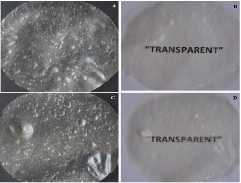
Aspect of the Films
The results of the analysis of the films regarding their aspect are shown in Figures 1 and 2. No major visual differences were observed between the films prepared with xanthan from the different sources, as shown in Figures 1A and 1B. The two formulations were relatively homogeneous before drying, initially presenting high concentrations of trapped air bubbles due to the high viscosity of the material; however, despite bubble coalescence apparently occurred during the drying process, the remaining bubbles did not cross the film thickness. When fully dried, the formulations resulted in thin, flexible films, with rippled surfaces, as described by Bellini et al. (2012) for films prepared with xanthan of analytical grade and chitosan at a 1:1 mass ratio. Both
formulations were translucent, but better
transparency was observed for the films
containing Xpilot instead of XKeltrol (Fig 1 C and D), and this particular feature was fairly important when considering application as dermal wound dressings, given that it would allow effective inspection of the wound bed during the recovery process without removing the film.
Bellini, M. Z. et al. 294
Typical results of the SEM analysis showed the presence of fibrillar structures homogeneously distributed over the surface of the films (Figs 2A and 2B). The integrity of the surface layer is critical in dermal dressings, allowing them to act as a barrier against the entry of infecting microorganisms in the skin lesions. The cross section analysis also showed that the films had lamellar structure, in accordance with observations for chitosan-alginate membranes (Wang et al. 2002; Rodrigues et al. 2008) and for C-X films prepared with xanthan gum of analytical grade (Bellini et al. 2012) and with XKeltrol obtained in different conditions (Veiga and Moraes 2012). Differences in the matrix packing along the thickness of the films can be also observed in Figures 2A and 2B.
Physico-Chemical Characterization of the
Films
Data on the mean thickness, swelling behavior and stability regarding mass loss in different aqueous solutions, mechanical properties and volumetric expansion of the films obtained are shown in Table 1. The dry films were 104.3 to 127.1 μm in thickness (Table 1). This is a relevant property in the architecture of the matrix, and may interfere not only with the comfort to the patient but also with the transport of gases such as O2, CO2, nutrients and metabolites in the wound region. Generally, devices used as dermal substitutes and in skin tissue engineering are thinner than the human dermis (Ma et al. 2001), which ranges between 500 and 2000 μm in thickness. In this context, both types of film formulations were suitable.
Figure 2 - SEM analysis of films obtained by combining chitosan to Xpilot (A) or XKeltrol (B).
Table 1 - Characteristics of membranes obtained from chitosan combined with xanthan gum of different sources: mean thickness values, mechanical properties, absorption and stability in aqueous solutions, and volumetric expansion in supplemented RPMI culture medium.
Property C-XMembrane formulation
pilot C-XKeltrol
Thickness (µm) 104.3 ± 15.1a 127.1 ± 5.0b
Mechanical properties of dry samples
Tensile strenght (MPa) 12.7 ± 2.0c 15.7 ± 5.0c Elongation at break (%) 2.1 ± 0.3d 2.1 ± 0.4d
Absorption of different aqueous solutions (g/g)
H2O (24 h) 58.5 ± 4.0
e
92.0 ± 2.7f
SS (24 h) 3.9 ± 0.3g 17.6 ± 0.7h
SBF (24 h) 13.0 ± 1.0i 8.9 ± 0.1i
RPMI (144 h) 6.9 ± 0.4j 5.2 ± 0.8j
Mass loss in different aqueous solutions (%)
H2O (24 h) 25.5 ± 1.9
k
20.7 ± 33.0k
SS (24 h) 15.7 ± 0.9m 11.2 ± 1.5m
SBF (24 h) 17.7 ± 1.3n 10.2 ± 0.4o
RPMI (144 h) 25.5 ± 3.4p 14.9 ± 1.0q
Volume expansion in RPMI culture medium (%) 96.5 ± 5.3r 84.5 ± 3.3r
SS - saline solution (NaCl at 0.9% w/v); SBF – simulated body fluid; RPMI – Roswell Park Memorial Institute culture medium
supplemented with com 0.3 g/L of L-glutamine, 2 g/L of D-glucose, 2 g/L of NaHCO3,10000 IU/L of penicillin, 0.05 g/L of
Properties of Films for Skin Lesions Therapy 295
Thin films and scaffolds with composition based in the use of polysaccharides are described in the literature, but only a few of them deal with the mixture chitosan-xanthan gum. Porous chitosan scaffolds with thickness between 60 and 80 μm
produced by lyophilization proved to be
appropriate for the in vitro culture of dermal fibroblasts, showing also to be potentially suitable for the use in skin tissue engineering (Ma et al. 2001). Chitosan-alginate membranes designed for treating skin lesions had thicknesses between 23.5
and 80 μm (Wang et al. 2001; Ragetly et al. 2010).
Films of chitosan complexed with xanthan gum of analytical grade at C:X mass ratios of 1:1 and 1.2:0.8 had mean thickness varying from 100 to
200 μm (Bellini et al. 2012), similar to membranes
prepared by combining C-XKeltrol in different conditions from those used in the present work, being, therefore, in the same range of the values reported herein.
In addition to proper thickness, modulus of elasticity and tensile strength compatible with the damaged tissue are important requirements of materials used in tissue regeneration. The tensile strength of the prepared films ranged from 12.7 to 15.7 MPa, while elongation at break was 2.1% (Table 1). The values of tensile strength obtained were lower than those of films prepared with chitosan and xanthan gum of analytical grade, equal to 25.1 MPa (Bellini et al. 2012). However, the attained values were higher than those of lamellar membranes of chitosan-alginate (6.9 MPa) (Rodrigues et al. 2008) and also of films prepared with chitosan and xanthan gum of the Keltrol® type at 1:1 mass ratio processed at different flow rates (2.2 to 5.2 MPa) (Veiga and Moraes 2012) or at the 1.2:0.8 proportion (8.7 MPa) (Bellini et al. 2012). Similarly, films prepared only with chitosan or with chitosan combined with chitin had low tensile strength, varying between 3.0 and 6.5 MPa (Dallan et al. 2007). As the values of tensile obtained for both
C-Xpilot and C-XKeltrol formulations were in the
range expected for normal skin (2.1 to 21 MPa) (Wang et al. 2002; Jussila et al. 2005), the films obtained seemed adequate for use as dermal dressings also considering this requirement. Regardless of the type of xanthan used, however, the elongation at break of the films was low (around 2%), not reaching the values reported (Hansen and Jemec 2002; Jussila et al. 2005) for normal skin, from 61 to 70%. The low elongation, however, did not disqualify biomaterials for
application in the regeneration of skin, only indicated that they would not be recommended for the regions with high mechanical requirements, such as knees and elbows. The low elongation of biomaterials prepared with chitosan and applicable as dermal dressings has been reported in the literature. Dressings and porous films prepared with chitosan and chitosan-alginate showed low elongations, between 0.8 and 3.8% (Lai et al. 2003; Kucharska et al. 2008; Bueno and Moraes 2011). Membranes prepared with chitosan and analytical grade xanthan gum also had low elongation at break values, ranging between 1.1 and 2.0% (Bellini et al. 2012), as well as C-XKeltrol films prepared at different flow rates (1.2 to 2.5%, according to Veiga and Moraes 2012). In addition, moisture can significantly increase the elongation capacity of the films due to the plasticizer effect of the water in contact with the polysaccharides, circumventing the problem at least partially. Suitable absorption and stability in the presence of
physiological fluids are also important
characteristics of biomaterials intended for skin regeneration. Water or saline can be employed, for example, to hydrate the films before their use on the patient and, when in contact with exudative lesions, the dressing should be able to quickly absorb the liquid without losing weight due to disintegration. If the films are considered to be used as scaffolds for skin tissue engineering, appropriate behavior of the biomaterials in culture media commonly used for this purpose would also be expected.
High absorption of water was noticed for the films of both formulations (Table 1). However, the
biomaterial obtained with XKeltrol showed
significantly higher uptake of water (92.0 g H2O/g dry film) than the one prepared with Xpilot (58.5 g/g), behaving similarly to membranes prepared alike but with analytical grade xanthan gum (Bellini et al. 2012) (85.5 g/g). High water absorption was also reported for the hydrogels prepared with chitosan and xanthan aiming at the controlled release of drugs (64 g of water per gram of hydrogel) (Popa et al. 2010) and for lamellar films prepared at a C:X mass ratio of 1.2:0.8 (48.9 g H2O per gram of dry material) (Bellini et al. 2012). Although the material prepared with Xpilot absorbed less water than the ones obtained with
XKeltrol, the performance obtained was satisfactory
Bellini, M. Z. et al. 296
2000; Dallan et al. 2007; Rodrigues et al. 2008), varying from 9.5 to 19.2 g H2O/g dry film, and also of C-X films prepared at different flow rates (Veiga and Moraes 2012) (between 15.9 and 39.4 g H2O/g dry film) and of chitosan-xanthan porous films (Bellini et al. 2012) prepared in the presence of the surfactants Tween 80 and Pluronic F68 (34.1 and 29.8 g H2O/g dry film, respectively). The absorption of NaCl and SBF is not as high as that observed for water, but no less important. Higher approximation between the chains of polysaccharides is expected in the solutions containing salts due to their high ionic strength, which would hinder the penetration of water into the film structure (Bueno and Moraes 2011). Appropriate absorption of cell culture medium by a dressing or bioactive scaffold is also desirable, since it would allow higher availability of nutrients to the cells grown therein. The films studied showed statistically similar supplemented RPMI culture medium uptake values (Table 1), equal to 6.9 and 5.2 g of medium per gram of dry film, respectively for membranes obtained with Xpilot and XKeltrol. These values were directly comparable to that observed for membranes produced with the combination of chitosan and xanthan gum of 6.3 g supplemented RPMI per gram of dry film (Bellini et al. 2012). However, more limited capacity of
three-dimensional expansion in this culture
medium was noted for the devices made with xanthan of analytical grade (77.1%) (Bellini et al. 2012) when compared to the samples prepared with Xpilot (96.5%) or XKeltrol (84.5%). These results indicated important advantages achieved
with these formulations, namely increased
availability of space for the accommodation of the cells inoculated in the biomaterials produced with
Xpilot and XKeltrol and potential for reducing the
mass transport limitations related to both the supply of nutrients and removal of metabolites toxic to the cells.
One of the differences between the types of xanthan gum used is their viscosity. While a 1% aqueous solution of Xpilot has a viscosity of 1600 cP, an equivalent solution of XKeltrol presents a viscosity of 1480 cP. In comparison to the analytical grade xanthan, which has a viscosity between 800 and 1200 cP for a solution of the same concentration (data from the supplier), both alternative biopolymers are constituted of large chains, what would explain the higher capacity of
Xpilot and XKeltrol-containing membranes to expand
in culture medium, a property of paramount
importance in a scaffold (Yang et al. 2010).
Once in contact with living organisms,
biodegradable scaffolds may suffer severe
degradation, therefore, the analysis of the stability of the biomaterial exposed to different aqueous media and also to cell culture medium is of great relevance. Table 1 showed that the films analyzed remained reasonably stable when in contact with the aqueous solutions tested. The film prepared with Xpilot showed the highest tendency to disintegrate, with maximum mass loss in water and culture media around 26%, in comparison to the formulation containing XKeltrol, which had a mass loss of 21% in water and of 15% in culture medium, values similar to those obtained for a formulation consisting of chitosan complexed with analytical grade xanthan (Bellini et al. 2012), equal to 14% in when exposed to water and 17% in culture medium. These values were also close to those reported by Rodrigues et al. (2008) for chitosan-alginate membranes in water (maximum mass loss of 20%) and by Veiga and Moraes (2012) for C-XKeltrol membranes (maximum loss of 13% in SBF, 10% in water and 4% in saline solution).
The larger mass losses were observed in water and culture medium, perhaps due to the pH variation within the matrix. The pH of the culture medium and of water were close to seven and given that the average pKa values of chitosan and xanthan were 6.3 (Malafaya et al. 2007) and 2.9, respectively (Rodd et al. 2001), it could be presumed that in these conditions, the films were structurally weakened. At neutral pH, the amine residues of chitosan would remain mostly deprotonated, not being available to interact with the free negatively charged carboxyl groups of xanthan, resulting in greater mass loss of the membrane. However, weight losses of up to 35% after six days of incubation in cell culture medium is acceptable and even desirable for absorbable devices (Bellini et al. 2012), because at this degradation rate, the biomaterial would remain stable in the body long enough to allow concomitant tissue regeneration. Mass transport of large and small molecular species to and from the inner structure of hydrated xanthan-chitosan membranes could be related to mass transfer in gels, given their high absorption of some of the fluids tested, especially water. Since the mixture of chitosan and xanthan
promotes the immediate formation of a
Properties of Films for Skin Lesions Therapy 297
gels internally reticulated with calcium ions. Also, given that the kinetics of binding of chitosan amino groups to xanthan carboxyl groups is fast, inhomogeneous molecular distribution might be somehow expected. Different mass transport mechanisms can be observed in gels and other similar structured materials, such as hydrodynamic flow, capillary flow, and molecular self-diffusion (Hermansson 2008; Schuster et al. 2014), depending on their structure. While hydrodynamic flow is observed in large and open structures, being driven by forces such as gravity and differences in chemical potential, capillary flow is noticed in gels with smaller pores and channels. In gels with pores of nanometric dimensions, diffusion is the dominant mechanism for the transport of water and other small molecules. If the gel has an open structure network not significantly obstructed by loose segment chains or molecules, the diffusion rate of small molecules in the matrix is not considerably affected. However, if pore obstruction is comparable to the dimension of the diffusing molecules, the transport rate may be significantly reduced (Hermansson 2008).
Based only on the endpoint experimental data of absorption and mass loss determined for the membranes exposed to the different physiological solutions, it is not possible to infer about the mass transport mechanisms of the tested molecular species in and out of the membranes. Further tests to elucidate the molecular structure of the membranes would be required for that. However, given that the membranes present high initial rates of solution absorption in the first min of exposure to the aqueous media, but stable plateau values are reached only after several hours, it can be assumed that the mass transfer mechanism of species going into the membranes probably changes with time due to modifications in the internal structure of the matrix during the swelling process. A similar assumption can be drawn from the mass loss data: the transference rate of loose polysaccharide chains from the interior of the matrix to the bulk solution potentially changed with time as a result of alterations in membrane molecular packing and organization.
Characterization of the Films in vitro
Cytotoxicity
Besides the physico-chemical characterization of a bioactive dressing or scaffold, it is necessary to evaluate the cytotoxicity of the biomaterial, since during application it would be in direct contact
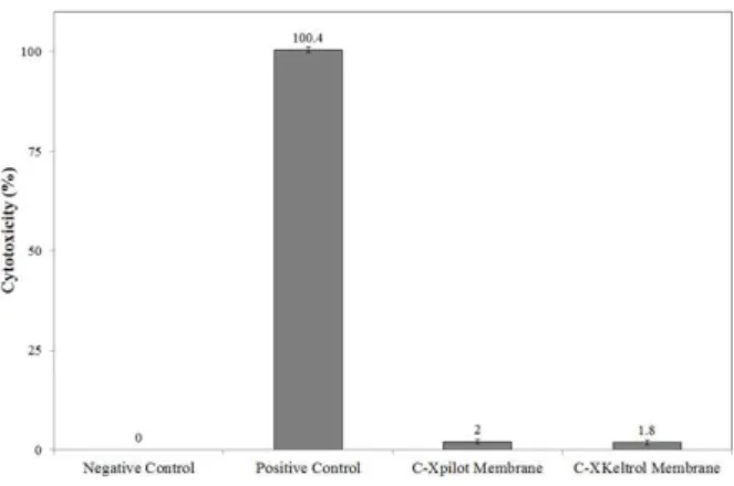
with the cells. Figure 3 shows the results obtained in the in vitro toxicity tests of extracts prepared from C-Xpilot and C-XKeltrol films to L929 cells. The films produced showed minimal cytotoxic effects regardless of the type of xanthan used, in agreement with observations previously reported in the literature regarding biocompatibility and safety of the complex chitosan-xanthan gum in the form of microspheres for controlled drug release (Chellat et al. 2000) and even in comparison with dense or porous formulations prepared with analytical grade xanthan (Bellini et al. 2012).
These results indicated that xanthan from
fermentation processes performed with different microbial strains and culture media could be more explored for the production of high added value materials such as those with direct use in the area of human health. Efforts in this direction would contribute to the expansion of the application fields of this biopolymer beyond the already well-established ones in various industrial sectors.
Figure 3 - In vitro cytotoxicity of the extracts of the films to L929 cells (0.05 g of C-X film per milliliter of supplemented RPMI culture medium). Extracts of culture plate fragments and of latex (in the form of amber tourniquet tubes regularly used in blood drawing) were used as negative and positive controls of cytotoxicity, respectively, and culture medium without cells was used as blank.
CONCLUSIONS
In this work, films prepared by combining chitosan to xanthan gum of different sources were
analyzed and their physico-chemical and
Bellini, M. Z. et al. 298
did not significantly affect most of the properties of the biomaterial obtained, even when the films were compared with the membranes of chitosan and xanthan gum of analytical grade. Thus, despite
polymers of natural origin might present
considerable variability depending on their
provenience, the production of xanthan-gum based biomaterials seemed to show improved flexibility regarding the choice of raw materials and their
suppliers, consequently expanding the
opportunities for more economically attractive processes and biomaterials.
ACKNOWLEDGEMENTS
The authors thank the São Paulo Research Foundation (FAPESP, Brazil) and the Ibero-American Programme for Science, Technology and Development (CYTED, Spain) for the support to this work, as well as the Coordination for the Improvement of Higher Educational Personnel (CAPES, Brazil) and the National Council for Scientific and Technological Development (CNPq, Brazil) for M. Z. Bellini’s and Â. M. Moraes’ fellowships, respectively.
REFERENCES
American Society for Testing and Materials ASTM D882–95a: Standard test methods for tensile properties of thin plastic sheeting, 1995.
Argin-Soysal S, Kofinas P, Lo YL. Food Hydrocolloids. 2014; 40: 138-144.
Bejenariu A, Popa M, Cerf DL, Picton L. Stiffness xanthan hydrogels: synthesis, swelling characteristics and controlled release properties. Polym Bull. 2008; 61: 631-641.
Bellini, MZ, Pires, ALR, Vasconselos MO, Moraes AM. Comparison of the Properties of Compacted and Porous Lamellar Chitosan–Xanthan Membranes as Dressings and Scaffolds for the Treatment of Skin Lesions. J Appl Polym Sci. 2012; 125: E421-E431. Bueno C, Moraes AM. Development of porous lamellar
chitosan-alginate membranes: effect of different surfactants on biomaterial properties. J Appl Polym Sci. 2011; 122: 624-631.
Carignatto RRC, Oliveira KSM, Lima VMG, Oliva-Neto P. New culture medium to xanthan production by Xantomonas campestris pv. campestris. Indian J Microbiol. 2011; 51(3): 283-288.
Ceulemans J, Vinckier I, Ludwig A. The use of xanthan gum in an ophthalmic liquid dosage form: Rheological characterization of the interaction with mucin. J Pharm Sci. 2002 ; 91(4) : 1117-1127.
Chellat F, Tabrizian M, Dumitriu S, Chornet E, Magny P, Rivard CH, et al. In vitro and in vivo biocompatibility of chitosan-xanthan polyonic complex. J Biomed Mater Res. 2000; 51(1): 107-116. Chen KY, Liao WJ, Kuo SM, Tsai FJ, Chen YS, Huang CY, Yao CH. Asymmetric Chitosan Membrane Containing Collagen I Nanospheres for Skin Tissue Engineering. Biomacromolecules. 2009; 10(6): 1642-1649.
Coma V. Polysaccharide-based biomaterials with antimicrobial and antioxidant properties. Polímeros. 2013; 23(3): 287-297.
Dallan PRM, Moreira PL, Petinari L, Malmonge SM, Beppu MM, Genari SC, et al. Effects of chitosan solution concentration and incorporation of chitin and glycerol on dense chitosan membrane properties. J Biomed Mater Res Part B. 2007; 80B(2): 394-405. Diniz DM, Druzian JI, Audibert S. Production of
xanthan gum by Xanthomonas campestris strains native from bark cocoa or whey. Polímeros. 2012; 22(3): 278-281.
Dyondi D, Webster TJ, Banerjee R. A nanoparticulate injectable hydrogel as a tissue engineering scaffold for multiple growth factor delivery for bone regeneration. Int J Nanomed. 2013; 8: 47-59. Eftaiha AF, El-Barghouthi MI, Rashid IS, Al-Remawi
MM. Compressibility and compactibility studies of chitosan, xanthan gum, and their mixtures. J Mater Sci. 2009; 44: 1054-1062.
Fernandes LL, Resende CX, Tavares DS, Soares GA, Castro LO, Granjeiro JM. Cytocompatibility of chitosan and collagen-chitosan scaffolds for tissue engineering. Polímeros. 2011; 21(1): 1-6.
Fukuda H, Kikuchi Y. Polyelectrolyte complexes of chitosan with carboxymethyldextran. Bull Chemical Soc Japan. 1978; 511: 1142-1144.
Hansen B, Jemec GB. The mechanical properties of skin in osteogenesis imperfecta. Arch Dermatol. 2002; 138(7): 909-911.
Hermansson, A-M. Structuring water by gelation. In: Aguilera JM, Lillford P. Food Materials Science - Principles and Practice. New York: Springer Science + Business Media, 2008. 255-280.
Izawa H, Nishino S, Maeda H, Morita K, Ifuku S, Morimoto M, Saimoto H, Kadokawa JI. Mineralization of hydroxyapatite upon a unique xanthan gum hydrogel by an alternate soaking process. Carbohydr Polym. 2014; 102: 846-851. Jayakumar R, Prabaharan M, Sudheesh Kumar PT, Nair
SV, Tamura H. Biomaterials based on chitin and chitosan in wound dressing applications. Biotechnol Adv. 2011; 29(3): 322-337.
Jussila J, Leppäniemi A, Paronen M, Kulomäki E. Ballistic skin simulant. Forensic Sci Int. 2005; 150(1), 63-71.
Properties of Films for Skin Lesions Therapy 299
Biomed Mater Res. 1990; 24(6): 721-734.
Kucharska M, Niekraszewicz A, Wišniewska-Wrona
M, Brzoza-Malczewska K. Dressing sponges made of chitosan and chitosan-alginate fibrids. Fibres & Textiles in Eastern Europe, 2008; 16(3): 109-113. Kumar AS, Mody K, Jha B. Bacterial
exopolysaccharides - a perception. J Basic Microbiol. 2007 ; 47(2): 103-117.
Lai HL, Abu’khalil A, Craig DQM. The preparation and characterization of drugloaded alginate and chitosan sponges. International Journal of Pharmaceutics. 2003; 251: 175-181.
Liu H, Mao J, Yao K, Yang G, Cui L, Cao Y. A study on a chitosan-gelatin-hyaluronic acid scaffold as artificial skin in vitro and its tissue engineering applications. J Biomater Sci Pol Ed. 2004; 15(1):25-40.
Ludwig A. The use of mucoadhesive polymers in ocular drug delivery. Adv Drug Delivery Rev. 2005; 57: 1595-1639.
Ma J, Wang H, He B, Chen J. Biomaterials. 2001; 22(4): 331-336
Ma L, Gao C, Mao Z, Zhou J, Shen J, Hu X, et al. Collagen/chitosan porous scaffolds with improved biostability for skin tissue engineering. Biomaterials. 2003; 24(26): 4833-4841.
Malafaya PB, Silva GA, Reis RL. Natural–origin polymers as carriers and scaffolds for biomolecules and cell delivery in tissue engineering applications. Adv Drug Delivery Rev. 2007; 59: 207-233.
Mayer L, Vendruscolo CT, Silva WP, Moura AB. Production, rheological properties and chemical composition of xanthan produced by Xanthomonas axonopodis pv. phaseoli. Rev Bras Tecnol Agroind. 2008; 2: 87-94.
Oliva-Neto P, Lima VMG, Carignatto CRR, Carvalho AFA. Produção biotecnológica do biopolímero goma xantana. Microbiologia in foco. 2011; 4: 14-22. Oliveira LHS, Dias FG, Duarte ICS, Oliva-Neto P,
Cruz R, Moreira AS, et al. Isolamento e caracterização de bactérias produtoras de goma xantana. Rev Científica Plural. 2000; 1: 115-120. Oliveira PD, Vendruscolo CT, Borges CD, Michel RC,
Lomba RT. Comparative evaluation of xanthans properties produced by pathovar pruni and clairana with commercial xanthan to predict their uses. Polímeros. 2013; 23(3): 417-424.
Pajoum-Shariati SR, Shokrgozar MA, Vossoughi M, Eslamifar A. In vitro co-culture of human skin keratinocytes and fibroblasts on a biocompatible and biodegradable scaffold. Iran Biomedical J. 2009; 13(3): 169-177.
Popa N, Novac O, Profire L, Lupusoru CE, Popa MI. Hydrogels based on chitosan-xanthan for controlled release of theophylline. J Mater Sci Mater Med. 2010; 21: 1241-1248.
Ragetly GR, Griffon DJ, Chung YS. The effect of type II collagen coating of chitosan fibrous scaffolds on
mesenchymal stem cell adhesion and chondrogenesis. Acta Biomater. 2010; 6: 3988-3997
Rodd AB, Dunstan DE, Ross-Murphy SB, Boger DV. Dependence of linear viscoelastic critical strain and stress values on extent of gelation for a thermally activated gelling system. Rheol Acta. 2001; 40:23-29. Rodrigues AP, Sanchez SEM, Costa AC, Moraes AM.
The influence of preparation conditions on the characteristics of chitosan-alginate dressings for skin lesions. J Appli Polym Sci. 2008; 109: 2703-2710. Santos JE, Soares JP, Dockal ER, Campana Filho SP,
Cavalheiro ETG. Characterization of commercial chitosan from different suppliers. Polímeros. 2003; 13(4): 242-249.
Schuster E, Eckardt J, Hermansson A-M, Larsson A, Lorén N, Altskär A, et al. Microstructural, mechanical and mass transport properties of isotropic and capillary alginate gels. Soft Matter. 2014; 10: 357-366.
She Z, Jin C, Huang Z, Zhang B, Feng Q, Xu V. Silk fibroin/chitosan scaffold: preparation, characterization, and culture with HepG2 cell. J Mat Sci Mater Med. 2008; 19: 3545-3553.
Silva GA, Ducheyne P, Reis RL. Materials in particulate form for tissue engineering. Basic concepts. J Tissue Eng Regener Med. 2007; 1(1): 4-24.
Silva MLA, Crawford A, Mundy JM, Correlo VM, Sol P, Bhattacharya M, et al. Chitosan/polyester-based scaffolds for cartilage tissue engineering: assessment of extracellular matrix formation. Acta Biomater. 2010; 6: 1149-1157.
Veiga IG, Moraes AM. Study of the swelling and stability properties of chitosan-xanthan membranes. J Appl Polym Sci. 2012;124: E154-E160.
Wang L, Khor E, Lim LY. Chitosan-alginate-CaCl2 system for membrane coat application. Journal of Pharmaceutical Sciences, J Pharm Sci. 2001; 90(8): 1134-1142.
Wang L, Khor E, Wee A, Lim LY. Chitosan-alginate PEC membrane as wound dressing: Assessment of incisional wound healing. J Biomed Mater Res. 2002; 63(5): 610-618.
Yan X, Khor E, Lim LY. PEC films prepared from chitosan-alginate co-acervates. Chem Pharm Bull. 2000; 48(7): 941-946
Yang B, Yin Z, Cao J, Shi Z, Zhang Z, Song H, et al. In vitro cartilage tissue engineering using cancellous bone matrix gelatin as a biodegradable scaffold. Biomed Mater. 2010: 5(4): 1-8.
Yin Y, Li Z, Sun Y, Yao K. A preliminary study on chitosan/gelatin polyelectrolyte complex formation. J Mater Sci. 2005; 40: 4649-4652.
Zeng X, Ruckenstein E. Control of pore sizes in macroporous chitosan and chitin membranes. Ind Eng Chem Res. 1996; 35(11): 4169-4175.