UNIVERSIDADE DA BEIRA INTERIOR
Ciências da Saúde
Development of new biomaterials with
antibacterial properties for future application in
regenerative medicine
Tânia Sofia dos Santos Vieira
Master Degree Thesis in
Biomedical Sciences
(2
ndcycle of studies)
Supervisor: Prof. Ilídio Joaquim Sobreira Correia (PhD)
UNIVERSIDADE DA BEIRA INTERIOR
Ciências da Saúde
Desenvolvimento de novos biomateriais com
propriedades antibacterianas para futuras
aplicações em medicina regenerativa
Tânia Sofia dos Santos Vieira
Dissertação para obtenção do Grau de Mestre em
Ciências Biomédicas
(2
ºciclo de estudos)
Orientador: Prof. Ilídio Joaquim Sobreira Correia (PhD)
iv
“Valeu a pena? Tudo vale a pena Se a alma não é pequena.
Quem quer passar além do Bojador Tem que passar além da dor.
Deus ao mar o perigo e o abismo deu, Mas nele é que espelhou o céu.”
v
Acknowledgments
First of all, I would like to thank my supervisor Professor Ilídio Correia for the opportunity to develop this project and for all the support, guidance and help. Furthermore, I would like to thank him for whatever he did to ensure all the necessary conditions for the development of this project.
I would like to thank to Eng. Ana Paula from the Optics center of Universidade da Beira Interior for the help in acquiring the lots of scanning electron microscopy images of the produced nanoparticles.
In addition, I would like to thank to all of my group colleagues for all the teaching and support during these last months. Their friendship was really important to overcome all the difficulties faced.
I also thank to all my friends that always give me support and advices and that had lots of patience with me in the bad or in the good moments of life, not only during the academic life, but also during my entire life. They all marked in a special way my life.
Finally, special thanks from the bottom of my heart to my parents and sister for granted me the possibility to make this master degree thesis. I thank for all the education, the advices, the support and the love. Thank you for always believe in me even when myself did not. Thank you for always help me in discover my way.
vii
Abstract
Bacterial infections have been a constant threat to human health throughout the history. Bacterial colonization of biomedical devices and implants causes enormous problems for healthcare systems worldwide, costs and increases patient’s suffering. Silver has been known, since the antiquity, by their antimicrobial properties and was used to produce reservoirs of food and with medical purposes. With the development of nanotechnology, silver nanoparticles have attracted the attention of different researchers due to their properties, as antimicrobial properties and high surface to volume ratio. However, these nanoparticles can form aggregates, which have toxic effects to the human cells. Recently, silver nanoparticles have been stabilized with several polymers and surfactants in order to avoid these problems.
In this work, silver nanoparticles were produced and stabilized with chitosan/dextran. The produced nanoparticles were characterized by Scanning Electron Microscopy, Ultraviolet-Visible and Fourier Transform Infrared spectroscopy. Furthermore, the antibacterial activity of the produced nanoparticles was evaluated and it was found that they are effective in the prevention of the growth of Escherichia coli through a minimum inhibitory concentration. These particles were also studied in contact with human osteoblast cells in order to ascertain if the particles that had an antibacterial effect to the bacteria do not have a toxic effect for human cells. The results herein obtained revealed that the nanoparticles can be used in a near future as a coating material of medical devices in order to avoid their bacterial colonization.
Keywords
Antibacterial mechanism; Chitosan/dextran nanoparticles; Escherichia coli; Nanotechology; Silver nanoparticles.
ix
Resumo
As Infecções bacterianas têm constituído uma preocupação constante para a saúde humana. A colonização da superfície dos dispositivos biomédicos e implantes pelas bactérias é a causa de algumas infecções sofridas pelos pacientes, contribuindo para o agravamento dos custos e do sofrimento do paciente. A prata é conhecida desde a antiguidade pelas suas propriedades antibacterianas e foi utilizada na produção de dispositivos de armazenamento de comida e para o tratamento de algumas doenças. Com o desenvolvimento da nanotecnologia, as nanoparticulas de prata têm atraído a atenção de diferentes investigadores devido às propriedades que apresentam, como propriedades antimicrobianas e elevada razão entre a área de superfície e o volume. Contudo, estas nanoparticulas podem sofrer um processo de agregação e produzir efeitos tóxicos para as células humanas. De modo a superar estes problemas, as nanoparticluas de prata têm sido estabilizadas por diversos polímeros e surfactantes.
Neste trabalho, as nanoparticulas de prata foram produzidas e estabilizadas com quitosano/dextrano de modo a evitar a agregação das mesmas. As nanoparticulas produzidas foram caracterizadas por Microscopia Electrónica de Varrimento, Espectroscopia do Ultravioleta-Visível e Espectroscopia de Infravermelho. A actividade antibacteriana das nanoparticulas produzidas foi avaliada na prevenção do crescimento de Escherichia coli, através dos valores da determinação da concentração inibitória mínima. O perfil de citotoxicidade das nanoparticulas foi caracterizado através da utilização de osteoblastos humanos. Estas partículas na concentração inibitória mínima não apresentam efeito tóxico para os osteoblastos humanos, devido ao crescimento e proliferação das células na presença destas nanoparticulas. Os resultados obtidos revelam que estas nanopartículas podem ser usadas no revestimento de dispositivos médicos, prevenindo a sua colonização por bactérias.
Palavras-chave
Escherichia coli; Mecanismo antibacteriano; Nanoparticulas de prata; Nanoparticulas de quitosano/dextrano; Nanotecnologia.
xi
Table of Contents
1. Introduction ... 2
1.1. Bacterial infections that affect human beings ... 2
1.1.1. Bacterial infections caused by biomaterials implantation ... 3
1.2. Nanotechnology ... 5
1.3. Silver Nanoparticles ... 7
1.3.1. Applications of silver nanoparticles ... 9
1.3.2. Mechanisms of action of silver nanoparticles ... 11
1.3.3. Bacterial silver nanoparticles resistance ... 13
1.3.4. Toxicity of silver nanoparticles ... 14
1.3.5. Combination of silver nanoparticles with other materials ... 15
1.3.5.1. Dextran ... 16
1.3.5.2. Chitosan ... 17
1.4. Objectives ... 19
2. Materials and Methods ... 21
2.1. Materials ... 21
2.2. Methods ... 21
2.2.1. Preparation of silver nanoparticles ... 21
2.2.2. Preparation of the Chitosan/Dextran nanoparticles ... 21
2.2.3. Preparation of the Chitosan/Dextran nanoparticles with silver ... 22
2.2.4. Nanoparticles Scanning Electron Microscopy Analysis ... 22
2.2.5. Nanoparticles Ultraviolet-Visible Spectroscopy Analysis ... 22
2.2.6. Nanoparticles Fourier Transform Infrared Spectroscopy Analysis ... 23
2.2.7. Determination of antibacterial activity ... 23
2.2.7.1. Minimum Inhibitory Concentration and Minimal Bactericidal Concentration . 23 2.2.8. Proliferation of cells in the presence of the produced nanoparticles ... 24
2.2.9. Evaluation of the cytotoxic profile of the produced nanoparticles ... 24
2.2.10. Statistical Analysis of MTS results ... 25
3. Results and Discussion ... 27
3.1. Characterization of Particles Morphology ... 27
3.2. Nanoparticles Ultraviolet-Visible Spectroscopy Analysis ... 33
3.3. Nanoparticles Fourier Transform Infrared Spectroscopy Analysis ... 35
xii 3.5. Evaluation of the cytotoxic profile of the produced nanoparticles ... 42
4. Conclusions and future perspectives ... 50 5. Bibliography ... 53
xiv
List of Figures
Chapter I – Introduction
Figure 1 – Applications of silver and AgNPs in medicine. ... 10
Figure 2 – Different mechanisms of action of AgNPs against bacteria... 13
Figure 3 – Representation of the dextran chemical structure. ... 17
Figure 4 – Representation of the chitosan chemical structure ... 18
Chapter III – Results and Discussion
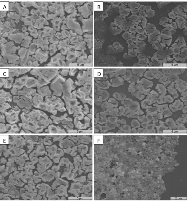
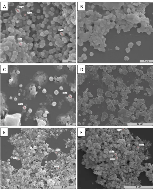
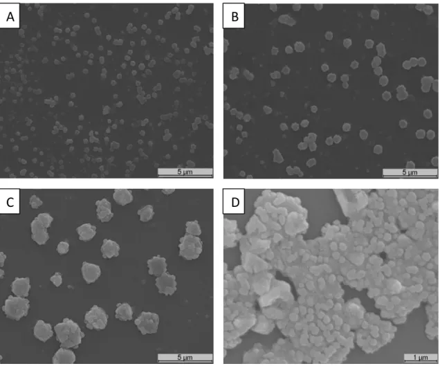
Figure 5 – SEM images of AgNPs ... 28Figure 6 – SEM images of chitosan/dextran nanoparticles produced with low molecular weight chitosan... 29
Figure 7 – SEM images of chitosan/dextran nanoparticles produced with high molecular weight chitosan... 31
Figure 8 – SEM images of chitosan/dextran nanoparticles with AgNPs and AgNPs produced in chitosan/dextran nanoparticles with NaBH4 and with C6H8O6. ... 33
Figure 9 – Ultraviolet-Visible spectra of the produced AgNPs . ... 35
Figure 10 – FT-IR spectra of chitosan, dextran, and chitosan/dextran nanoparticles ... 37
Figure 11 – FT-IR spectra of chitosan/dextran nanoparticles with AgNPs and AgNPs produced in chitosan/dextran nanoparticles with NaBH4 and with C6H8O6 ... 38
Figure 12 – Determination of MIC and MBC by the microdilution method in microplate. ... 40
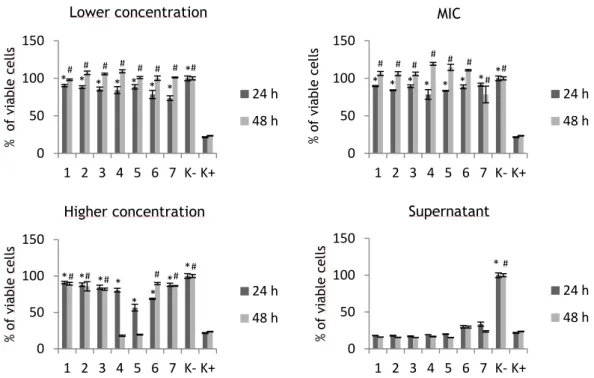
Figure 13 – Inverted Light Microscope Images of human osteoblast cells in contact with nanoparticles of lower and MIC concentrations after 24 and 48 h ... 45
Figure 14 – Inverted Light Microscope Images of human osteoblast cells in contact with nanoparticles of higher concentrations and their supernatants after 24 and 48 h ... 46
Figure 15 – Cellular activities measured by the MTS assay after 24 and 48 h in contact with nanoparticles in lower, MIC, higher concentrations and their supernantants ... 48
xvi
List of Tables
Chapter I – Introduction
Table 1 – Commercially available medical products containing AgNPs. ... 11
Chapter III – Results and Discussion
Table 2 – Formation of chitosan/dextran nanoparticles for different ratios. ... 32 Table 3 – MIC obtained for the different tested nanoparticles. ... 39 Table 4 –MBC obtained for the different tested nanoparticles. ... 40 Table 5 – Different nanoparticles and their concentrations to perform the cytotoxic assays. .... 44
xviii
List of Acronyms
Ag0 Metallic silver
Ag+ Silver ions
[Ag(NH3)2]+ Diamminesilver ions
AgNO3 Silver nitrate
AgNPs Silver nanoparticles ATP Adenosine triphosphate
ATSDR Agency for Toxic Substances and Disease Registry CFU Colony-forming unit
C6H8O6 Ascorbic acid
DMEM-F12 Dulbecco’s modified eagle’s medium DNA Deoxyribonucleic acid
E. coli Escherichia coli
EtOH Ethanol
FBS Fetal bovine serum
FT-IR Fourier Transform Infrared HIV Immunodeficiency virus
K- Negative control
K+ Positive control
LB Luria Bertani
LPS Lipopolysaccharides
MBC Minimum Bactericidal Concentration MIC Minimum Inhibitory Concentration
MTS 3-(4,5-dimethylthiazol-2-yl)-5-(3-carboxymethoxyphenyl)-2-(4-sulfophenyl)-2H-tetrazolium
NaBH4 Sodium borohydride
Na3C6H5O7 Sodium citrate
NCCLS National Committee for Clinical Laboratory Standards PBS Phosphate buffered saline
PEG Poly(ethylene glycol) PEI Poly(ethylene-imine) PMS Phenazine Methosulfate PVA Poly(vinyl alcohol) PVP Poly(vinylpyrrolidone) ROS Reactive oxygen species S. aureus Staphylococcus aureus SDS Sodium dodecylsulfate SEM Scanning Electron Microscopy
xix SPR Surface Plasmon Resonance
Chapter I
Introduction
Chapter I - Introduction
2
1. Introduction
1.1. Bacterial infections that affect human beings
Bacterial infections have been a constant threat to human health throughout history (Vasilev et al. 2010). Human beings are often infected by different microorganisms such as bacteria, molds, yeasts, and viruses (Shahverdi et al. 2007; da Silva Paula et al. 2009). In the beginning of the 20th century, infectious diseases were the main cause of death worldwide (Huh
et al. 2011). Examples of these diseases are the bubonic plague, tuberculosis, malaria, and the acquired immunodeficiency syndrome pandemic caused by the human immunodeficiency virus (HIV) that have affected a substantial number of patients worldwide, causing significant morbidity and mortality (Tenover 2006). In the middle of the 20th century, the development of
new antibiotics and other methods to control infections helped the humans to prevent and treat from several diseases (Tenover 2006). All these advances began when Flemming, in 1928, discovered the first antibiotic that he called penicillin (Ligon 2004). Antibiotics are defined as chemical substances that are produced by a microorganism, that have the capacity, in dilute solutions, to selectively inhibit the growth of or to kill other microorganisms (Collier 2004). The penicillin was extracted from a plant of the genus Penicillium (Ligon 2004) and their commercial production began in the late 1940s (Kalishwaralal et al. 2010; Huh et al. 2011). The use of antibiotics had a great success in 70th and 80th decades of the 20th century (Huh et al. 2011;
Prucek et al. 2011), when newer and even strong antibiotics were developed (Huh et al. 2011). However, the development of antimicrobial drugs contribute to the current crisis in fighting against multi-drug resistance bacterial strains (Huh et al. 2011), where initially susceptible populations of bacteria become resistant to an antibacterial agent and proliferate and spread under the selective pressure of use of that agent, leading to the need of development of new antibiotics (Tenover 2006; Xu et al. 2011). The mechanisms of antibiotics resistance are spread in a variety of bacterial genera (Tenover 2006). These mechanisms are the result of the acquisition of genes encoding enzymes by the organism, such as β-lactamases, that destroy the antibacterial agent before it can have an effect. Furthermore, the bacteria may acquire efflux pumps that extrude the antibacterial agent from the cell, before it can reach its target site and exert its effect. Finally, bacteria may acquire several genes for a metabolic pathway which produces altered bacterial cell walls that do not present the binding site of the antimicrobial agent, or bacteria may acquire mutations that limit the access of the antimicrobial agents to the intracellular target site via downregulation of porin genes (Stewart et al. 2001; Tenover 2006; Xu et al. 2011). Currently, the treatment of bacterial infections using classical antibiotics is becoming a serious global health problem (Sondi et al. 2004; Kim et al. 2007; Ghosh et al. 2010; Liu et al. 2010; Potara et al. 2011; Prucek et al. 2011) due to the fact that most of the prominent infectious disease agents are resistant to all the antibiotics presently available (Xu et
Chapter I - Introduction
3 al. 2011). As an example, almost all known antibiotics are ineffective against the MDM-1 bacteria, which was discovered recently (Prucek et al. 2011). So far, many efforts have been done to develop effective and safe antibacterial drugs against bacteria (Potara et al. 2011). Although a large number of natural and synthetic antibiotics have already been reported in the literature (Liu et al. 2010), most of them are not effective against bacteria or have safety concerns (Liu et al. 2010). As an example, the Grepafloxacin and Trovafloxacin were removed from the market in several countries due to their secondary effects to humans (Liu et al. 2010).
1.1.1. Bacterial infections caused by biomaterials implantation
Over the past half century, advancements in the use of natural and synthetic biomaterials and the improvement of surgical techniques have led to an increase in the demand of biomaterials to be used in implants and medical devices production (Simchi et al. 2011). The biomaterials market is estimated to be worth more than 300 billion US Dollars and to be increasing 20% per year (Simchi et al. 2011). Medical specialists treat millions of patients every year using different implanting devices, like pacemakers, artificial hip joints, breast implants, dental implants, hearing devices and skin substitutes (Simchi et al. 2011). In orthopedic surgery, different implant materials have been used (Campoccia et al. 2010). This implanted materials ranging from internal to percutaneous and from resorbable to long-term ones (Campoccia et al. 2010). They included prostheses, soft moldable or injectable cavity-fillings, hard and heavy weight-bearing metallic, ceramic or polymeric biomaterials, allogeneic bone, viable tissue grafts, and also include the recent tissue engineering products, such as scaffolds, drug delivery systems, among others (Campoccia et al. 2010). In 2010 more than 4.4 million people had one internal fixation device, and 1.3 million people have an artificial joint (Simchi et al. 2011).
Even though very significant advances in microbiology field (Vasilev et al. 2010), one problem that is common to all medical devices (Campoccia et al. 2010; Fernebro 2011), is their colonization with bacteria or fungi, which cause infections in the host (Nava-Ortiz et al. 2010). Depending upon the location and type of the medical device, bacterial infections may result in morbidity or even in patient dead (Jones et al. 2008; Nava-Ortiz et al. 2010). Thus, sometimes prosthesis removal and replacement is the only option to definitively eradicate severe infections and avoid patient dead (Campoccia et al. 2006). An additional problem is the risk of re-infection in a second implantation, which usually occurs in 1-2% of the patients, depending on type of prosthetic implant, patient condition, clinical setting, and surgical procedure (Campoccia et al. 2010). As an example, the infection rate for total hip arthroplasties has been reported to occur in 0,5-3% of the cases, and the rate of reinfection, after revision of infected hip prostheses, is up to 14% (Campoccia et al. 2010). These drastic interventions bear obvious implications in terms of attendant patient trauma, prolonged hospitalization as well as in terms of health and social costs (it has been estimated that the treatment of each single episode of infected arthroplasty costs more than $50,000) (Campoccia et al. 2006; Vasilev et al. 2010).
Chapter I - Introduction
4 Implant-associated infections are the result of bacteria adhesion to an implant surface and subsequent biofilm formation at the implantation site (Jones et al. 2008; Simchi et al. 2011). Biofilms are communities of microbial cells that attach to a surface and secrete a hydrated extracellular polymeric matrix (Gurjala et al. 2011). The organisms become embedded in this matrix, which is composed of polysaccharides, proteins, glycoproteins, glycolipids, and extracellular deoxyribonucleic acid (DNA) (Gurjala et al. 2011). The matrix polymers support microcolonies of cells, allows cell–cell communication, forms water channels, retains and concentrates nutrients, and can support gene transfer through conjugation, transformation, and transduction (Edwards et al. 2004; Martin et al. 2009). The matrix is thereby responsible for the maintenance of the structural integrity of the biofilm and provide an ideal matrix for bacterial cell growth (Monteiro et al. 2009). The biofilm formation is described as a sequence of different steps. In the first one, microbial cells adhere to the biomaterial surface, through exopolysaccharides that are synthesized by the bacteria (Speranza et al. 2004; Kalishwaralal et al. 2010). Surface adhesion is a critical step in the pathogenesis of implant-related infections and represents the beginning of the colonization of biomaterial surfaces (Montanaro et al. 2011). Thereafter, it follows the accumulation in multiple cell layers, biofilm maturation, and detachment of cells from the biofilm into a planktonic state to initiate a new cycle of biofilm formation elsewhere (Speranza et al. 2004; Montanaro et al. 2011).
The biofilm allows microbes to survive to host immune defenses and systemic antibiotic therapies (Campoccia et al. 2010), which is the main reason for the high prevalence of infections (Fernebro 2011). Therefore, extraordinary antibiotic resistance is a general feature of biofilm which is caused by several factors (Simchi et al. 2011). These factors include the compact nature of biofilm structures, the presumed reduced rates of cellular growth and cellular respiration of the bacteria in the biofilm and the protection conferred by biofilm matrix polymers (Simchi et al. 2011). The antibiotic resistance of biofilms is also related to the fact that in the extracellular polymeric substance cells are allowed to change their proteome (up to 50% of the proteome may differ from the same microorganism in a planktonic state) to their existence in a sessile state (Nava-Ortiz et al. 2010). This state has low metabolic levels and downregulated cell activity, which leads a decreased antimicrobial susceptibility compared with planktonic cells (Nava-Ortiz et al. 2010). Furthermore, the alteration of the proteome makes that bacteria in a biofilm express different sets of genes than those that are expressed in its planktonic form (Martin et al. 2009). This have important implications for clinical therapeutics, since the antimicrobials does not reach the bacterial cells in the biofilm (Martin et al. 2009). Thus, in the biofilm, after bacteria colonization, the resistance to antimicrobial agents is dramatically increased (up to 1000-fold) and even the antimicrobial agents that are effective against planktonic cells are ineffective against the same bacteria growing in a biofilm (Jones et al. 2008; Martin et al. 2009; Monteiro et al. 2009; Simchi et al. 2011). Thereby, biofilm leads to an undesirable and deteriorative impact to several fields, such as in medicine, industry, and commercial products (Inphonlek et al. 2010).
Chapter I - Introduction
5 Due to these facts, it is important the use of antibacterial agents to inhibit bacterial adhesion, in order to prevent implant-associated infections (Kim et al. 2007; Inphonlek et al. 2010; Simchi et al. 2011). In order to achieve this purpose, medical devices with different antibiotics incorporated such as gentamicin, norfloxacin, nitrofurazone, minocycline, and rifampin have been produced, to avoid biofilm formation (Dave et al. 2011). However, most of these coatings only allow short release profiles, making them inappropriate for relatively long-term use (Dave et al. 2011). Furthermore, some antimicrobial agents are extremely irritant and toxic to the human being (Sondi et al. 2004), which emphasizes the need to develop antimicrobial materials to be applied in health and biomedical device, food, and personal hygiene industries (Häntzschel et al. 2009; Vasilev et al. 2010). These materials must be cost-effective, avoid bacterial resistance for them, have ability to act against a wide spectrum of bacteria, have high levels of bactericidal and bacteriostatic activity, be safe for the environment and be biocompatible for eukaryotic cells (Kim et al. 2007; Chaloupka et al. 2010; Fayaz et al. 2010; Inphonlek et al. 2010).
Since the antibiotic resistance is growing up (Nagy et al. 2011) and this has become a major issue in public healthcare (Mohammed Fayaz et al. 2009), there is a renewed interest in the development of products containing silver, since these have antimicrobial properties (Arora et al. 2008; Monteiro et al. 2009; Madhumathi et al. 2010; Nagy et al. 2011). In fact, the antibiotic-resistant pathogens has led to the resurgence of silver-based materials with antibacterial agents purpose(da Silva Paula et al. 2009; Fayaz et al. 2010; Kalishwaralal et al. 2010; Huh et al. 2011; Nagy et al. 2011), due to their antimicrobial activity against a large number of microorganisms and far lower propensity to induce microbial resistance than that of antibiotics (Arora et al. 2008; Fayaz et al. 2010). Thus, the incorporation of silver in topical dressings or as coating material on medical products may therefore play an important role in the era of antibiotic resistance (Ip et al. 2006). However, silver has high toxicity for the human being and in order to solve this problem, nanoscale materials have emerged as novel effective alternative to be used as antimicrobial agents (Sondi et al. 2004; Rai et al. 2009).
1.2. Nanotechnology
Nanotechnology is an area of science that appeared in the 20th century (Lu et al. 2008). It
is emerging as a rapid growing field with applications in Science and Technology at the nanoscale level (Rai et al. 2009). The term Nanotechnology was created by Professor Norio Taniguchi of Tokyo Science University in 1974, to describe precision of manufacturing materials at the nanometer level (Rai et al. 2009). But the concept of Nanotechnology was given previously, in 1959, by physicist Professor Richard P. Feynman in his lecture “There’s plenty of room at the Bottom” (Rai et al. 2009). The term Nanotechnology is derived from the word “nano” (Rai et al. 2009). “Nano” is a Greek word synonymous to dwarf meaning extremely small, used to indicate one billionth of a meter or 10−9 m (Rai et al. 2009). Nanoscale is taken to include active
Chapter I - Introduction
6 components or objects in the size range of 1–100 nm (Cumberland et al. 2009; Rai et al. 2009; Kurek et al. 2011).
Bionanotechnology has emerged up as integration between biotechnology and nanotechnology, to allow the development of biosynthetic and environmental-friendly technology for synthesis of nanomaterials (Rai et al. 2009). Nanotechnology allowed the development of several materials, devices and systems (Öztürk et al. 2008; Türkmen et al. 2009). Among the three areas mentioned above, the area of nanomaterials is the most advanced at present, both at the scientific level and for commercial applications (Öztürk et al. 2008; Cumberland et al. 2009; Türkmen et al. 2009). Nowadays nanomaterials are used for the development of novel devices that can be used in various physical applications as biophotonics, biosensors, fuel cells, photovoltaic devices, semiconductor nanowires, solar energy conversion, and also in catalysis, water treatment, and biological, biomedical and pharmaceutical applications (Raffi et al. 2008; Babu et al. 2010). Nanomaterials display unique and superior properties, like higher surface to volume ratio, increased percentage of atoms at the grain boundaries and the predominance of quantum effects instead of gravitational ones. This distinct properties are unavailable in conventional macroscopic materials (Raffi et al. 2008).
In the medical field, nanomaterials may provide a reliable and effective tool to treat diseases at a molecular level (Chouhan et al. 2009). This fact is important since their dimensions are close to that of the cellular components and biological molecules (Hung et al. 2007). Among the various types of nanomaterials, nanoparticles have attracted much attention in the present century due to the defined chemical, optical and mechanical properties (Rai et al. 2009). They need to be formulated with improved bioavailability and release rates, which can decrease required dosages while increasing safety and reducing side effects (Wong et al. 2009). However, on the other hand, several studies suggested that nanoparticles can cause injuries in the biological systems (Yen et al. 2009), since the similar size of nanoparticles to the cellular components make them bypass the natural barriers, such as the cell membranes, causing harmful effects to living cells (Wong et al. 2009; Yen et al. 2009). There is no concern, until now, about the interaction of nanoparticles with the living cells and the results in this field are very controversial (Arvizo et al. 2012). Nevertheless, nanoparticles have been used as drug delivery systems, as biomolecular sensing molecules, as targeted imaging, and as thin film coatings (Hung et al. 2007). The advances in the use of nanostructured materials for medical applications are possible due to the availability of novel techniques of processing, characterization and modeling and also the technology for manipulation and manufacturing of the nanostructured materials (Brigmon, Berry et al. 2010). Moreover, the production of devices with specific functionalities is obtained by the specific interactions between biological structures (e.g., tissues and other cellular processes) and nanostructured materials (Brigmon et al. 2010).
Inorganic nanoparticles, of either simple or composite nature, are a type of nanoparticles that have been receiving considerable attention as a result of their unique properties like chemical, electronic, magnetic, optical, and physical, including also antimicrobial and catalytic activity (Shahverdi et al. 2007; Guzman et al. 2011), which are different from those of the bulk
Chapter I - Introduction
7 materials (Yin et al. 2005; Guidelli et al. 2011). These special and unique properties could be attributed to their small sizes and large specific surface area (Guzman et al. 2011). All of this makes the inorganic nanoparticles adequate for applications in biomedicine, catalysis, electronics, energy science, magnetic, mechanics, optics, and so on (Shahverdi et al. 2007; Fayaz et al. 2010). A number of recent works in this field, describe the possibility of generating new types of nanostructured inorganic materials with designed surface and structural properties (Sondi et al. 2004). Thus, the preparation, characterization, surface modification, and functionalization of nanosized inorganic particles open the possibility to formulate a new generation of bactericidal materials to avoid microbial biofilm formation on biomaterials surface (Sondi et al. 2004; Lipovsky et al. 2011). Nanoparticles with antibacterial properties offer many distinctive advantages with respect to the therapy with antibiotics since they allow the reduction of in vivo toxicity, overcoming the problem of resistance to the antibiotics, and lowering the cost associated with their production (Huh et al. 2011).
Among the different types of nanoparticles, the metallic ones are the most promising candidates for this purpose, since they show good antibacterial properties (Ruparelia et al. 2008; Rai et al. 2009), due to their high specific surface area, high fraction of surface atoms (Hung et al. 2007; Shahverdi et al. 2007; Martin et al. 2011), and small size. These properties allow nanoparticles to interact closely with cellular membranes of the bacteria. In addition to these characteristics, this kind of nanoparticles release metal ions in solution, which increases the antibacterial properties (Ruparelia et al. 2008). Another properties of these nanoparticles are the long life and the heat resistance (Potara et al. 2011). Well-known metallic nanoparticles with these properties are the silver nanoparticles (AgNPs).
1.3. Silver Nanoparticles
The unique antimicrobial properties of silver in the treatment of infections have been known for a long time (Gurunathan et al. 2009; Häntzschel et al. 2009; Mohammed Fayaz et al. 2009). Since 1000 BC, Egyptians, Greeks, Romans and other ancient civilizations used silver vessels to store perishable foods, to produce silver cutlery, glassware and dishes (Vasilev et al. 2010). Silver was also used with medical purposes for rheumatism, tetanus, gonorrhea and wound healing treatment (Vertelov et al. 2008). In the 18th century, silver nitrate (AgNO
3) was used for
the treatment of venereal diseases, fistulae from salivary glands, bone abscesses (Rai et al. 2009), and ulcers (Neal 2008). Dilute solutions of AgNO3 have been used since the 19th century in
treatment of infections and burns (Ip et al. 2006). Due to the successful of the registered cases, in 1920s silver was recognized by the United States Food and Drug Administration for its antimicrobial activity and was regulated for wound management (Neal 2008). In 1940s, after penicillin was introduced in the market, the use of silver for the treatment of bacterial infections was reduced (Rai et al. 2009). Silver reappear again in the 1960s when Moyer introduced the use of 0.5% AgNO3 for the treatment of burns as previously done in the 19th
Chapter I - Introduction
8 century (Rai et al. 2009). In 1990s, silver was introduced in a colloidal form (i.e. AgNPs) in ointments that could be applied to open wounds, in order to kill bacteria (Arora et al. 2008). Recently, due to the emergence of antibiotic-resistant bacteria and limitations associated with the use of these medicines, doctors have restarted to use silver, mainly in the form of AgNPs in order to fight different types of infections affecting humans (Rai et al. 2009).
AgNPs are nano-sized structures formed from silver atoms that are metallically bonded together and have a size from approximately 1 nm to 100 nm (Chaloupka et al. 2010; Songsilawat et al. 2010). It has been shown that they can be used in medicine and health-related areas (Cumberland et al. 2009), due to their interesting optical and catalytic properties, high resistance to oxidation and high thermal conductivity (Vertelov et al. 2008; Wong et al. 2009; Prucek et al. 2011). Furthermore, their tunable size, shape and surface chemistry allow them to be designed with specific properties that are critical for several applications (Potara et al. 2011), most typically antimicrobial and sterile applications (Marambio-Jones et al. 2010). Taking into account the antibacterial activity of silver, it is better for AgNPs than for other silver forms, as bulk silver (Potara et al. 2011; Xu et al. 2011), silver ions (Ag+) and other silver salts (Li et al.
2011), even when applied in lower concentrations (da Silva Paula et al. 2009; Domingos et al. 2011). The higher antibacterial activity is due to high specific surface area and high fraction of surface atoms (more than 1000 atoms in one 5 nm particle (Vertelov et al. 2008)) in AgNPs (da Silva Paula et al. 2009), which allows a better contact with microorganisms (Ghosh et al. 2010; Juan et al. 2010; Li et al. 2010; Guzman et al. 2011). Therefore, smaller-sized particles with 1/1000 of the bacterium size (since AgNPs have a nanometer size and bacteria a micrometer size) show stronger antibacterial activity (Guzman et al. 2011; Kurek et al. 2011; Shameli et al. 2011). Furthermore, the nanoparticles release Ag+ in aqueous solutions, which enhance their
bactericidal activity (da Silva Paula et al. 2009; Chaloupka et al. 2010; Juan et al. 2010).
AgNPs due their antimicrobial properties, are capable of kill several microorganisms responsible for 650 types of different diseases (Raffi et al. 2008). These nanoparticles have revealed bactericidal activity against as many as 16 bacteria species (Sondi et al. 2004) either gram-positive or gram-negative (Monteiro et al. 2009; Sheikh et al. 2010), including Escherichia coli (E. coli), Staphylococcus aureus (S. aureus), Bacillus subtilis, Streptococcus mutans, Staphylococcus epidermidis (Li et al. 2011) and highly multiresistant strains such as methicillin-resistant S. aureus (Fayaz et al. 2010). Moreover, they also showed antifungal activity (Kacarevic-Popovic et al. 2007; Häntzschel et al. 2009; Monteiro et al. 2009) against Candida albicans, Candida glabrata, Candida parapsilosis, Candida krusei, and Trichophyton mentagrophytes (Li et al. 2011). More recently, it has also been reported that AgNPs can inactivate virus (Kacarevic-Popovic et al. 2007; Monteiro et al. 2009; Li et al. 2010) like hepatitis B virus, herpes simplex, monkeypox, respiratory syncytial virus (Li et al. 2011), and also exhibit antiviral properties against HIV infected cells (Panác ek et al. 2006; Shameli et al. 2010; Shameli et al. 2011), via preferential binding of the AgNPs to the gp120 glycoprotein knobs through the sulfur-bearing residues of glycoprotein amino acids, thus inhibiting the virus from binding to the target cell membrane receptor (Thomas et al. 2007).
Chapter I - Introduction
9 Besides its antimicrobial activity, it has been recently found that AgNPs reduce cytokine release (Guidelli et al. 2011), decreasing lymphocyte and mast cell infiltration and also induce apoptosis of inflammatory cells (Chaloupka et al. 2010). These characteristics of AgNPs are responsible for the anti-inflammatory effect and contributes for accelerating the epithelialization by over 40% and, as a consequence, accelerate wound healing (Lu et al. 2008; Chaloupka et al. 2010; Kalishwaralal et al. 2010).
1.3.1. Applications of silver nanoparticles
The properties presented by AgNPs make them good candidates to be used in different applications, like in electronics and in sensor design based on the surface-enhanced Raman spectroscopy (SERS) (Badawy et al. 2010; Parashar et al. 2011). Moreover, these nanoparticles have been used in a number of medical applications (Arora et al. 2009), due to the antimicrobial activity owned by the silver-based compounds containing ionic silver or metallic silver (Panác ek et al. 2006; Arora et al. 2008).
Some applications of silver and AgNPs are presented in figure 1. In the case of silver, it is used in the form of AgNO3 solutions to perform cauterization, in order to stop epistaxis and the
growth of post-traumatic granulomas (Chaloupka et al. 2010). AgNO3 is also used to prevent
some infections and to promote an anti-inflammatory effect in the procedure of pleurodesis (Chaloupka et al. 2010). Silver is also used in the form of silver sulfadiazine cream to apply in ulcers and burns to promote the skin regeneration (Chaloupka et al. 2010).
AgNPs have been used as coating material for medical purposes, orthopedic, vascular or dental graft materials (Panác ek et al. 2006; Ruparelia et al. 2008; Vertelov et al. 2008), indwelling catheters (Vasilev et al. 2010), and arthroplasty (Panác ek et al. 2006). AgNPs can be impregnated in wound dressings (Xu et al. 2011), in diabetic ulcers (Ip et al. 2006; Li et al. 2011), in chronic ulcers, and in traumatic injuries in order to prevent infections and enhance wound repair (Ip et al. 2006). Some medical products containing AgNPs available in the market, as wound dressings and catheters, are presented in table 1 (Arora et al. 2009; Chaloupka et al. 2010).
Moreover, silver can also be employed to eliminate microorganisms on textile products, food storage containers, cosmetics in the form of nanogels and nanolotions, contraceptive devices, and they can be used for water filtration too (Monteiro et al. 2009; Shameli et al. 2010; Vasilev et al. 2010; Mirzajani et al. 2011).
Chapter I - Introduction
10 Figure 1 – Applications of silver (right-hand size) and AgNPs (left-hand size) in medicine (Chaloupka et al. 2010).
Now, silver is an additive of consumer products (Chaloupka et al. 2010), like socks, shirts, shoes, water filters, antiperspirants, combs, paints, washing machines (Chaloupka et al. 2010; Nagy et al. 2011), dishwashers, refrigerators, toilet seats (Li et al. 2011), antibacterial sprays, cosmetics, dietary supplements, cell phones, laptop keyboards, and children’s toys, among other products, which purportedly exploit the antimicrobial properties of silver nanomaterials (Marambio-Jones et al. 2010).
Chapter I - Introduction
11 Table 1 – Commercially available medical products containing AgNPs (Chaloupka et al. 2010).
Product Company Description Clinical uses
ActicoatTM
Smith & Nephew Nanocrystalline silver wound dressings
Dressing for a range of wounds including burns and ulcers; prevents bacterial infections and improves wound healing.
Silverline® Spiegelberg
Polyurethane ventricular catheter impregnated with
AgNPs
Neurosurgical drain of cerebrospinal fluid for hydrocephalus. Also can be adapted for use as shunts. Antibacterial AgNPs coating prevents catheter associated infections.
SilvaSorb® Medline Industries and AcryMed
Antibacterial products: hand gels, wound dressings,
cavity filler
Wound dressings and cavity filler prevent bacterial infection. Hand gels used to disinfect skin in clinical and personal hygiene purposes.
QN-Q Silver Soaker TM
I-Row Corporation AgNPs coated catheter for drug delivery
Delivery of medication (e.g. local anesthetics or analgesics) per-, peri-, post-operatively for pain management or for antibiotic treatment.
Thus, a quite a large amount of AgNPs are manufactured worldwide to be used in several different applications from research, academia and industry to even households (Parashar et al. 2011).
Due to all properties and applications of AgNPs, it is fundamental to know their mechanisms of action and their issues related with resistance and toxicity of them.
1.3.2. Mechanisms of action of silver nanoparticles
The mechanism of action of AgNPs against the bacteria has not been fully elucidated (Martinez-Castanon et al. 2008; Li et al. 2010; Fuertes et al. 2011; Mirzajani et al. 2011; Xu et al. 2011). However, their mechanism and the mechanism of Ag+ (Nagy et al. 2011), which are
Chapter I - Introduction
12 released by AgNPs in aqueous solution (Martinez-Castanon et al. 2008; Vertelov et al. 2008; Juan et al. 2010) that enhance their bactericidal activity (Rai et al. 2009; Juan et al. 2010; Kurek et al. 2011), have been explored extensively (Nagy et al. 2011). Several mechanisms of how AgNPs act against bacteria and allow their destruction have been proposed (Ruparelia et al. 2008).
Among the hypotheses that have been proposed to explain the mechanism of antimicrobial activity of AgNPs, it is believed that Ag+ interact with the bacterial cell wall peptidoglycans
(sulfate, oxygen and nitrogen), promoting bacterial lysis through the potassium release from bacteria (Rai et al. 2009). AgNPs can be incorporated through the cell membrane by the same mechanism of Ag+ (Lu et al. 2008; Maneerung et al. 2008; Ruparelia et al. 2008; Rai et al. 2009).
Nanoparticles may attach on the surface of the cell membrane and disturbs its power function, such as electron transport chain and permeability (Martinez-Castanon et al. 2008; Raffi et al. 2008; Gurunathan et al. 2009; Li et al. 2010; Fuertes et al. 2011). A damage in the membrane permeability affects the transport through the plasma membrane, like the efflux of reducing sugars and proteins as well as the depletion of the levels of intracellular adenosine triphosphate (ATP) (Raffi et al. 2008; Xu et al. 2011). This makes the bacterial cells incapable of properly regulate the transport through its membrane, resulting in cell dead (Ruparelia et al. 2008). In Gram-negative species, like E. coli, AgNPs are responsible for the formation of irregular shaped “pits” in the outer membrane of the bacteria. Such “pits” are accountable for the increase of the cell wall permeability by progressive release of lipopolysaccharides (LPS) molecules and membrane proteins (Raffi et al. 2008; Mirzajani et al. 2011) resulting in the collapse of the cell membrane potential (Xu et al. 2011). In addition, it is believed that silver binds to functional groups of proteins, resulting in protein desnaturation (Raffi et al. 2008).
In addition, cell membrane disruption also allows the passage of AgNPs into cytoplasm (Ruparelia et al. 2008; Li et al. 2010; Kurek et al. 2011; Potara et al. 2011). Subsequently, AgNPs interact with phosphates of DNA (Thomas et al. 2007) and it loses its replication ability (Martinez-Castanon et al. 2008; Raffi et al. 2008; Vertelov et al. 2008). In a study performed by Raffi and colleagues, they reported that DNA may have lost its replication ability and cellular proteins became inactive, after cells being treated with AgNPs (Raffi et al. 2008). The entrance of such nanoparticles inactivate their enzymes, generate hydrogen peroxide and cause bacterial cell death (Raffi et al. 2008).
Other important factor that is involved on antimicrobial mechanism of AgNPs is the formation of reactive oxygen species (ROS) (Lu et al. 2008; Kurek et al. 2011; Potara et al. 2011). The formation of ROS is one of the primary mechanisms of nanoparticle toxicity, and these are thought to result in damage of proteins and DNA, as well as perturb cell membrane integrity (Kurek et al. 2011; Nagy et al. 2011). Furthermore, the ROS facilitate the interactions of AgNPs with the bacteria through the membrane lipid peroxidation (Kurek et al. 2011).
Chapter I - Introduction
13 Figure 2 – Different mechanisms of action of AgNPs against bacteria. In general, these mechanisms include: photocatalytic production of ROS that damage cellular and viral components, compromising the bacterial cell wall/membrane, interruption of energy transduction, and inhibition of enzyme activity and DNA synthesis. Adapted from (Chaloupka et al. 2010; Huh et al. 2011).
1.3.3. Bacterial silver nanoparticles resistance
The probability of AgNPs induce microorganism resistance is much lower than that of conventional antibiotics (Xu et al. 2011) or than other antimicrobial materials (Li et al. 2011). This ability to promote minimal (Ip et al. 2006) or no resistance in microorganisms (Prucek et al. 2011) allows to postulate their use for replace some of the antibiotics presently in use (Sheikh et al. 2010). This ability is due the fact that the metal attacks a broad range of targets in the organisms, which means that they would have to develop a host of mutations simultaneously to protect themselves from the AgNPs (Pal et al. 2007). Furthermore, the presence of this multiple bactericidal mechanisms that act in synergy against bacteria, makes more difficult the acquisition of resistance by bacteria to AgNPs (Chaloupka et al. 2010).
In fact, resistance to silver is rare, but not unknown (Atiyeh et al. 2007). In the literature, there are two forms of resistance described: cells can bind to silver and form an intracellular complex, or they can be excreted from microorganisms, by using cellular efflux systems (Atiyeh et al. 2007). Li and collaborators showed that resistance was induced using low concentrations of silver (Li et al. 1997). Bactericidal levels of silver do not produce resistance, however, minimum inhibitory concentration (MIC) (2–4 mg Ag+/L) and sub-MIC levels can allow the development of
resistance by bacteria (Atiyeh et al. 2007). This occurs due to halide ions that act as precipitating agents, for example, the chloride remove Ag+ by precipitating in the form of silver
Silver ions cause destruction of the peptidoglycan bacterial cell wall and lysis of the cell membrane.
Silver ions bind to DNA bases. This causes DNA to condense and lose its ability to replicate, thereby preventing bacterial reproduction via binary fission.
Silver ions may denature ribosomes, thereby inhibiting protein synthesis and causing degradation of the cell membrane. ROS production that
damage cellular and viral components
Bacterial cell wall
Silver ions
Plasmid DNA 70S ribosome
Chapter I - Introduction
14 chloride (Silver 2003), which decreases silver bioavailability and increases bacterial silver resistance (Marambio-Jones et al. 2010). Resistant bacteria also have modified plasmids that confer this resistance (Silver 2003), in this case, resistant cells appeared to develop reduced permeability to silver combined with an upgraded active efflux mechanism that pumps silver out of the cell and protecting the cytoplasm against toxic concentrations of silver (Parikh et al. 2008). It is therefore clear that non-controlled use of silver in sublethal levels may result in development of resistance by the bacteria, like to other antibiotics (Atiyeh et al. 2007).
1.3.4. Toxicity of silver nanoparticles
Silver toxicity for different organisms is known for a long period (Cumberland et al. 2009) and has been described by different researchers (Huh et al. 2011). Silver toxicity can cause an irreversible skin pigmentation (arygria - a permanent disorder caused by silver deposition in the skin’s micro vessels in patients who are exposed to high quantities of silver, 50,000-300,000 ppm) and pigmentation in the eyes (agyroses) (Rai et al. 2009; Huh et al. 2011). In addition, other toxic effects include organ damage, (e.g., the deposition of silver in liver and kidney), irritation (e.g., eyes, skin, respiratory, and intestinal tract), and changes in the blood cell counts (Arora et al. 2008; Huh et al. 2011; Kurek et al. 2011). Beyond the toxicity for the several organisms, heavy metal accumulation in the environment has been mentioned by the United States Agency for Toxic Substances and Disease Registry (ATSDR) as well as by the European Commission as a concern (Monteiro et al. 2009). Nevertheless, silver has not been cited amongst the most prevalent heavy metals, and it is not in the priority list of hazardous substances for public health (Monteiro et al. 2009).
Conversely, the citotoxicity of AgNPs is not fully characterized (Sheikh et al. 2010). AgNPs have an advantage over ionic silver because of their efficacy at low concentrations (Pal et al. 2007; Gurunathan et al. 2009) show reduced toxicity and higher antibacterial potential (Kurek et al. 2011; Prucek et al. 2011). The toxicity of AgNPs is concentration-dependent (Monteiro et al. 2009), and therefore, it is prudent to incorporate a minimum amount of silver in the organism, for example on implant surfaces in order to reduce bacterial adhesion as well as minimizing tissue cytotoxicity (Chen et al. 2006). Moreover, several studies reported that AgNPs significantly decreased the function of mitochondria and induced cell necrosis or apoptosis in several cell types (Yen et al. 2009; Huh et al. 2011), via the production of ROS, which leads to cell death (Atiyeh et al. 2007). It was also observed liver function abnormalities, following acute silver toxicity (50 µg/mL) due to nanocrystalline silver (Atiyeh et al. 2007).
A consensus about the detailed molecular mechanism of action of AgNPs that is responsible for its toxicity is still missing (Mohammed Fayaz et al. 2009; Arvizo et al. 2012). It is possible to state that a lack of physical barriers for nanoparticle diffusion into cells, determines their generalized (bio)availability, with the risk of a massive uptake by eukaryotic cells, which eventually leads to their death (Mohammed Fayaz et al. 2009). In fact, the issue of possible adverse effects and toxicity of nanoparticles for the human body is progressively recognized as a
Chapter I - Introduction
15 central issue and, although the increasing number of studies, they are still limited (Mohammed Fayaz et al. 2009; Arvizo et al. 2012). Li and collaborators reported that the AgNPs with the size from 6 to 20 nm can induce the mitochondrial dysfunction and ROS production (Chaloupka et al. 2010; Li et al. 2011) that can induce DNA damage and chromosomal aberrations (Li et al. 2011). Another study published by Rosas-Hernández and colleagues reported that the AgNPs with a distribution size from 10 to 90 nm, have selective and specific effects on the vascular endothelium in a concentration-dependent manner (Rosas-Hernández et al. 2009). In general, the toxicity is associated with the size of the AgNPs. The small AgNPs (<5 nm) are more toxic than the largest ones (Atiyeh et al. 2007) and than any other form of silver, as metallic silver or silver solutions (Songsilawat et al. 2010). Other studies demonstrated that, beyond the size, morphology, aggregation and surface functionality are also critical factors that influence the toxicity and the biologic responses to the presence of these type of nanoparticles (Li et al. 2011).
1.3.5. Combination of silver nanoparticles with other materials
At the nanometric scale, due to small interparticle distances, aggregation of the nanoparticles can occur owing to van der Waals forces (Domingos et al. 2011). Furthermore, the high surface area to volume ratio of the nanoparticles results in high reactivity that can also lead to particle aggregation (Badawy et al. 2010). This on the other hand, affects their toxicity (Kvítek et al. 2008; Songsilawat et al. 2010), since in a non-agglomerated and well dispersed form, AgNPs do not present toxicity for cells (Tomsic et al. 2009) and do not lost their antibacterial activity (Kvítek et al. 2008). Beyond of agglomeration, AgNPs can also undergo precipitation and oxidation (Radziuk et al. 2007). Therefore, all these phenomena make AgNPs to lose their peculiar properties associated with the nanoescale, producing toxic effects to the human being and make them to lose their strong antibacterial activity (Mohammed Fayaz et al. 2009).
Consequently the preparation and stabilization of metal nanoparticles represents a great challenge (Mohammed Fayaz et al. 2009; Domingos et al. 2011). For this purpose, different polymers and surfactants in small concentration, like polyphosphate, polyacrylate, sodium dodecyl sulfate, poly(vinyl-sulfate), poly(ethylene-imine) (PEI), poly(allylamine), poly(vinylpyrrolidone) (PVP), poly(ethylene glycol) (PEG), poly(vinyl-alcohol) (PVA), polyacylamides, polyurethanes, poly(oxyethylene–oxypropylene)-monoamine, and chitosan have been used, for nanoparticles stabilization and preventing of the formation of aggregates (Radziuk et al. 2007; Kvítek et al. 2008; Mohammed Fayaz et al. 2009; Marambio-Jones et al. 2010; Lee et al. 2011; Lin et al. 2012). Particularly, in the case of AgNPs, the most prevalent capping agents are citrate, PVP, chitosan, and PVA (Badawy et al. 2010). The stabilization of metal nanoparticles is explained by the electronic interaction of the polymer functional groups with the metal particles (Mohammed Fayaz et al. 2009) providing electrostatic, steric, or electrosteric repulsive forces between particles, avoiding particles aggregation (Levard et al.
Chapter I - Introduction
16 2012). Protective polymers can coordinate metal ions before reduction (Mohammed Fayaz et al. 2009), forming a polymer-metal ion complex, which can then be reduced to form zerovalent metal colloids (Radziuk et al. 2007; Domingos et al. 2011). This process allows the production of nanoparticles with a narrower size distribution, than those obtained without protective polymers (Mohammed Fayaz et al. 2009). Once the reduction occurs, particles are attached to the much larger protecting polymers that cover or encapsulate the metallic particles and thus stabilize them to be used in biomedical field (Mohammed Fayaz et al. 2009). In order to be applied in the referred field, is also required that both the stabilizing and the reducing agents must not represent a biological hazard (Mohammed Fayaz et al. 2009).
Besides the problem of aggregation, as already described, typically AgNPs are likely to be toxic for cells under physiological conditions, which limits their applications in biological systems (Potara et al. 2011). The polymers and surfactants, used to stabilized AgNPs, are also used to solve the cytotoxic problems (Ghosh et al. 2010) and can also lead to synergistic antibacterial agents with new, improved optical, electrical and catalytic properties, unavailable in the individual components themselves (Maneerung et al. 2008; Potara et al. 2011). Thus, the use of the stabilizing agents in order to avoid aggregation, also provide a protective interfacial barrier between the metal core and cells, which is especially important for preventing damage to the surrounding healthy tissues (Schrand et al. 2008). Moreover, it was demonstrated that the incorporation of AgNPs into polymers create a protective interfacial barrier that do not affect the antibacterial properties of the nanoparticles and may increase them, as mentioned before (Schrand et al. 2008). All these advantages have been widely employed in a vast number of engineering and technical areas, especially in medical field, to produce biomedical devices with specific properties (Prashantha et al. 2006; Liu et al. 2008; Maneerung et al. 2008).
Chitosan and dextran were used in this work to stabilize AgNPs in order to widening its applications.
1.3.5.1.
Dextran
Dextran (Figure 3) is a bacterial-derived polysaccharide generally produced by enzymes from certain strains of Leuconostoc or Streptococcus (Xiao et al. 2009), with good biodegradability and biocompatibility (Wang et al. 2011). It is built by glucose molecules containing 17-20% sulfur coupled into long branched chains, mainly through 1,6-glucosidic and some through 1,3-glucosidic linkages (Tiyaboonchai et al. 2007; Hwang et al. 2010; Anitha et al. 2011; Saboktakin et al. 2011; Wang et al. 2011).
Moreover, it is also colloidal, water-soluble, and inert to biological systems (Hwang et al. 2010; Jeong et al. 2011). Due to these properties, dextran has been studied to be used as a carrier system for a variety of therapeutic agents including antidiabetic, antibiotic, anticancer, peptides, and enzymes (Hwang et al. 2010). It has been also investigated to be used as an antiviral agent, in the treatment of hypolipidemia, and for the prevention of free radical damage, among other applications (Saboktakin et al. 2011).
Chapter I - Introduction
17 Furthermore, it is the most widely used polysaccharide since it is cheap (when compared to hyaluronan or heparin), available (when compared to glucomannans for instance) and the presence of the sulfate groups ensures strong electrostatic interactions with other the positive polymers, like chitosan (Delair 2011).
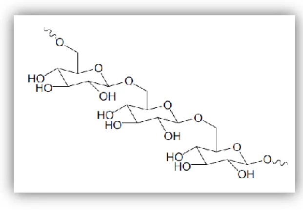
Figure 3 – Representation of the dextran chemical structure(Liu et al. 2009).
1.3.5.2.
Chitosan
Chitosan (figure 4) is a deacetylated form of chitin, that is the major compound of the exoskeletons of crustaceans shells such as crabs, shrimps and lobsters and is also found in some microorganisms, as yeasts and fungi (Sarmento et al. 2007; Tiyaboonchai et al. 2007; Nagpal et al. 2010). It is composed of randomly distributed β-(1-4)-linked D-glucosamine (deacetylated unit) and N-acetyl-D-glucosamine (acetylated unit) (Sundar et al. 2010; Anitha et al. 2011; Saboktakin et al. 2011). The commercially available chitosan has a deacetylation degree between 66 and 95% and has molecular weights ranging from 3.8 to 2000 kDa (Sundar et al. 2010).
The solubility of chitosan (pKa 6.5) is dependent on protonation of the amino groups of their molecules; therefore it is often solubilised in acids at pH lower than 6.5 including formic, acetic, tartaric, and citric acid (Chen et al. 2003; Tiyaboonchai et al. 2007). It is a weak base and has a positive charge (Chen et al. 2007), which allows chitosan to react with negatively charged surfaces (via mucoadhesion) and materials, including polymers (alginate, dextran, PVA) and DNA (Chen et al. 2007; Sundar et al. 2010).
These features, and other properties like its biodegradability in vivo by lysozyme (Sundar et al. 2010) and others hydrolytic enzymes (Carmen Rodríguez-Argüelles et al. 2011), low toxicity, good biocompatibility, improving wound healing and blood clotting, absorption of liquids in order to form protective films and coatings, make it versatile and attractive to be used in biomedical and pharmaceutical formulations (Chen et al. 2007; Babu et al. 2010). Chitosan has been used for the preparation of microparticles and nanoparticles (Sundar et al. 2010) to be used in the regeneration of different types of tissues, especially skin, bones and in many other
Chapter I - Introduction
18 biomedical and pharmaceutical applications (Carmen Rodríguez-Argüelles et al. 2011). Furthermore, it also presents very important biological properties among which antimicrobial, anti-inflammatory, and antioxidant (Carmen Rodríguez-Argüelles et al. 2011).
Figure 4 – Representation of the chitosan chemical structure(Kumirska et al. 2011).
In this work, the chitosan aqueous solution was added to dextran sulfate polyanion aqueous solution. The formation of polycation–polyanion (polyelectrolyte) complex was mainly driven by an electrostatic mechanism where charge neutralization and possible local bridging (such as hydrogen bounding, Coulomb forces, van der Waals forces, and transfer forces) occurs (Yu et al. 2005; Meng et al. 2010). The advantages of chitosan/dextran nanoparticles are enhanced stability and increased mechanical strength compared with chitosan/tripolyphosphaste microparticles, whose lower stability and mechanical strength limit their application for drug delivery (Chen et al. 2007). It has also been reported that DNA and insulin structures are protected when dextran is used in the formulation of polyethylenimine/dextran nanoparticles (Chen et al. 2007). Dextran was also described as being capable of reducing the cationic charge-related cytotoxicity of PEI nanoparticles in vitro (Chen et al. 2007). Therefore, it is possible that the combination of chitosan and dextran as matrix materials, in an optimal charge ratio, may act synergistically to incorporate and protect proteins and drugs, in order to reduce the toxicity of chitosan caused by its cationic charge (Kean et al. 2010).
Chapter I - Introduction
19
1.4. Objectives
In the present study different nanoparticles with antibacterial properties were produced in order to be applied in several biomedical products like bone implants or skin substitutes. The specific objectives of the workplan herein presented are the following:
- Development of AgNPs stabilized with chitosan/dextran;
- Determination of the antibacterial activity of the produced nanoparticles;
- Characterization of the different nanoparticles by different methods: Fourier Transform Infrared (FT-IR), Ultraviolet-Visible (UV-Vis), and Scanning Electron Microscopy (SEM);
Chapter II
Materials and Methods
Chapter II – Materials and Methods
21
2. Materials and Methods
2.1. Materials
Human fibroblasts cells (Normal Human Dermal Fibroblasts adult, criopreserved cells) were purchased from PromoCell (Spain), bacterial strain Escherichia coli DH5α (ATCC 68233) was purchased from ATCC (United States).
Fetal bovine serum (FBS) was purchased from Biochrom AG (Berlin, Germany). 3-(4,5-dimethylthiazol-2-yl)-5-(3-carboxymethoxyphenyl)-2-(4-sulfophenyl)-2H-tetrazolium reagent (MTS) and electron coupling reagent phenazine methosulfate (PMS) were purchased from Promega. Amphotericin B, ascorbic acid (C6H8O6), Dulbecco’s Modified Eagle Medium-F12
(DMEM), ethanol (EtOH), high molecular weight chitosan, L-glutamine, LuriaBertani (LB) Broth, penicillin G, phosphate-buffered saline (PBS), rezazurin sodium salt, sodium borohydride (NaBH4), sodium citrate (Na3C6H5O7), and trypsin were purchased from Sigma. Dextran sulfate
500,000 was purchased from Amresco. AgNO3 was purchased from Panreac (Spain). LB agar was
purchased from Pronadise.
2.2. Methods
2.2.1. Preparation of silver nanoparticles
The AgNPs were produced based on the method previously developed by Mafune and collaborators (Mafune et al. 2000). Briefly, the procedure consists on the rapid injection of 0.5 ml of NaBH4 (10 mM) into an aqueous solution, with continuous stirring, containing 0.5 ml of
AgNO3 (0.1 M, 0.01 M, and 0.001 M) and 20 ml of Na3C6H5O7 (0.001 M). The resultant solution was
stirred for 1 h and aged for 2 h. Moreover, the NaBH4 solution was replaced by C6H8O6 (10 mM)
and this component was added to a 0.01 M AgNO3 solution. The resulting nanoparticles were
washed three times with distilled water and centrifuged at 75000 g, during 30 min. Subsequently, a dried powder of particles was obtained by freeze-drying the particles overnight.
2.2.2. Preparation of the Chitosan/Dextran nanoparticles
Chitosan/dextran nanoparticles were prepared by the ionotropic gelation of chitosan and dextran as described before by Chen and colleagues (Chen et al. 2007). A 0.1 % (m/v) chitosan solution was prepared by dissolving chitosan in aqueous acetic acid 0.2 % (v/v) and the pH was adjusted to 3.5. A 0.1 % (m/v) dextran solution was prepared by dissolving the dextran in water.