Resumo
Introdução: O transtorno do humor bipolar (THB) é uma condição debilitante que afeta aproximadamente 1,3% das pessoas em todo o mundo, embora alguns estudos relatem uma prevalência acumulada de até 3,9% e de 4-6% em adultos quando os critérios diagnósticos mais abrangentes são aplicados.
Objetivo: Comparar as diferenças nos volumes totais de substância branca (SB), corpo caloso (CC) e volume total de substância cinzenta (SC) em pacientes com THB tipo I em estágios iniciais e tardios em comparação com controles.
Métodos: Cinquenta e cinco sujeitos foram incluídos neste protocolo de estudo. O desenho de caso com duplo controle incluiu 14 pacientes com THB em estágio inicial; 15 pacientes com THB em fase tardia; e seus respectivos controles correspondentes (14 e 12 sujeitos).
Resultados: Os volumes do CC e total de SB foram significativamente
menores nos pacientes com THB nos estágios iniciais e tardios vs. controles. Não houve diferença para o volume total de SC no grupo em estágio inicial, mas em pacientes em fase tardia o volume total
de SC foi significativamente menor do que nos controles. A redução
do volume total de SC em pacientes em fase tardia está de acordo com a teoria da neuroprogressão do THB. A redução dos volumes de SB em SB total e no CC em fases precoces e tardias suporta a possibilidade de que um processo de desmielinização precoce poderia ocorrer subjacente à manifestação clínica de THB.
Conclusão: Nossos achados podem direcionar a investigação de anormalidades da SB em populações de alto risco para o desenvolvimento de THB, talvez como biomarcadores precoces antes da síndrome aberta.
Descritores: Transtorno humor bipolar, ressonância magnética, transtornos afetivos, estadiamento, biomarcadores.
Abstract
Introduction: Bipolar disorder (BD) is a debilitating mood condition that affects approximately 1.3% of people worldwide, although some studies report up to 3.9% lifetime prevalence and 4-6% in adults when broad diagnostic criteria are applied.
Objective: To compare differences in total white matter (WM), corpus callosum (CC) and total gray matter (GM) volumes in patients with type I BD at early and late stages compared with controls.
Methods: Fifty-five subjects were enrolled in this study protocol.
The double case-control design included 14 patients with BD at early stage; 15 patients at late stage; and their respective matched controls (14 and 12 subjects).
Results: CC and total WM volumes were significantly smaller
in patients with BD at early and late stages vs. controls. There was no difference for total GM volume in the early stage group,
but in patients at late stage total GM volume was significantly
smaller than in controls. The total GM volume reduction in patients at late stage is in agreement with the neuroprogression theory of BD. The reduction of WM volumes in total WM and in the CC at early and late stages supports the possibility that an early demyelination process could occur underlying the clinical manifestation of BD.
Conclusion: Our findings may direct to the investigation of WM
abnormalities in populations at high risk to develop BD, perhaps as early biomarkers before the overt syndrome.
Keywords: Bipolar disorder, magnetic resonance imaging, affective disorders, staging, biomarkers.
1 Departamento de Radiologia e Ressonância Magnética, Hospital de Clínicas de Porto Alegre (HCPA), Porto Alegre, RS, Brazil. 2 Tomoclínica, Canoas, RS, Brazil. 3
Laboratory of Molecular Psychiatry, National Science and Technology Institute for Translational Medicine (INCT-TM), HCPA, Universidade Federal do Rio Grande do Sul (UFRGS), Porto Alegre, RS, Brazil. 4 Departamento de Psiquiatria, Universidade Federal do Paraná (UFPR), Curitiba, PR, Brazil. 5 Laboratório de Farmacologia
e Fisiologia, Universidade de Caxias do Sul, Caxias do Sul, RS, Brazil. Submitted Mar 16 2017, accepted for publication Jan 11 2018.
Suggested citation: Duarte JA, Massuda R, Goi PD, Vianna-Sulzbach M, Colombo R, Kapczinski F, et al. White matter volume is decreased in bipolar disorder at early and late stages. Trends Psychiatry Psychother. 2018;40(4):277-284. http://dx.doi.org/10.1590/2237-6089-2017-0025
White matter volume is decreased in bipolar disorder at
early and late stages
O volume da substância branca está reduzido tanto nos estágios iniciais quanto
nos estágios avançados do transtorno bipolar
Juliana A. Duarte,1,2,3 Raffael Massuda,3,4 Pedro D. Goi,3 Mireia Vianna-Sulzbach,3 Rafael Colombo,3,4,5
Introduction
Bipolar disorder (BD) is a debilitating mood condition that affects approximately 1.3% of people worldwide,1
although some studies report up to 3.9% lifetime prevalence2 and 4-6% in adults when broad diagnostic
criteria are applied.3
Recurrent episodes influence the outcome of BD by increasing a patient’s vulnerability to subsequent episodes and reducing treatment response.4 An
episode-depen dent deterioration pattern has been widely described in serum biomarkers,5,6 brain imaging7,8 and
functioning.9-13
Alterations in brain structures have been widely reported in BD, including enlargement of the third and lateral ventricles and reduction in the gray matter (GM) volumes of the orbital and medial prefrontal cortex, ventral striatum and mesotemporal cortex.1-3 White
matter (WM) assessment through structural volumetric imaging has provided evidence of subtle abnormalities in patients with BD compared to healthy volunteers.14
The most-studied WM sub-region in BD is the corpus callosum (CC), the largest WM tract that connects the two hemispheres of the brain. Several studies have found that the CC is smaller in patients with BD.15-20
One of them has shown a decrease in volume of the posterior CC in late stage BD, but not in early stage.21
These network alterations have been associated with cognitive symptoms of BD, suggesting that WM alterations may occur as an early neuropathological process underpinning the overt cognitive decline.22
In summary, it is not clear whether WM alterations appear early in the course of BD or present an episode-dependent reduction like GM.
The aim of the present study was to compare differences in total WM, CC and total GM volumes in patients with type I BD at early and late stages compared with healthy controls.
Methods
Fifty-five subjects were enrolled in this study protocol: 29 patients and 26 controls matched for age, gender, education and body mass index (BMI). The double case-control design included 14 patients with BD at early stage (individuals who exhibit the same status in the interepisodic period as they did before the onset of BD); 15 patients with BD at late stage (individuals who are unable to maintain personal self-care and to live autonomously); and their respective matched controls (14 and 12 subjects). Patients at early stage had to present a score <36
on the Functioning Assessment Short Test (FAST),13
and those at late stage a score ≥36. The definition of
staging was in accordance with the BD staging model described elsewhere.23
All subjects were required to be at least 18 years old and no older than 60. Written informed consent was obtained from all subjects in accordance with the Declaration of Helsinki. The local ethics committee approved the study protocol.
Inclusion criteria for patients were: a) fulfilling Diagnostic and Statistical Manual of Mental Disorders, 4th edition (DSM-IV) criteria for BD I24; and b) meeting
remission criteria defined as a score <7 on the 17-item Hamilton-Depression Scale (HAM-D)25 and on the Young
Mania Rating Scale (YMRS),26 for at least one month
previous to the assessment. All patients received pharmacological treatment by their psychiatrist according to clinical protocols. Patients with severe clinical illnesses detected during clinical interviews or during review of medical records were excluded.
The control group consisted of healthy volunteers who had neither current or previous history nor first-degree family history of a major psychiatric disorder, including dementia or mental retardation assessed by the non-patient version of the Structured Clinical Interview for DSM-IV (SCID).
SCID Axis I and Axis II were administered to confirm diagnosis. Sociodemographic, clinical and pharmacological data were collected via a structured interview with the patient and examination of clinical records. The 17-item HAM-D and the YMRS were administered by trained raters to assess depressive and manic symptoms, respectively.
Magnetic resonance imaging (MRI) data were obtained using a Philips Achieva 1.5 Tesla scanner (Amsterdam, the Netherlands). T1 high resolution sagittal 3D magnetization-prepared rapid gradient-echo (MPRAGE) images were acquired with NEX=1, image matrix=256×232, flip angle=8 degrees, echo time=4 ms, repetition time=8.63 ms, and voxel size 1×1×1 mm³, yielding 160 slices.
Cortical and subcortical volumetric segmentations were performed with the FreeSurfer image analysis suite version 5.1.0 (http://surfer.nmr.mgh.harvard.edu/). The process includes motion correction, removal of non-brain tissue,27 automated Talairach transformation,
segmentation of subcortical WM and deep GM volumetric structures,28,29 intensity normalization,30 tessellation of
the GM/WM boundary, automated topology correction,31,32
class.33-35 Previous studies have shown that subcortical
segmentations performed with the FreeSurfer software are reliable when compared to manual segmentation.28,36
All images were processed and checked by the same researcher. Intracranial volume was regressed out from the volumes of CC, WM, and GM.
Demographic and clinical characteristics were analyzed using the chi-square, Mann-Whitney or Student’s t tests. Descriptive analyses are presented as mean (standard deviation) or median (interquartile range); p-values <0.05 were considered significant. Appropriate tests were used for parametric or nonparametric distribution.
Results
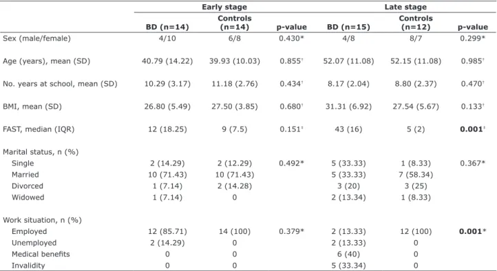
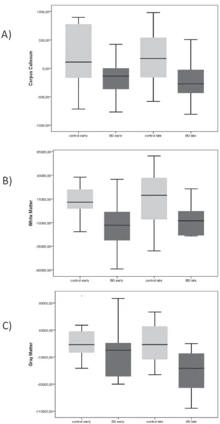
The subjects’ general characteristics are summarized in Table 1. Data of patients with BD are presented in Table 2. CC (p = 0.035 for early; p = 0.028 for late stage groups) and total WM volumes (p = 0.005 for early; p = 0.021 for late stage) were significantly smaller in patients with BD than in controls. There was no difference for total GM volume in the early stage
group (p = 0.306) vs. controls. Total GM volume was significantly smaller in patients with BD at late stage compared to controls (p = 0.001). Volumetric results are shown in Figure 1.
Discussion
To our knowledge, this is the first study to examine CC, total WM and total GM volumes in patients with BD at early and late stages compared to matched controls. CC and total WM volumes were decreased in patients with BD at early and late stages; total GM volume was decreased in patients at late stage, but not at early stage, when compared to matched controls.
The hypothesis of different patterns of changes in brain morphology over the course of BD37 is widely
supported by many authors.1-3,14,38-40 Illness progression
or neuroprogression,41 a term that explains the
pathological reorganization of the central nervous system (CNS) as a consequence of recurrent mood episodes and their influence on BD outcomes, has been used to explain vulnerability to subsequent episodes and changes in treatment response.4 The
Table 1 - Characteristics of healthy controls and patients with BD
Early stage Late stage
BD (n=14)
Controls
(n=14) p-value BD (n=15)
Controls
(n=12) p-value
Sex (male/female) 4/10 6/8 0.430* 4/8 8/7 0.299*
Age (years), mean (SD) 40.79 (14.22) 39.93 (10.03) 0.855† 52.07 (11.08) 52.15 (11.08) 0.985†
No. years at school, mean (SD) 10.29 (3.17) 11.18 (2.76) 0.434† 8.17 (2.04) 8.80 (2.37) 0.470†
BMI, mean (SD) 26.80 (5.49) 27.50 (3.85) 0.680† 31.31 (6.92) 27.54 (5.67) 0.133†
FAST, median (IQR) 12 (18.25) 9 (7.5) 0.151‡ 43 (16) 5 (2) 0.001‡
Marital status, n (%)
Single 2 (14.29) 2 (12.29) 0.492* 5 (33.33) 1 (8.33) 0.367*
Married 10 (71.43) 10 (71.43) 5 (33.33) 7 (58.34)
Divorced 1 (7.14) 2 (14.28) 3 (20) 3 (25)
Widowed 1 (7.14) 0 2 (13.34) 1 (8.33)
Work situation, n (%)
Employed 12 (85.71) 14 (100) 0.379* 2 (13.33) 12 (100) 0.001*
Unemployed 2 (14.29) 0 2 (13.33) 0
Medical benefits 0 0 6 (40) 0
Invalidity 0 0 5 (33.34) 0
BD = bipolar disorder; BMI = body mass index; IQR = interquartile range; SD = standard deviation. p-values in bold font are statistically significant.
neuroprogression pattern has been widely described in serum biomarkers,5,6 functioning,9-13 cognitive
performance,42,43 and brain imaging. 7,8,40 The classical
brain findings in BD are a result of GM reduction, including enlargement of the third and lateral ventricles and reduction in total GM volumes of the orbital and medial prefrontal cortex, ventral striatum and mesotemporal cortex.1-3 These changes also seem to be
related to illness progression.40
Conversely, a number of studies have shown that the CC may play a significant role in the pathophysiology of
BD38,39 and highlight the importance of WM alterations
in underpinning the clinical presentation of BD.37 The
CC is the main interhemispheric commissure and is crucial for interhemispheric communication and cognitive processes.38,39 CC abnormalities may lead to
altered interhemispheric communication, which could be relevant for the pathophysiology of the cognitive disturbances present in patients with BD.44-46 It is
also possible that reduced CC volume is secondary to abnormalities of glial cells.47 Glia, especially
oligodendrocytes and the myelin that they produce, are essential in achieving and maintaining optimal brain function.48 In humans, approximately 50% of
the WM volume is composed of myelin, demonstrating that changes in volume can have serious functional repercussions.49 Myelination can increase the speed
of propagation of electrical information by ~100 times. At the same time, there is a 30-fold reduction in the refractory period, increasing the number of action potentials propagated per unit time. Taken together, these changes increase connectivity and promote a 3000-fold increase in the brain’s information-processing capacity, which is essential for the maintenance of cognition.48,50 The continuum
of increasing myelin vulnerability resulting from the protracted myelination that underlies disease phenotypes have been reported in BD.51
Table 2 - Characteristics of patients with BD
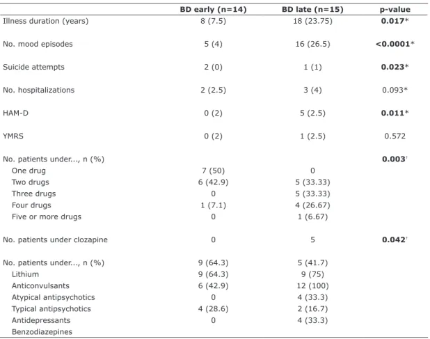
BD early (n=14) BD late (n=15) p-value
Illness duration (years) 8 (7.5) 18 (23.75) 0.017*
No. mood episodes 5 (4) 16 (26.5) <0.0001*
Suicide attempts 2 (0) 1 (1) 0.023*
No. hospitalizations 2 (2.5) 3 (4) 0.093*
HAM-D 0 (2) 5 (2.5) 0.011*
YMRS 0 (2) 1 (2.5) 0.572
No. patients under..., n (%) 0.003†
One drug 7 (50) 0
Two drugs 6 (42.9) 5 (33.33)
Three drugs 0 5 (33.33)
Four drugs 1 (7.1) 4 (26.67)
Five or more drugs 0 1 (6.67)
No. patients under clozapine 0 5 0.042†
No. patients under..., n (%) 9 (64.3) 5 (41.7)
Lithium 9 (64.3) 9 (75)
Anticonvulsants 6 (42.9) 12 (100)
Atypical antipsychotics 0 4 (33.3)
Typical antipsychotics 4 (28.6) 2 (16.7)
Antidepressants 0 4 (33.3)
Benzodiazepines
Data presented as median (interquartile range), unless otherwise specified.
BD = bipolar disorder; HAM-D = 17-item Hamilton-Depression Scale; YMRS = Young Mania Rating Scale. p-values in bold font are statistically significant.
Figure 1 - A) Box-plot of total corpus callosum size in patients at early and late BD and their matched controls. Median levels are indicated by horizontal lines (Mann-Whitney: control early vs. BD early, p = 0.035; control late vs. BD late, p = 0.028). B) Box-plot
of total white matter volume in patients with BD and their matched controls. Median levels are indicated by horizontal lines (Mann-Whitney: control early vs. BD early, p = 0.005; control late vs. BD late, p = 0.021). C) Box-plot of total gray matter volume in patients
WM in general and CC in particular seem to be especially vulnerable to perceived stress52 and have been
shown to be impaired by stressful events and trauma.53,54
Reductions in CC volume have been demonstrated in both children55 and adults with post-traumatic stress
disorder (PTSD).56,57 Although there is robust evidence
of gross WM involvement in BD, the origin of these abnormalities remains unclear. Evidence suggests that neuronal metabolic disorders, inflammation and mitochondrial dysfunction are associated with demyelination and functional deterioration in BD.41,58
Some of these changes may be mediated by decrease in the activity of protein kinase B (Akt) and increase
in the activity of glycogen synthase kinase 3β (GSK-3β). Activation of GSK-3 β increases inflammation
and promotes demyelination, while inactivation by phosphorylation of this protein acts in the opposite way.48 As a matter of fact, structural MRI studies have
found abnormalities in volume, signal intensity and microstructure in patients suffering from BD.16,59,60
The increasing attention devoted to WM disruption in other neuropsychiatric conditions, such as multiple sclerosis (MS), has allowed to draw an analogy considering the inflammatory processes and clinical progression of WM abnormalities. BD has a well-documented inflammatory component61-65 that could
exert deleterious effects on glia components, such as myelin,48,51,66,67 and also on WM, as mentioned
before. Current findings reinforce the presence of WM abnormalities at the first episode,37 differently
from GM findings, which show an episode-dependent deterioration pattern40 along with cognitive and
functioning decline.13,42
Our findings support the growing body of evidence that patients with BD have smaller total WM volume, particularly in the CC, compared to controls, in line with previous studies.16,59,60 A meta-analysis of MRI studies
showed reduction of total intracranial and WM volumes in patients with BD at first episode, but not of GM and whole brain volumes.37 These meta-analytical findings
are in line with our results of decreased CC and total WM volumes in patients with BD at early stage, but in contrast with others that report decrease of CC volume only in patients at late stage.21
Albeit innovative, the present study has important limitations that must be pointed out. First, the use of structural MRI to assess WM may not detect subtle alterations. The 1.5-Tesla equipment may not have been able to assess those volumes compared to higher definition scanners. The heterogeneous nature of BD could also highlight non-pathological volumetric particularities. Also, the reduced number of participants in each group is a crucial limitation. Larger groups could reach more
powerful results and differences among groups. For the same reason, this study was not able to investigate the characteristics of individual pharmacological treatments, which could contribute to volumetric differences. Another limitation of this study was that we could not evaluate the effect of psychotic symptoms.
In conclusion, even considering the limitations of the sample size and cross-sectional design of this study, the total GM volume reduction observed in patients at late stage is in agreement with the neuroprogression theory in BD. The reduction of WM volumes in total WM and in the CC at both early and late stages of the disease is consistent with the disconnection syndrome of frontal and subcortical regions described since the early stage of the illness.37 These findings on WM, added
to the current evidence, support the possibility that an early demyelination process could occur underlying BD symptoms. Our findings, together with current literature, direct to the need to investigate WM abnormalities in populations at high risk to develop BD, perhaps as early biomarkers before the overt syndrome.
Acknowledgements
This work was supported by Conselho Nacional de Desenvolvimento Científico e Tecnológico (CNPq) and Coordenação de Aperfeiçoamento de Pessoal de Nível Superior (CAPES; grants CNPq Universal 443526/2014-1 and 470326/20443526/2014-1443526/2014-1-5; CNPq Produtividade em Pesquisa 304443/2014-0) and by Fundação de Amparo à Pesquisa do Estado do Rio Grande do Sul (FAPERGS; PRONEM 11/2057-2).
Disclosure
Flavio Kapczinski has received grant/research support from AstraZeneca, Eli Lilly, Janssen-Cilag, Servier, CNPq, CAPES, NARSAD, and the Stanley Medical Research Institute; has been a member of the speakers’ boards of Astra-Zeneca, Eli Lilly, Janssen, and Servier; and has served as a consultant for Servier. Clarissa S. Gama has served as a paid speaker for Lundbeck and as a consultant/speaker for Roche, Pfizer, Daichii-Sankyo, and Actelion. No other conflicts of interest declared concerning the publication of this article.
References
and bipolar disorder within UK biobank: cross-sectional study of 172,751 participants. PLoS One. 2013;8:e75362.
2. Kessler RC, Berglund P, Demler O, Jin R, Merikangas KR, Walters EE. Lifetime prevalence and age-of-onset distributions of DSM-IV disorders in the National Comorbidity Survey Replication. Arch Gen Psychiatry. 2005;62:593-602.
3. Johnson KR, Johnson SL. Cross-national prevalence and cultural correlates of bipolar I disorder. Soc Psychiatry Psychiatr Epidemiol. 2014;49:1111-7.
4. Leow A, Ajilore O, Zhan L, Arienzo D, GadElkarim J, Zhang A, et al. Impaired inter-hemispheric integration in bipolar disorder revealed with brain network analyses. Biol Psychiatry. 2013;73:183-93.
5. Oertel-Knöchel V, Reinke B, Alves G, Jurcoane A, Wenzler S, Prvulovic D, et al. Frontal white matter alterations are associated with executive cognitive function in euthymic bipolar patients. J Affect Disord. 2014;155:223-33.
6. Torgerson CM, Irimia A, Leow AD, Bartzokis G, Moody TD, Jennings RG, et al. DTI tractography and white matter fiber tract characteristics in euthymic bipolar I patients and healthy control subjects. Brain Imaging Behav. 2013;7:129-39.
7. Benedetti F, Yeh PH, Bellani M, Radaelli D, Nicoletti MA, Poletti S, et al. Disruption of white matter integrity in bipolar depression as a possible structural marker of illness. Biol Psychiatry. 2011;69:309-17.
8. Wessa M, Houenou J, Leboyer M, Chanraud S, Poupon C, Martinot JL, et al. Microstructural white matter changes in euthymic bipolar patients: a whole-brain diffusion tensor imaging study. Bipolar Disord. 2009;11:504-14.
9. Wang F, Kalmar JH, He Y, Jackowski M, Chepenik LG, Edmiston EE, et al. Functional and structural connectivity between the perigenual anterior cingulate and amygdala in bipolar disorder. Biol Psychiatry. 2009;66:516-21.
10. Zanetti MV, Jackowski MP, Versace A, Almeida JR, Hassel S, Duran FL, et al. State-dependent microstructural white matter changes in bipolar I depression. Eur Arch Psychiatry Clin Neurosci. 2009;259:316-28.
11. Mahon K, Wu J, Malhotra AK, Burdick KE, DeRosse P, Ardekani BA, et al. A voxel-based diffusion tensor imaging study of white matter in bipolar disorder. Neuropsychopharmacology. 2009;34:1590-600. 12. Versace A, Almeida JR, Hassel S, Walsh ND, Novelli M, Klein CR, et
al. Elevated left and reduced right orbitomedial prefrontal fractional anisotropy in adults with bipolar disorder revealed by tract-based spatial statistics. Arch Gen Psychiatry. 2008;65:1041-52. 13. Rosa AR, Magalhães PV, Czepielewski L, Sulzbach MV, Goi PD,
Vieta E, et al. Clinical staging in bipolar disorder: focus on cognition and functioning. J Clin Psychiatry. 2014;75:e450-6. 14. Mahon K, Burdick KE, Szeszko PR. A role for white matter
abnormalities in the pathophysiology of bipolar disorder. Neurosci Biobehav Rev. 2010;34:533-54.
15. Arnone D, McIntosh AM, Chandra P, Ebmeier KP. Meta-analysis of magnetic resonance imaging studies of the corpus callosum in bipolar disorder. Acta Psychiatr Scand. 2008;118:357-62. 16. Atmaca M, Ozdemir H, Yildirim H. Corpus callosum areas in
first-episode patients with bipolar disorder. Psychol Med. 2007;37:699-704.
17. Brambilla P, Nicoletti MA, Sassi RB, Mallinger AG, Frank E, Kupfer DJ, et al. Magnetic resonance imaging study of corpus callosum abnormalities in patients with bipolar disorder. Biol Psychiatry. 2003;54:1294-7.
18. Coffman JA, Bornstein RA, Olson SC, Schwarzkopf SB, Nasrallah HA. Cognitive impairment and cerebral structure by MRI in bipolar disorder. Biol Psychiatry. 1990;27:1188-96.
19. Hauser P, Dauphinais ID, Berrettini W, DeLisi LE, Gelernter J, Post RM. Corpus callosum dimensions measured by magnetic resonance imaging in bipolar affective disorder and schizophrenia. Biol Psychiatry. 1989;26:659-68.
20. Walterfang M, Wood AG, Reutens DC, Wood SJ, Chen J, Velakoulis D, et al. Corpus callosum size and shape in first-episode affective and schizophrenia-spectrum psychosis. Psychiatry Res. 2009;173:77-82.
21. Lavagnino L, Cao B, Mwangi B, Wu MJ, Sanches M, Zunta-Soares GB, et al. Changes in the corpus callosum in women with late-stage bipolar disorder. Acta Psychiatr Scand. 2015;131:458-64. 22. Schneider MR, DelBello MP, McNamara RK, Strakowski SM,
Adler CM. Neuroprogression in bipolar disorder. Bipolar Disord. 2012;14:356-74.
23. Kapczinski F, Magalhães PV, Balanzá-Martinez V, Dias VV, Frangou S, Gama CS, et al. Staging systems in bipolar disorder: an
International Society for Bipolar Disorders Task Force Report. Acta Psychiatr Scand. 2014;130:354-63.
24. First MB, Spitzer RL, Gibbon M, Williams JB. Structured Clinical Interview for DSM-IV (SCID). New York: Department of Psychiatry, College of Physicians and Surgeons, Columbia University; 1988. 25. Hamilton M. A rating scale for depression. J Neurol Neurosurg
Psychiatry. 1960;23:56-62.
26. Young RC, Biggs JT, Ziegler VE, Meyer DA. A rating scale for mania: reliability, validity and sensitivity. Br J Psychiatry. 1978;133:429-35. 27. Ségonne F, Dale AM, Busa E, Glessner M, Salat D, Hahn HK,
et al. A hybrid approach to the skull stripping problem in MRI. Neuroimage. 2004;22:1060-75.
28. Lancaster JL, Rainey LH, Summerlin JL, Freitas CS, Fox PT, Evans AC, et al. Automated labeling of the human brain: a preliminary report on the development and evaluation of a forward-transform method. Hum Brain Mapp. 1997;5:238-42.
29. Fischl B, Salat DH, van der Kouwe AJ, Makris N, Ségonne F, Quinn BT, et al. Sequence-independent segmentation of magnetic resonance images. Neuroimage. 2004;23 Suppl 1:S69-84. 30. Sled JG, Zijdenbos AP, Evans AC. A nonparametric method for
automatic correction of intensity nonuniformity in MRI data. IEEE Trans Med Imaging. 1998;17:87-97.
31. Fischl B, Liu A, Dale AM. Automated manifold surgery: constructing geometrically accurate and topologically correct models of the human cerebral cortex. IEEE Trans Med Imaging. 2001;20:70-80.
32. Ségonne F, Pacheco J, Fischl B. Geometrically accurate topology-correction of cortical surfaces using nonseparating loops. IEEE Trans Med Imaging. 2007;26:518-29.
33. Dale AM, Fischl B, Sereno MI. Cortical surface-based analysis. I. Segmentation and surface reconstruction. Neuroimage 1999;9:179-94.
34. Dale AM, Sereno MI. Improved localizadon of cortical activity by combining EEG and MEG with MRI cortical surface reconstruction: a linear approach. J Cogn Neurosci. 1993;5:162-76.
35. Fischl B, Dale AM. Measuring the thickness of the human cerebral cortex from magnetic resonance images. Proc Natl Acad Sci U S A. 2000;97:11050-5.
36. Tae WS, Kim SS, Lee KU, Nam EC, Kim KW. Validation of hippocampal volumes measured using a manual method and two automated methods (FreeSurfer and IBASPM) in chronic major depressive disorder. Neuroradiology. 2008;50:569-81.
37. Vita A, De Peri L, Sacchetti E. Gray matter, white matter, brain, and intracranial volumes in first-episode bipolar disorder: a meta-analysis of magnetic resonance imaging studies. Bipolar Disord. 2009;11:807-14.
38. Gazzaniga MS. Cerebral specialization and interhemispheric communication: does the corpus callosum enable the human condition? Brain. 2000;123 (Pt 7):1293-326.
39. Sánchez MM, Hearn EF, Do D, Rilling JK, Herndon JG. Differential rearing affects corpus callosum size and cognitive function of rhesus monkeys. Brain Res. 1998;812:38-49.
40. Strakowski SM, DelBello MP, Zimmerman ME, Getz GE, Mills NP, Ret J, et al. Ventricular and periventricular structural volumes in first- versus multiple-episode bipolar disorder. Am J Psychiatry. 2002;159:1841-7.
41. Berk M, Kapczinski F, Andreazza AC, Dean OM, Giorlando F, Maes M, et al. Pathways underlying neuroprogression in bipolar disorder: focus on inflammation, oxidative stress and neurotrophic factors. Neurosci Biobehav Rev. 2011;35:804-17.
42. Lebowitz BK, Shear PK, Steed MA, Strakowski SM. Verbal fluency in mania: relationship to number of manic episodes. Neuropsychiatry Neuropsychol Behav Neurol. 2001;14:177-82. 43. Burdick KE, Ketter TA, Goldberg JF, Calabrese JR. Assessing
cognitive function in bipolar disorder: challenges and recommendations for clinical trial design. J Clin Psychiatry. 2015;76:e342-50.
44. Borkowska A, Rybakowski JK. Neuropsychological frontal lobe tests indicate that bipolar depressed patients are more impaired than unipolar. Bipolar Disord. 2001;3:88-94.
45. El-Badri SM, Ashton CH, Moore PB, Marsh VR, Ferrier IN. Electrophysiological and cognitive function in young euthymic patients with bipolar affective disorder. Bipolar Disord. 2001;3:79-87.
46. Wilder-Willis KE, Sax KW, Rosenberg HL, Fleck DE, Shear PK, Strakowski SM. Persistent attentional dysfunction in remitted bipolar disorder. Bipolar Disord. 2001;3:58-62.
48. Haroutunian V, Katsel P, Roussos P, Davis KL, Altshuler LL, Bartzokis G. Myelination, oligodendrocytes, and serious mental illness. Glia. 2014;62:1856-77.
49. Miller DJ, Duka T, Stimpson CD, Schapiro SJ, Baze WB, McArthur MJ, et al. Prolonged myelination in human neocortical evolution. Proc Natl Acad Sci U S A. 2012;109:16480-5.
50. Grydeland H, Walhovd KB, Tamnes CK, Westlye LT, Fjell AM. Intracortical myelin links with performance variability across the human lifespan: results from T1- and T2-weighted MRI myelin mapping and diffusion tensor imaging. J Neurosci. 2013;33:18618-30.
51. Watkins CC, Sawa A, Pomper MG. Glia and immune cell signaling in bipolar disorder: insights from neuropharmacology and molecular imaging to clinical application. Transl Psychiatry. 2014;4:e350. 52. Li H, Li W, Wei D, Chen Q, Jackson T, Zhang Q, et al. Examining
brain structures associated with perceived stress in a large sample of young adults via voxel-based morphometry. Neuroimage. 2014;92:1-7.
53. Jackowski AP, Douglas-Palumberi H, Jackowski M, Win L, Schultz RT, Staib LW, et al. Corpus callosum in maltreated children with posttraumatic stress disorder: a diffusion tensor imaging study. Psychiatry Res. 2008;162:256-61.
54. Rinne-Albers MA, van der Wee NJ, Lamers-Winkelman F, Vermeiren RR. Neuroimaging in children, adolescents and young adults with psychological trauma. Eur Child Adolesc Psychiatry. 2013;22:745-55.
55. De Bellis MD, Keshavan MS, Shifflett H, Iyengar S, Beers SR, Hall J, et al. Brain structures in pediatric maltreatment-related posttraumatic stress disorder: a sociodemographically matched study. Biol Psychiatry. 2002;52:1066-78.
56. Kitayama N, Brummer M, Hertz L, Quinn S, Kim Y, Bremner JD. Morphologic alterations in the corpus callosum in abuse-related posttraumatic stress disorder: a preliminary study. J Nerv Ment Dis. 2007;195:1027-9.
57. Villarreal G, Hamilton DA, Graham DP, Driscoll I, Qualls C, Petropoulos H, et al. Reduced area of the corpus callosum in posttraumatic stress disorder. Psychiatry Res. 2004;131:227-35. 58. Bartzokis G, Lu PH, Heydari P, Couvrette A, Lee GJ, Kalashyan
G, et al. Multimodal magnetic resonance imaging assessment of white matter aging trajectories over the lifespan of healthy individuals. Biol Psychiatry. 2012;72:1026-34.
59. Brambilla P, Nicoletti M, Sassi RB, Mallinger AG, Frank E, Keshavan MS, et al. Corpus callosum signal intensity in patients
with bipolar and unipolar disorder. J Neurol Neurosurg Psychiatry. 2004;75:221-5.
60. Yurgelun-Todd DA, Silveri MM, Gruber SA, Rohan ML, Pimentel PJ. White matter abnormalities observed in bipolar disorder: a diffusion tensor imaging study. Bipolar Disord. 2007;9:504-12. 61. Kapczinski F, Dal-Pizzol F, Teixeira AL, Magalhaes PV,
Kauer-Sant’Anna M, Klamt F, et al. Peripheral biomarkers and illness activity in bipolar disorder. J Psychiatr Res. 2011;45:156-61. 62. Kapczinski F, Dal-Pizzol F, Teixeira AL, Magalhaes PV,
Kauer-Sant’Anna M, Klamt F, et al. A systemic toxicity index developed to assess peripheral changes in mood episodes. Mol Psychiatry. 2010;15:784-6.
63. Maes M, Smith R, Scharpe S. The monocyte-T-lymphocyte hypothesis of major depression. Psychoneuroendocrinology. 1995;20:111-6.
64. Stertz L, Magalhães PV, Kapczinski F. Is bipolar disorder an inflammatory condition? The relevance of microglial activation. Curr Opin Psychiatry. 2013;26:19-26.
65. Haarman BC, Riemersma-Van der Lek RF, de Groot JC, Ruhé HG, Klein HC, Zandstra TE, et al. Neuroinflammation in bipolar disorder - A [(11)C]-(R)-PK11195 positron emission tomography study. Brain Behav Immun. 2014;40:219-25.
66. Pfaffenseller B, Fries GR, Wollenhaupt-Aguiar B, Colpo GD, Stertz L, Panizzutti B, et al. Neurotrophins, inflammation and oxidative stress as illness activity biomarkers in bipolar disorder. Expert Rev Neurother. 2013;13:827-42.
67. Gos T, Schroeter ML, Lessel W, Bernstein HG, Dobrowolny H, Schiltz K, et al. S100B-immunopositive astrocytes and oligodendrocytes in the hippocampus are differentially afflicted in unipolar and bipolar depression: a postmortem study. J Psychiatr Res. 2013;47:1694-9.
Correspondence:
Clarissa S Gama
Hospital de Clínicas de Porto Alegre Rua Ramiro Barcelos, 2350
90035-903 - Porto Alegre, RS - Brazil Tel.: +55 (51) 33598845