Silencing the
gip
gene of
Phytophthora cinnamomi
by iRNA
and studying the subcellular localization of GIP and NPP1
proteins
ABDESSALEM CHAHED
Dissertation submitted to Escola Superior Agrária de Bragança to obtain the Degree of Master in Biotechnological Engineering
Supervised by
Altino Choupina Maria João De Sousa
Noureddine Chatti
Bragança
Dissertation made under the agreement of Double Diploma
between the Escola Superior Agrária de Bragança|IPB and the
i
ACKNOWLEDGEMENTS
There are so many people who have helped me and have made my time at the Polytechnic Institute of Bragança so enjoyable and productive.
First and foremost, I would like to thank my supervisor Pr. Altino Choupina for his guidance, support, and infinite patience during this year. I would like also to express my sincere appreciation to my co-supervisor Pr. Maria João De Sousa for encouragement and sharing her valuable expertise and advice. Also thank, to Pr. Anabela Martins the coordinator of the Biotechnological Engineering, Masters Program for supporting me all through those difficult moments in Bragança.
I also want to acknowledge the effort of Rodrigo Costa for his excellent support in the laboratorial experiments. You have been really helpful and gave all necessary information for the successful completion of this thesis.
I am grateful to all the teaching and non-teaching staff of the Polytechnic Institute of Bragança. A special thanks to Pr Elsa Ramalhosa, Pr Antonio Peres and Pr Paula Rodrigues for their kindness and friendship.
I would also like to thank all researchers in the Mountain Research Center (CIMO) and all my friends who have been there for me all through my stay in Bragança specially Vitor, Taofiq and Sana.
iii
TABLE OF CONTENT
INDEX OF FIGURES ... vii
INDEX OF TABLES ... ix
ABBREVIATIONS LIST ... xi
ABSTRACT ... xv
RESUMO ... xvii
1-INTRODUCTION ... 1
1.1-The problem of ink disease ... 1
1.2-The genus Phytophthora ... 3
1.2.1-Phytophthora species ... 3
1.2.2-Phytophthora cinnamomi ... 4
1.2.3-Asexual reproduction of Phytophthora ... 4
1.2.4-Sexual reproduction of Phytophthora ... 5
1.2.5-Mechanism of infection ... 5
1.3-Traditional methods used to control ink disease ... 6
1.4-Gene gip ... 7
1.4.1-Gene gip in Phytophthora species ... 7
1.4.2-Gene gip in Phytophthora cinnamomi... 7
1.5-Gene npp1 ... 8
1.5.1-Gene npp1 in Phytophthora species ... 8
1.5.2-Gene npp1 in Phytophthora cinnamomi ... 8
1.6-RNA interference ... 8
1.6.1-RNA interference discovery ... 8
1.6.2-RNA interference silencing mediators ... 9
1.6.3-RNA interference mechanism ... 10
1.6.4-Biological functions of RNAi... 11
1.6.5-Applications... 12
1.7-The green fluorescent protein (GFP)... 13
2-OBJECTIVE ... 15
3-MATERIALS AND METHODS ... 17
3.1-Biological materials ... 17
iv
3.2.1-Culture medium used for E. coli ... 17
3.2.2-Culture media for P. cinnamomi ... 18
3.3-Culture conditions and maintenance of microorganisms ... 18
3.4-Vectors ... 19
3.5-DNA extraction ... 21
3.6-Visualisation and purification of nucleic acids in agarose gels ... 21
3.7-Design of silencing cassette (shRNA based vector) for gip gene ... 22
3.8-Transformation of E. coli with the silencing construct ... 24
3.9-Extraction of the plasmid DNA of the transformed E. coli ... 25
3.10-Cloning the silencing cassette into the pTH210 vector ... 25
3.11-Transformation of Phytophthora cinnamomi zoosporeswith the recombinant pTH210 vector ... 26
3.12-Genomic analysis of the transformed Phytophthora cinnamomi (PCR & DNA sequencing) ... 26
3.13-Castanea sativa infection with P. cinnamomi strains ... 27
3.14-Fusion of the ORF’s into the pTOR-eGFP vector ... 27
3.14.1-Primer design to amplify the ORF’s of gip and npp1 genes of Phytophthora cinnamomi by PCR ... 27
3.14.2-PCR amplification of gip and npp1 ... 29
3.14.3-Enzymatic digestion and dephosphorylation of pTOR-eGFP ... 29
3.14.4-Fusion of the PCR products into the pTOR-eGFP plasmid ... 30
3.15-E. coli Transformation with the recombinant pTOR-eGFP vector ... 30
3.16-Extraction of the plasmid DNA of the transformed E. coli ... 30
3.17-Prediction of the subcellular localization of the GIP and NPP1 proteins ... 31
4-RESULTS AND DISCUSSION ... 33
4.1-DNA extraction of Phytophthora cinnamomi ... 33
4.2-Synthesis and preparation of the gip silencing cassette ... 33
4.3-Transformation of E. coli with the gip silencing cassette ... 35
4.4-Cloning the silencing cassette into the pTH210 expression vector ... 36
4.5-Confirmation of P. cinnamomi transformation by culture in medium containing hygromycin antibiotic ... 38
4.6-Genotypic confirmation of P. cinnamomi transformation... 39
4.7-Sequencing of the transformed Phytophthora... 40
v
4.9-Amplification of gip and npp1 genes ... 45
4.10-Cloning the ORF’s of gip and npp1 genes into the pTOR-eGFP vector ... 47
4.11-Prediction of the subcellular localization of the GIP and NPP1 proteins ... 48
5-CONCLUSIONS AND FUTURE RESEARCH ... 53
vii
INDEX OF FIGURES
Figure 1.Symptoms of ink disease in infected Castanea sativa ... 1
Figure 2.Distribution map of ink disease on Castanea sativa ... 2
Figure 3.A diagram depicting the life cycle of Phytophthora cinnamomi ... 6
Figure 4.Mechanism of RNAi ... 11
Figure 5.Linearised map of the pTOR-eGFP vector ... 19
Figure 6.Map of the pUC57 vector ... 20
Figure 7.Map of the pTH210 vector ... 20
Figure 8.The DNA ladders used to determine the size of bands in agarose gels ... 21
Figure 9.Structure of the silencing cassette ... 22
Figure 10.Methodology for shRNA silencing cassette design ... 24
Figure 11.The sequence of gip gene ... 28
Figure 12.Visualization of Phytophthora cinnamomi genomic DNA in agarose gel 0.8 % (w/v) ... 33
Figure 13.The sense,loop and antisense of the gip silencing cassette ... 34
Figure 14.Visualization of the synthetized cassette on agarose gel ... 34
Figure 15.Screening of transformed E.coli resistant to ampicillin antibiotic ... 35
Figure 16.Enzymatic digestion of pTH210 plasmid and the recombinant pUC57 plasmid with ApaI ... 36
Figure 17.Enzymatic digestion of the recombinant pTH210 vector with ApaI, PstI and SmaI ... 37
Figure 18.Screening of P. cinnamomi transformed with the recombinant pTH210 vector resistant to hygromycin antibiotic ... 38
Figure 19.Visualization of the silencing cassette and the hygromycin fragment PCR products ... 39
viii
Figure 21.Alignment between the sequenced hygromycin PCR product and the hpt
gene sequence ... 41
Figure 22.Alignmnet between the sequenced gip silencing cassette and the gip gene sequence ... 42
Figure 23.Chestnuts in bottles with sterile vermiculite ... 43
Figure 24.Aspect of chestnuts after infection with non-transformed and transformed P. cinnamomi ... 44
Figure 25.Amplification of gip gene ... 46
Figure 26.Amplification of npp1 gene ... 46
Figure 27.Visualization of the recombinant pTOR-eGFP vector digestion ... 47
Figure 28.Subcellular localization prediction of GIP protein ... 49
ix
INDEX OF TABLES
Table 1.List of primers used for gip silencing cassette construction ... 23
Table 2.List of primers used for PCR of the cassette and the hygromycin fragment .. 26
xi
ABBREVIATIONS LIST
% (w/v): percentage expressed in weight by volume BLAST: Basic Local Alignment Search Tool bp: base pairs
CaCl2: Calcium chloride CaCO3: Calcium carbonate cDNA: Complementary DNA
CECT: Spanish Type Culture Collection CIAP: Calf Intestinal Alkaline Phosphatase CPMP: coat protein mediated protection DNA: Deoxyribonucleic acid
dNTP: Deoxynucleotide solution mix dsRNA: double stranded RNA
EDTA: Ethylenediaminetetraacetic acid EGase: endoglucanases
EMBL-EBI: European Molecular Biology Laboratory-European Bioinformatics Institute ER: endoplasmic reticulum
Exp5: exportin 5 g: gram
gfp: green fluorescent protein gene
GFP: green fluorescent protein gip: glucanase inhibitor protein gene
GIP: glucanase inhibitor protein HIV: human immunodeficiency virus hpt: Hygromycin phosphotransferase gene
xii Hsp70: 70 kilodalton heat shock proteins
Kb: kilo base pairs kD: kiloDalton L: liter
LB: Luria-Bertani medium LGC-1: low glutenin content-1 M: molar
Map kinases: mitogen activated protein kinases miRNA: micro RNA
mRNA: messenger RNA NaCl: Sodium chloride
NCBI: National Center for Biotechnology Information NLP: Necrosis-inducing proteins
npp1: gene for necrosis-inducing protein
NPP1: P.cinnamomi necrosis inducing protein ORF: open reading frame
PCR: polymerase chain reaction PDA: Potato dextrose agar PR: Pathogenesis-Related
PTGS: post transcriptional gene silencing qRT- PCR: quantitative real time PCR RISC: RNA-induced silencing complex RNA: Ribonucleic acid
xiii siRNA: small interfering RNA
SOC: Super Optimal broth with Catabolite repression TAE: buffer solution of Tris-Acetate and EDTA Tail-PCR: Thermal Asymmetric Interlaced PCR TGS: Transcriptional Gene Silencing
Tm: melting temperature Tris-HCL: Tris-Hydrochloride U: enzyme unit
V: volt
xv
ABSTRACT
Ink Disease is considered one of the most important causes of the decline of chestnut orchards. The break in yield of Castanea sativa Mill is caused by two species: Phytophthora cinnamomi and Phytophthora cambivora, being the first one the foremost pathogen of ink disease in Portugal. P. cinnamomi is one of the most aggressive and widespread plant pathogen with nearly 1,000 host species. This oomycete causes enormous economic losses and it is responsible for the decline of many plant species in Europe and worldwide. Up to now no efficient treatments are available to fight these pathogens. Because of the importance of chestnut at economical and ecological levels, especially in Portugal, it becomes essential to explore the molecular mechanisms that determine the interaction between Phytophthora species and host plants through the study of proteins GIP (glucanase inhibitor protein) and NPP1 (necrosis-inducing Phytophthora protein 1) produced by P. cinnamomi during the infection. The technique of RNA interference was used to knockdown the gip gene of P. cinnamomi. Transformants obtained with the silenced gene have been used to infect C. sativa, in order to determine the effect of gene silencing on the plant phenotype. To know more about the function of GIP and NPP1 involved in the mechanism of infection, the ORF’s of gip and npp1 genes have been cloned to the pTOR-eGFP vector for a future observation of P. cinnamomi transformants with fluorescent microscopy and determination of the subcellular localization. Moreover the prediction by bioinformatics tools indicates that both GIP and NPP1 proteins are secreted. The results allow to predict the secretory destination of both GIP and NPP1 proteins and confirm RNAi as a potential alternative biological tool in the control and management of P. cinnamomi.
xvii
RESUMO
A doença de tinta do castanheiro é considerada uma das mais importantes causas do declínio dos soutos. A quebra da produção de Castanea sativa Mill é causada por duas espécies: Phytophthora cambivora e Phytophthora cinnamomi, sendo o principal causador da doença da tinta em Portugal. P.cinnamomi é um dos patógenos de plantas mais agressivo e generalizado, com cerca de 1.000 espécies de hospedeiros. Este oomiceta provoca enormes perdas económicas e é responsável pelo declínio de muitas espécies de plantas na Europa e no mundo. Até ao momento não se conhecem tratamentos eficazes para combater esses patógenos. Devido à importância do castanheiro a níveis económico e ecológico, especialmente em Portugal, é importante explorar os mecanismos moleculares que determinam a interação entre espécies de Phytophthora e plantas hospedeiras através do estudo das proteínas GIP (proteína inibidora de glucanases) e NPP1 (proteína indutora de necroses em Phytophthora) produzidos por P.cinnamomi durante a infeção. A técnica de RNA de interferência foi usada para o silenciamento do gene gip de P.cinnamomi. Os transformantes obtidos com o gene silenciado foram utilizados para infectar C. sativa, a fim de determinar o efeito de silenciamento de genes no fenótipo da planta. Para saber mais sobre a função de GIP e NPP1 no mecanismo da infeção, estes genes foram clonados no vector pTOR-eGFP para uma posterior observação de transformantes de P.cinnamomi com microscopia de fluorescência a fim de determinar a sua localização subcelular, apesar de, a previsão com ferramentas bioinformática indicarem que tanto GIP como NPP1 são proteínas secretadas para o meio extracelular. Os resultados permitem prever o destino celular de ambas as proteínas e confirmar a técnica de silenciamento de genes por iRNA como uma potencial ferramenta biológica alternativa no controle de P. cinnamomi.
1
1-INTRODUCTION
1.1-The problem of ink disease
Ink disease is one of the most ruinous diseases altering Castanea sativa. It leads to root and collar rot of adult trees and of seedlings in nurseries, plantations and forests. Manifestation of the disease on adult trees comprises: chlorotic leaves shrinking, thinning of the crown, and immature peel stick around the tree after leaf-fall.
Generally, it is the large roots that are affected; they generate a black exudate that tints the surrounding soil. After trees debarking, dark necrosis are visible on the collar, but for young trees with fine bark, the necrosis is visible without debarking.
An extensive necrosis of the tap-root that reaches the lateral roots is observable in the root system and reaches the stem for some centimeters. A rapid or gradual withering of the leaves is noticeable in infected plants (Figure 1) (Vannini and Vettraino, 2001).
Figure 1.Symptoms of ink disease in infected Castanea sativa (Professor A. Choupina)
2
countries, however information from several European countries has provided an updated distribution map of ink disease in Europe. Ink disease has been recorded in Italy, Spain, Portugal, United Kingdom, France, Greece, Switzerland, Turkey, Hungary, Macedonia, Slovakia, Romania and Azerbaijan (Figure 2) (Vannini and Vettraino, 2001).
Figure 2.Distribution map of ink disease on Castanea sativa .Countries where the disease has been registered are marked in grey. (Vannini and Vettraino, 2001)
.
The existence of ink disease coincides with the distribution maps of Phytophthora cinnamomi and Phytophthora cambivora the two main species responsible for the induction of the disease; generally, they can provoke the death of adult trees in one to three years (Vannini and Vettraino, 2001).
3
1.2-The genus Phytophthora 1.2.1-Phytophthora species
Phytophthora derives from the Greek word ‘‘phyto’’ (plant) and ‘‘phthora’’ (destroyer). Phytophthora genus was so named by Anton de Baryin 1876, when Phytophthora infestans was described as the type specie of this genus (Cortizo et al, 1999).
Phytophthora species are a diverse group of filamentous, eukaryotic plant pathogens that are associated to other stramenopile (heterokont) eukaryotes within the chromalveolate super-group. The stramenopile group contains golden-brown algae, diatoms, brown algae, and oomycetes, only lately after phylogenetic analysis, these organisms which vary from autotrophic algae to pathogenic, fungus-like oomycetes were arranged together. The oomycete lineage (non-photosynthetic stramenopiles, including the genus Phytophthora) was demonstrated to be evolutionarily ancient. The oomycota lineage is believed to have diverged from the photosynthetic stramenopiles in the ochrophyta lineage near the Proterozoic-Phanerozoic boundary about 570 million years ago (Baldauf, 2003; Brown and Sorhannus, 2010; Fahlgren et al, 2013).
Phytophthora includes some of the most ravaging plant pathogens and contains more than 100 formally described species. Practically all eudicot plants and various monocot species can be contaminated by Phytophthora species, which are dependent on conditions of humidity for their survival but capable of adopting various strategies to adapt to climate change (Blair et al, 2008; Fry, 2008; Hardham, 2005).
For a long time, the genus Phytophthora was included in the fungi kingdom as they use similar infection strategies, are heterotrophic organisms, having hyphal growth polarization and possess vegetative spores adapted to dispersion by currents of air or by water. On the other hand, Phytophthora genus has many characteristics that cannot be found in fungus: - It has zoospores that produce a cell wall during the process of encystment required to germinate and provoke infection
- The cell wall is formed by β-glucans and some cellulose unlike fungi whose main component is chitin
- The zoospores possess two flagella with different morphology, they are diploid during the vegetative phase, unlike the fungi that are haploid
4
- Accumulate as a substance of reserve, the β 1-3-glucans and mannitol while in fungi the main substance of reserve is mannitol
- Cannot synthesize sterols and for that reason are not sensitive to the fungicides that interfere with its biosynthesis (Gouveia, 2004).
1.2.2-Phytophthora cinnamomi
P. cinnamomi Rands was first isolated in 1922, on the island of Sumatra, from the tree of cinnamon (Cinnamomum burmanii). The pathogen is believed to have emerged near Papua New Guinea but now has a worldwide distribution in over 70 countries and has almost 1000 hosts which are predominantly woody plants. The main hosts include the avocado, eucalyptus, pineapple, chestnut, several pine species, many ornamental plants, and over 2500 Australian native species. P. cinnamomi was introduced in Western Australia, America, Western Europe and Africa (Hardham, 2005; Roberts and Boothroyd, 1972; Zentmyer, 1981).
P. cinnamomi can grow saprophytically in the soil for long periods, rapidly capitalizing on the advent of advantageous conditions to sporulate and produce vast numbers of asexual, biflagellate zoospores. The motile zoospores are attracted to suitable infection sites, where they attach and invade the plant. Within a few days, the hyphae ramify throughout the tissues of susceptible plants, forming sporangia on the plant surface and rapidly expanding the disease inoculum. P. cinnamomi is known to survive for as long as 6 years in moist soil, and it
is obvious that moisture is a key factor in the establishment, spread and longevity of P. cinnamomi diseases. Asexual sporulation needs a liquid environment, both for the
formation of sporangia and for the release and activity of motile zoospores. Disease development is enhanced after heavy rain and in waterlogged soils (Hardham, 2005; Reglinski et al, 2009; Zentmyer and Mircetich, 1966).
Phytophthora species present two types of reproduction in their life cycle: asexual reproduction (with formation of chlamydospores and sporangia containing zoospores) and sexual reproduction (with production of oospores).
1.2.3-Asexual reproduction of Phytophthora
The coenocytic hyphae is the basic component of asexual reproduction in P. cinnamomi, by the cleavage of its cytoplasm, hyphae produce chlamydospores and
5
In the presence of moisture and optimum temperature between 20 to 28ºC the sporangium germinate directly through a germ tube or indirectly through a process known as zoosporogenesis. This requires the cleavage of multinucleate sporangium producing uninucleate zoospores cells (Hyde et al, 1991).
Chlamydospores can be also produced during the asexual phase by Phytophthora which represent a component of resistance and survival. Generally, chlamydospores are hyaline but they change to yellowish or slightly brownish marks with the passing of time. In conditions of ideal temperature, chlamydospores can germinate by cleavage of its cytoplasm and generate numerous germinative tubes or produce sporangia (Erwin et al, 1983).
1.2.4-Sexual reproduction of Phytophthora
P. cinnamomi is heterothallic in normal conditions and needs the presence of mycelium of opposite sexual compatibility (A1/A2) to have sexual reproduction with the formation of oospore. The process starts by sexual contact and penetration of the antheridium precursor by the precursor of oogonium without fusion of cytoplasm between gametes. After that the expansion phase of the oogonium takes place due to the flow of cytoplasm via oogonium rod that remains open and functional. When the expansion phase ends, the rod of oogonium is jammed with material similar to the cell wall. After that a diploid oospore is formed which will develop into sporangia and the cycle will continue as is would asexually (Gouveia, 2004).
1.2.5-Mechanism of infection
The mycelium, the oospore or the chlamydospore, have the ability to survive for a long time in soil, or remain in dead plant tissue in the absence of the host. They can germinate in the presence of moisture and produce sporangium (Erwin et al, 1983).
The sporangium can germinate directly or indirectly, releasing zoospores that will follow the course of water in soil, find the plant tissue and perforate the root of the host (Figure 3). The attraction of zoospores by chemical stimulus (chemotaxis) of the host roots presents the first step in the process of infection (Khew and Zentmyer, 1973).
6
Figure 3. A diagram depicting the life cycle of Phytophthora cinnamomi (Diagram Professor A Hardham, The Australian National University, Canberra, A.C.T)
1.3-Traditional methods used to control ink disease
Traditional fungicides are not effective in controlling Phytophthora diseases, because
there is a significant evolutionary distance between Phytophthora and the true fungi, so it is
possible to have a chemical compound that can inhibit fungi and with no effect on
Phytophthora. Therefore to control Phytophthora diseases it is better to develop other approaches since such root pathogens can survive as chlamydospores in soil and in the roots of symptomless plants (Hardham, 2005).
Phosphite is a valuable inhibitor of the pathogen spread, but its potency fluctuates with
different P. cinnamomi isolates (Wilkinson et al, 2001a) and environmental conditions, such
as the phosphorus levels in the soil (Hardham, 2005; Guest and Grant, 1991). On the other hand phosphite normally shows low phytotoxicity, recent research has proved that in some plant phosphite can cause foliar damage, reduction of pollen viability and pollen tube growth,
an increase in the frequency of abnormal mitotic and meiotic cell divisions and a decrease in
root growth (Barrett et al, 2002; Fairbanks et al, 2001; Hardham, 2005; Nartvaranant et al,
7
In the best cases, phosphite can just reduce zoospores production and indeed the
pathogen’s fecundity may not be altered as many researches have proved; that zoospore
production is possible on phosphite treated hosts (Hardy et al, 2001; Wilkinson et al, 2001b). The development of resistant varieties and the resort to chemical methods has been shown to be useless by which the development of alternative ways to control the disease assumes great importance (Abreu et al, 1995; Salesses et al, 1993).
Recently, the research at the level of molecular biology has given much information that can help to understand the molecular mechanisms responsible for pathogenicity and to eradicate the diseases caused by this pathogen.
1.4-Gene gip
1.4.1-Gene gip in Phytophthora species
There are many strategies used by plants to defend against microorganism pathogens, the development of defense mechanisms like the secretion of hydrolytic enzymes that degrade the polysaccharides cell wall represent an essential role in reducing the invasion of pathogens. The interaction between plants and pathogens has as important characteristic; the secretion of glucanase inhibitor proteins (GIP) by the pathogens as a response to plants hydrolytic proteins, endo-β-1,3-glucanases (Hein et al, 2009; Kamoun, 2006).
Because of the great interest of GIPs at the level of pathogenicity and response of the host plant, it becomes required to understand the mechanism of action of these proteins.
1.4.2-Gene gip in Phytophthora cinnamomi
The gip gene of P. cinnamomi (GenBank GIP, code AM259384) presents a total size of 1171 bp, an ORF of 810 bp and 269 amino acids. The amplification of the gene was achieved using the generic primers designed on the basis of homology of gip’s (Phytophthora sojae) ORF and by using the technique of TAIL-PCR (Michiels et al, 2003).
8
and key residues of the inhibitor proteins that contribute to the specificity of recognition (Damasceno et al, 2008; Martins et al, 2014).
1.5-Gene npp1
1.5.1-Gene npp1 in Phytophthora species
Phytophthora species secrete the necrosis-inducing protein (NPP1) recently identified among a class of a necrosis-inducing proteins, known as Nep1-like proteins (NLPs). The function of NLPs is not well understood, but it is believed that they act as positive virulence factors, accelerating disease and pathogen growth in plant hosts. This protein has been discovered in many other plant pathogens which raise the interest of their study (Gijzen and Nurnberger, 2006; Martins et al, 2010).
1.5.2-Gene npp1 in Phytophthora cinnamomi
The npp1 gene of P. cinnamomi (GenBank NPP1, code AM403130.1) presents a total size of 1328 bp, an ORF comprising 770 bp encoding a 256 amino acids protein with a molecular weight of approximately 25 kD. Polymerase chain reaction was utilized to amplify a fragment of the npp1 gene, using degenerate oligonucleotides which were designed taking into account the homology of previous published Phytophthora species NLP’s sequences. NPP1 is believed to cause necrosis on leaf and roots of the plant, leading to the plant death and it is produced later during the nectrophic phase (unlike the protein GIP produced during the infection phase) (Kanneganti et al, 2006; Martins et al, 2010; Rose et al, 2002).
Plant cells respond to NLPs by entering into a hyper-defensive state prior to death. They liberate ethylene, enhance the activity of MAP kinases, produce phytoalexins, induce PR gene transcription, increase cytoplasmic Ca2+ levels, and show many other rapid and prolonged changes in physiology and gene expression (Gijzen and Nurnberger, 2006).
1.6-RNA interference
1.6.1-RNA interference discovery
9
loss of the target mRNA. Since then, the use of double stranded RNA to silence gene has been called RNA interference (Fire et al, 1998; Guo and Kemphues, 1995).
RNAi discovery was preceded by observations of transcriptional inhibition by antisense RNA expressed in transgenic plants and more directly from reports on the surprising results of experiments performed in 1990s when in order to produce more intense purple colored Petunias, Richard Jorgensen and colleagues introduced additional copies of a transgene encoding chalcone synthase (a key enzyme for flower pigmentation). Surprisingly, the experiment produced an unpredictable result, in which the Petunias instead of the darker flower were either variegated or completely white. This phenomenon was called co-suppression of gene expression (Mittal et al, 2012; Napoli et al, 1990).
Similar post transcriptional gene silencing (PTGS) like effect was also seen in fungi Neurospora crassa (Quelling). Another RNAi related phenomenon; coat protein mediated protection (CPMP) in plants gave insight into the mechanism of PTGS. In 1995 Guo and colleagues first studied RNA silencing in animals. They used an antisense RNA technique to silence par1 mRNA expression in C. elegans, but found that par1 mRNA itself suppressed par1 gene and deduced that both sense and antisense RNA could cause silencing. Their observation inspired the experiment of Fire, Mello and colleagues (Mittal et al, 2012).
1.6.2-RNA interference silencing mediators
RNAi is mediated through a number of molecules including; microRNA (miRNA),
small interfering RNA (siRNA) and short hairpin RNA (shRNA).
miRNA
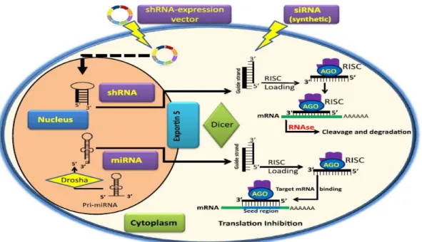
These are an endogenous single-stranded RNAs that are expressed in all higher eukaryotes with 19-25 nucleotides; they are generated in the nucleus and converted from 70 nucleotides hairpin precursors by Rnase III nuclease Dicer. They can induce gene silencing through destruction of homologous mRNA in plants or blocking its translation in plants and animals (Cullen, 2004; Mittal et al, 2012; Novina and Sharp, 2004).
siRNA
The mechanism of RNAi can be activated via the delivery of chemically synthesized exogenous siRNA to the cell, or by the introduction of dsRNA (Dorsett and Tuschl, 2004).
The dsRNA inserted is converted by Dicer into duplexes of 21 to 24 nucleotides
10
synthetic siRNA does not go through Dicer processing and it is directly incorporated into the
RISC. RISC unwinds the duplex siRNA and one strand bind to the target mRNA causing specific gene silencing (Singh et al, 2011).
shRNA
shRNAs can be generated from plasmids or viral vectors, exogenous vectors encoding a
shRNA construct is transfected into the cell, where it translocates to the nucleus and integrate
the genome. After that the pri-shRNA will be converted into pre-shRNA by Drosha. The
transfer of pre-shRNA to the cytoplasm is accomplished by the nuclear membrane protein
Exp5 then Dicer transforms it to siRNA capable of incorporating into a RISC to degrade or
block the target mRNA (De fougerolles et al, 2007; Paddison et al, 2002).
1.6.3-RNA interference mechanism
The process of RNAi can be divided into four stages: (i) double-stranded RNA cleavage,
(ii) silencing complex formation, (iii) silencing complex activation
(iv) mRNA degradation and recycling of the RISC complex
The cleavage of dsRNA into double-stranded fragments of 21-24 nucleotides long is the first step generating siRNAs. The integration of siRNAs into the protein complex called RISC is the second step of the process, however this complex is inert in this form and cannot induce RNAi.
11
Figure 4.Mechanism of RNAi (Singh et al, 2011)
1.6.4-Biological functions of RNAi
RNAi protects against viral infections and silences mobile elements
The information collected until now indicate that RNA silencing derive from an ancestral mechanism that controlled nucleic acids invasions. In different eukaryotic kingdoms RNA silencing is responsible for transposons, viruses and/or transgenes silencing. This supposition was verified by researches showing that silent transposons are reactivated in PTGS or RNAi impaired Arabidopsis, C. elegans and Chlamydomonas mutants (Hirochika et al, 2000; Ketting et al, 1999; Kuliñska- Szweykowska et al, 2003; Wu-Scharf et al, 2000).
RNAi-like mechanisms repress protein synthesis and regulate the development of organisms
Lately other type of endogenous RNA, called microRNA present in many organisms was discovered and led to many investigations on the nature and mechanism of these RNA molecules. These miRNAs bind to mRNA and regulate the gene expression by mRNA degradation or suppression of translation.
12
represents a new major principle of gene regulation. (Daneholt, 2006; Lagos-Quintana et al, 2001; Lee and Ambros, 2001).
RNAi-like mechanisms keep chromatin condensed and suppress transcription
It was proved after plant studies that gene silencing could occur at the transcriptional level (TGS). After the discovery of RNAi, it was indicated that TGS in plants operates via RNAi-like mechanisms. In the fission yeast Schizosaccharomyces pombe, and next on in Drosophila and vertebrates, it was discovered that similar processes keep heterochromatic regions condensed and transcriptionally suppressed (Daneholt, 2006).
1.6.5-Applications Functional genomics
The technique of RNAi is used to discover novel genes involved in disease processes. In order to study the function of genes, siRNAs are used to knock down/ knockout the expression of these specific genes. Various investigations have been undertaken to clarify the role of specific genes in basic cellular processes like DNA damage response and cell cycle control general cell metabolism, signaling, the cytoskeleton and its rearrangement during mitosis, membrane trafficking transcription and, DNA methylation. Thus, this method has become the most commonly used technique for gene annotation (Mittal et al, 2012).
Genetic improvement of crop plants
In order to improve the quality of crop plants the RNAi technique is used to enhance nutritional values or make plants resistant to pests; like the barley, crop that establishes a resistance against the barley yellow dwarf virus after utilization of RNAi (Wang et al, 2000).
RNAi has been confirmed to improve rice plants by decreasing its glutenin level and producing low glutenin content-1. The product LGC-1 is a low-protein rice and it is helpful for patients with kidney disease whose protein intake is restricted (Angaji et al, 2010).
13 RNA interference as a novel therapeutic agent
The ability to operate this native RNAi pathway has been identified as one the most exciting biotechnology advances in the last decade. RNAi is a way to control the development of diseases earlier in their process. The field of RNAi is progressing at an impressive rate, generating good results and their use has been documented for many diseases (Mittal et al, 2012).
In cancer research, various oncologic targets have been published in the literature, such as the employment of transferin with nanoparticles to target Ewing's sarcoma cells in a mouse xenograph model. This study confirmed the feasibility of using non-lipid based nanoparticles for the targeted delivery of siRNAs in a cancer model, and gives a powerful proof of principle of systemic delivery of siRNAs to a metastatic cancer (Ambesajir et al, 2012; Aagaard and Rossi, 2007).
HIV infection can also be stopped by targeting either viral genes (such as; gag, rev, tat and env) or human genes (for example, cd4, the principal receptor for HIV) that are implicated in the HIV life cycle. The strategy was used to suppress the central structural protein in the virus, p24, and the human protein CD4, which the virus needs to enter the cells (Mittal et al, 2012; Paddison et al, 2002).
1.7-The green fluorescent protein (GFP)
The gene encoding GFP was cloned from the jellyfish Aequoria victoria which is being used as a marker for gene expression and protein targeting in intact cells and organisms (Tsien, 1998). GFP’s normal function is the conversion of the blue chemiluminescence of the Ca+2-sensitive photoprotein aequorin into green light emission (Cody et al, 1993).
GFP is a relatively small protein of only 238 amino acids that does not interfere with biological processes, GFP was often used in transgenic investigations because it is a small and inert molecule that does not affect any biological process. The active protein does not need a cofactor or exogenous substrates to fluoresce, and it does not require the destruction or the fixation of organisms to be visualized in living cells (Chalfie, 2009; Ward et al, 1980).
15
2-OBJECTIVE
The main goal of this work is to contribute to a better understanding of the molecular interactions between P. cinnamomi and C. sativa by examining the effect of pathogen gene silencing on the host phenotype and studying the subcellular localization of GIP and NPP1 proteins involved in the mechanism of infection, which is essential for the implementation of control strategies.
To reach this aim, these specific objectives will be performed:
1/ Design of gene constructs for silencing the gip gene by RNAi and transformation of P. cinnamomi by the genetic construct
2/ C. sativa infection with P. cinnamomi transformed and non-transformed in order to determine the effect of gene gip silencing
3/ Fusion of the ORFs of gip and npp1 genes into the pTOR-eGFP plasmid for subsequent P. cinnamomi transformation and observation of protein subcellular localization using fluorescent microscopy
17
3-MATERIALS AND METHODS 3.1-Biological materials
Phytophthora cinnamomi
Phytophthora cinnamomi were isolated from soil samples associated with Castanea sativa Mill trees, affected by ink disease in Trás-os-Montes region (northeast of Portugal),
characterized by molecular methods and deposited in the Spanish Type Culture Collection
(CECT) with CECT 20919 code.
Escherichia coli
The NZY5α competent Escherichia coli cells (NZYTech, Portugal) which have similar properties to DH5α were used throughout this work. The NZY5α competent cells allow high efficiency transformation in a wide variety of application.
The NZY5α competent cells offer the following benefits:
-Increased performance in extracting plasmids due to endA1 mutation -Blue/White screening capability due to lacz M15
-Ensure the stability of the insert due to ecA1 mutation. Castanea sativa (Chestnuts)
Chestnuts of one year age have been used. The stratification of chestnuts was made in advance in the sand, and maintained in humid conditions in order to have germination. The germinated Chestnuts are planted in alveoli trays with a size of 6 cm x 6 cm x 20 cm for normal growth of the plants.
3.2-Culture media
The media were prepared in ultra-filtered water. Solid media were prepared by adding agar to the corresponding liquid media. All media were autoclaved at 120 °C for 20 minutes. In order to select clones resistant to antibiotics, media were supplemented with the corresponding antibiotic in a suitable concentration before pouring into plates.
3.2.1-Culture medium used for E. coli
18
The solution was then autoclaved. After cooling, the respective antibiotic was added at a concentration of 100 μg/ml, then the solution was aliquoted in sterile bottles, 5 ml in each, covered with the lid and stored at room temperature.
For agar plates, 1.5% (w/v) of agar was added, and the plates were poured while the medium was liquid. Then the plates were kept at room temperature until the agar set and then they were stored at 4 °C.
3.2.2-Culture media for P. cinnamomi V8 liquid medium
V8 medium was prepared by adding to 330 ml of V8 juice (Campbell Soup Co, Camden, NJ), 4.5 g of calcium carbonate (CaCO3) and stirred for 30 min. Culture media was transferred to 1000 ml centrifuge flasks and centrifuged at 2500 x g for 15 min at 20 °C.
The supernatant was then, poured into a new flask without disturbing the pellet. The cleared V8 juice (V8 broth) was then, diluted 10 fold with ultra-filtered water and
autoclaved at 120 °C for 20 min. V8 solid medium
It was prepared as aforementioned, but 15 g of agar per 1000 ml was added to the diluted V8 broth before autoclaving. After sterilization, the culture media was poured into Petri dishes, allowed to solidify at room temperature and kept at 4 °C.
YEPD (Yeast Extract Peptone Dextrose Medium)
The liquid YEPD medium was prepared by adding the following: yeast extract 1%, peptone 2% and D-glucose 2%.
The solid YEPD medium was prepared as above, but adding 2.0% Agar. PDA medium
PDA (Potato Dextrose Agar – HIMEDIA) was prepared by adding 39 g of commercial PDA powder to 1 L of ultra-filtered water, then autoclaved at 120 °C for 20 min.
3.3-Culture conditions and maintenance of microorganisms
19
under 150-200 rpm agitation in orbital shaker (Stuart®, S150). Additionally, to maintain viable strains for a long time, the microorganisms were stored in glycerol 15-30% at low temperatures -70 ° C.
The bacterial strain used throughout this work was stored at -70 °C, in 2 ml cryovials in their culture medium with 1/3 volume glycerol 100%. After subcultures in Petri dishes in agar LB medium E. coli were stored at a temperature of 4 °C.
3.4-Vectors
The vectors used were : pTOR-eGFP vector
Howard S. Judelson (University of California, Riverside, USA) has made this vector with approximately 7 kb in size, containing the neomycin resistance gene. This vector is regulated by promoters and terminators from the hsp70 and ham34 genes of Bremia lactucae. This vector was used to clone the PCR products of gip and npp1 genes for further transformation of P. cinnamomi and observation of the subcellular localization of GIP and NPP1 proteins under fluorescent microscopy (Figure 5).
Figure 5.Linearised map of the pTOR-eGFP vector
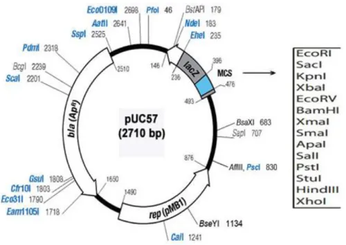
pUC57 vector
20
Figure 6.Map of the pUC57 vector
pTH210 vector
This expression vector was made by Howard S. Judelson with 5030 bp driven by hsp 70 promoter and containing the hygromycin resistance gene. This vector was used in order to insert the gip silencing cassette into the genome of P. cinnamomi (Figure 7)
Figure 7.Map of the pTH210 vector
21
3.5-DNA extraction
There are several methods for the extraction of genomic DNA (depending on the type of the biological sample), although all methods involve cell lysis, followed by deproteinisation and DNA purification.
For the extraction of P. cinnamomi’s genomic DNA, the oomycete has been grown in PDA medium, covered with aluminum foil at 25°C and after six days of mycelial growth, DNA was extracted.
The process of extraction consisted in the use of a Lysis Solution, 200 mM Tris-HCL; 25 mM EDTA; 250 mM NaCl and SDS 0.5 % (w/v) followed by a deproteinisation with phenol / chloroform / isoamyl alcohol (25: 24: 1) and precipitation of DNA by washing with ethanol (100 % - 70 %) at -20°C, and the DNA pellet was then dissolved in ultra-filtered water. The treatment of DNA was done with RNase 5 mg/ml for 5 minutes at 37 ºC.
3.6-Visualisation and purification of nucleic acids in agarose gels
The visualization and separation of DNA fragments were performed by electrophoresis in agarose gel low melting 0.8 % (w/v) in TAE (Tris-Acetate 40 mM, 1 mM of EDTA),
with 0.5 μg/ml GreenSafe Premium (NZYtech,Portugal), for 40 minutes at room
temperature, at 80 V.
After irradiation of the gel using the ChemiDoc™ XRS+ imaging system (BioRad) it was possible to view the size and intensity of the desired band by comparison with DNA ladders (Figure 8).
22
The Isolation and purification of DNA fragments from agarose gel were made by cutting the agarose band containing the desired fragment from gel prepared in TAE with a clean razor blade. DNA molecules present in the band were purified by the QIAquick® Gel Extraction Kit (QIAGEN, Germany) following the manufacturer's instructions.
3.7-Design of silencing cassette (shRNA based vector) for gip gene
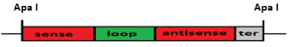
Several methods are used for constructing silencing cassettes; including annealed oligonucleotide and PCR-based cloning. The PCR-based method has been chosen because it is cheaper and reliable. The silencing shRNA construct was made by joining a 5’sequence fragment to another fragment from the 3’ end of the same sequence in an inverted orientation (by performing two separate PCR reactions) separated by a spacer or loop DNA (Figure 9). Transcripts generated from such constructs will have regions of self-complementarity that have the potential to form shRNA duplexes that generate a siRNA in the cell capable of degrading specific sequences of mRNA.
Figure 9.Structure of the silencing cassette (red for sense and antisense sequences, green for the loop sequence, grey for terminator).
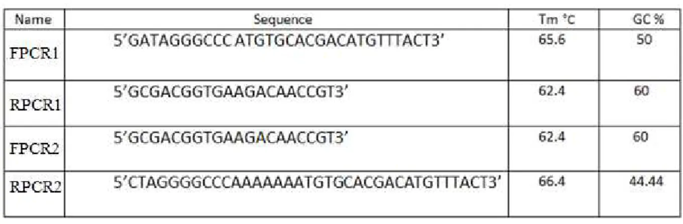
The amplification of the sense sequence selected within the ORF of gip gene has been done using specific primers designed following Tiscornia et al protocol (Table 1) (Tiscornia et al, 2007).
The forward primer includes: (1) 4–5 extra nucleotides to assist in digestion following PCR; (2) a 5' restriction site recognition sequence in order to insert the PCR product into the pUC57 vector; and (3) approximately 20 nucleotides of 5' sense strand sequence, the reverse primer contains only 20 nucleotides complementary of the 3' antisense strand sequence.
23
solution are retained on the silica membrane then with a washing solution the components of PCR mix are eliminated. Finally, the DNA is eluted in a nuclease free water.
The ligation to the loop sequence (provided by Professor Sophien Kamoun - The Sainsbury Laboratory (TSL), Norwich, UK) was done using T4 DNA Ligase (Promega) and following the manufacturer's instructions.
For the amplification of the antisense sequence; forward primer with approximately 20 nucleotides and a reverse primer including: (1) 4–5 extra nucleotides to assist in digestion; (2) 3' restriction site recognition sequence; (3) AAAAA termination sequence complementary strand; and (4) 20 nucleotides complementary of 3' antisense strand sequence were used. The PCR product was purified as aforementioned and ligated with T4 DNA Ligase (Promega) to the fragment containing the sense sequence and the loop.
Table 1.List of primers used for gip silencing cassette construction
The construct with approximately 551 bp length, was purified after ligation and then digested with ApaI restriction enzyme in a final volume of 30 μl with a reaction buffer (1x) (Promega), 1.5 uL (7U / microl) of ApaI enzyme, 1 ug / μl DNA, for 4 hours at a temperature of 37 °C.
In order to approximate the concentration comparing with the 500 bp band of the DNA ladder (Promega 100 bp ladder), the digested construct was after that visualized on a 1% agarose gel, excised with a clean razor blade and purified using the QIAquick® Gel
Extraction Kit according to manufacturer’s instructions (QIAGEN, Germany) so that can be
24
Figure 10.Methodology for shRNA silencing cassette design
3.8-Transformation of E. coli with the silencing construct
25
3.9-Extraction of the plasmid DNA of the transformed E. coli
The bacterial colonies, that have grown during the incubation period and which were expected to contain recombinant plasmids, have been picked off and transferred to the LB liquid media containing 100 μg/ml of ampicillin antibiotic. The cultures were incubated overnight on the shaker at a shaking rate of 200 rpm at 37 ºC.
The plasmid miniprep has been made, using the NZYMiniprep kit (NZYtech,Portugal) according to manufacturer’s instructions which is based on alkaline lysis of bacterial cells followed by adsorption of DNA onto silica. Afterward, the recombinant plasmid was digested by ApaI restriction enzyme then, the digested products were subjected to electrophoresis in agarose gel. The insert was purified from the gel by QIAquick® Gel Extraction Kit according to manufacturer’s instructions (QIAGEN, Germany).
3.10-Cloning the silencing cassette into the pTH210 vector
The pTH210 vector was extracted from E. coli previously transformed, digested with ApaI enzyme; as described for the cassette digestion and then dephosphorylated with Calf Intestinal Alkaline Phosphatase (CIAP).
Dephosphorylation was performed to remove phosphate groups from the 5 'ends of the linearized plasmid and prevent recircularization. The linearized plasmid was incubated with CIAP, "Calf Intestine Alkaline Phosphatase" (Promega,USA) (0.01u/μl) with 5 µl of the appropriate buffer CIAP 10X and ultra-filtered water to a final volume of 50 µl for 30 minutes at 37 ° C.
The fragments of the silencing cassette and the vector pTH210 were excised with a clean razor blade and purified using QIAquick® Gel Extraction Kit according to
manufacturer’s instructions (QIAGEN, Germany) then ligated using T4 DNA Ligase enzyme.
To clone the cassette into the pTH210 vector the ligation of DNA fragments was carried out in a 10 μl standard ligation reaction. The reaction contained DNA fragments with compatible ends, 100 ng of plasmid were used for a typical ligation reaction, 0.5 μl of T4 DNA ligase, and a 1μl of ligation mix buffer (Promega). Reactions were incubated at 4 °C overnight.
26
3.11-Transformation of Phytophthora cinnamomi zoosporeswith the recombinant pTH210 vector
The production of zoospores was made according to an adaptation of the protocol prepared by Edgar Huitema (Huitema et al, 2011). The zoospores used for transformation were subjected to centrifugation and the pellet was resuspended in 1.0 M mannitol / V8 medium (4/1) and regenerated for 16 hours at 25 °C in the dark.
After incubation, the zoospores were collected again by centrifugation at 3300 xg for 5 minutes, washed with a regenerating solution, and the recombinant pTH210 vector was added together with the zoospores.
After achieving the transformation procedures, P. cinnamomi with the recombinant pTH210 vector was grown in medium containing hygromycin (200 ug / ml), the growth was incubated for 10 days. At the same time non-transformed P. cinnamomi was cultured as a negative control in medium containing hygromycin (200 ug / ml) for the same incubation time.
3.12-Genomic analysis of the transformed Phytophthora cinnamomi (PCR & DNA sequencing)
The mycelium of transformed P. cinnamomi was selected in order to extract the genomic DNA and make a screening PCR to confirm the integration of the pTH210 into P. cinnamomi genomic DNA. The screening PCR was performed with the primers listed in
Table 2.
Table 2.List of primers used for PCR of the cassette and the hygromycin fragment
27
sequencing with respective primers and using an automated sequencer ABI PRISM 377W that perform the electrophoretic separation and detection of DNA fragments labeled with fluorescence.
Four different colors of fluorescence can identify the four dideoxynucleotide incorporated into the extension reaction (A, G, T or C). After analyses with BioEdit program the nucleotide sequences were blasted on (www.ncbi.nlm.nih.gov/BLAST/) to investigate the ID of the sequenced genes. The alignment of similar genes, was analysed and compared using the Muscle tool server (CLUSTAL multiple sequence alignment) from EMBL-EBI database (http://www.ebi.ac.uk/Tools/msa/muscle/).
3.13-Castanea sativa infection with P. cinnamomi strains
Infection of C. sativa roots was performed using mycelium of P. cinnamomi
transformed and non-transformed in order to compare the effect of gene silencing on the plant
phenotype. The roots were covered with fully colonized V8 medium, the plants were placed in sterile vermiculite and incubated for 72 hours at 25ºC.
3.14-Fusion of the ORF’s into the pTOR-eGFP vector
3.14.1-Primer design to amplify the ORF’s of gip and npp1 genes of Phytophthora cinnamomi by PCR
For the amplification of gip’s ORF, primers were designed with adapters having enzyme restriction sites to allow the insertion of DNA fragments in the pTOR-eGFP vector.
For gip gene, NotI (gcggccgc) and AatII (gacgtc) restriction sites were chosen to be incorporated in primers sequences since this restriction sites are present in the vector pTOR-eGFP.
28
Figure 11.The sequence of gip gene (in black the non coding sequence of the gene , in red the ORF of gene , in blue based sequences for primers design)
The specific characteristics of the primers such as the melting temperature (Tm) and the efficiency of primers were determined with the program FastPCR.
The Restriction Mapper website (http://www.restrictionmapper.org/) was used to assure the absence of restriction sites for NotI and AatII in the ORF since both of these restriction enzymes will be used for cloning the PCR product into the pTOR-eGFP vector.
29 Table 3.List of primers used for PCR of gip and npp1 genes
3.14.2-PCR amplification of gip and npp1
The PCR technique is a methodology that is based in in-vitro amplification based on an in-vivo process (replication). This allows an exponential and selective synthesis of a reduced amount of DNA. The technique consists in a cycle of three phases, which comprises the DNA denaturation, hybridisation (annealing) of primers and polymerization of DNA by Taq DNA polymerase enzyme.
The thermal cycler ''my cycler'' (Bio-Rad) was used to amplify a 810 bp fragment of the gip gene, with the primers designed as described in Table 3. The PCR cycling conditions were; 94 ºC /5 min, followed by 36 cycles of 94 ºC /1 min; 61 ºC /1 min; 72 ºC /30 s, and ending with 72 ºC /5 min. Each 50 µl PCR contained 0.8 mM dNTP, 0.2 mM of each primer, 100 ng genomic DNA, and 1 U Taq DNA polymerase in the appropriate buffer.
The PCR of npp1’s ORF was done as mentioned for gip gene but with 66 °C as annealing temperature.
3.14.3-Enzymatic digestion and dephosphorylation of pTOR-eGFP
Digestion with restriction enzymes is an essential step for the construction of recombinant vectors. Enzymes were selected according to the restriction sites in the DNA sequence, or from the plasmids map.
30
The first digestion of gip’s ORF was done in a final volume of 30 µl with a reaction buffer (1x) (Promega), 1.5 µl (7U / microl) of NotI enzyme, 1 ug / µl DNA, for 4 hours at a temperature of 37 °C. After incubation and purification, the insert was digested with AatII enzyme as described for NotI enzyme.
The ORF of npp1 gene was digested first by NotI enzyme, purified and digested again with SacII enzyme.
On the other hand the plasmid pTOR-eGFP was extracted from E.coli previously transformed, digested separately with respective restriction enzymes, and then dephosphorylated with CIAP as described in section 3.10.
The insert and the plasmid were after that visualized on agarose gel 0.8% (w /v) in order to approximate the concentration of both fragment. The estimation of the insert and the plasmid concentrations is a very important step for the efficiency of the ligation reaction. The approximation was done after visualization on agarose gel and comparing with the 500 bp band of the 100 bp, DNA ladder (Promega) which has a known concentration, (the same volume was used for the DNA ladder, the vector and the insert in electrophoresis).
The amount of the insert in nanograms required for the reaction was estimated according to the following ligation equation :
ng of insert = (ng of vector × kb size of insert)kb size of vector × (molar ratio of vector )insert
3.14.4-Fusion of the PCR products into the pTOR-eGFP plasmid
The insert and the plasmid were excised from agarose gel with a clean razor blade, purified using the QIAquick® Gel Extraction Kit and then allowed to ligate at 3:1 ratio by incubating the reaction mixture as reported in section 3.10 overnight at 4 °C.
3.15-E. coli Transformation with the recombinant pTOR-eGFP vector
This task was done as described in section 3.8, but using the neomycin antibiotic.
3.16-Extraction of the plasmid DNA of the transformed E. coli
31
recombinant plasmid with npp1 gene, the digestion was made using NotI and SacII enzymes and then, the digested products were subjected to electrophoresis in agarose gel.
3.17-Prediction of the subcellular localization of the GIP and NPP1 proteins
Protein subcellular localization prediction foretells the destination of a protein in the cell through computational methods that take as input the protein sequence for example and produce a prediction of the protein localization as output.
There are several softwares publicly available using different methods for predicting the localization of proteins (Amino Acid Composition, N-peptide Composition, Physico-chemical Composition … ) which is a very important part of bioinformatics based prediction of protein function and genome annotation.
The softwares used for the protein localisation predictions can be accessed through the URL addresses as follows:
-SignalP 3.0: (http://www.cbs.dtu.dk/services/SignalP-3.0/) -Cello: (http://cello.life.nctu.edu.tw/)
-LOCtree: (https://rostlab.org/services/loctree2/)
33
4-RESULTS AND DISCUSSION
4.1-DNA extraction of Phytophthora cinnamomi
After six days of mycelial growth in the dark at 25°C, the genomic DNA of Phytophthora cinnamomi was extracted to design the gip silencing cassette and to amplify the
ORF’s of gip and npp1 genes (Figure 12).
Figure 12.Visualization of Phytophthora cinnamomi genomic DNA in agarose gel 0.8 % (w/v) M) DNA ladder of 1 kb (Promega); 1) genomic DNA of Phytophthora cinnamomi
The gel showed that the extracted DNA is concentrated, not degraded and does not contain proteins neither RNA molecules.
4.2-Synthesis and preparation of the gip silencing cassette
In order to synthesize an efficient conventional silencing cassette that targets the corresponding mRNA of the gip gene, many measures should be taken into consideration: -The potential construct sequence should not have a perfect match of more than 16 nucleotides to an off-target gene of P. cinnamomi (verifying by BLAST search) (http://www.ncbi.nlm.nih.gov/BLAST/).
-The construct should not have an internal repeat or palindromes and must include low-to-medium G/C content (Cheng and Chang, 2007).
34
The sense and anti-sense sequences were produced by performing two separate PCR reactions then the PCR products were ligated by T4 DNA ligase (Figure 13).
Figure 13.The sense,loop and antisense of the gip silencing cassette
The generated cassette was purified, digested with ApaI enzyme and then visualized on 1% agarose gel, as shown in figure 14.
35
The gel showed a specific band of 543 bp length with a good DNA quality and confirmed the success of producing a silencing cassette that can trigger a post transcriptional gene silencing.
RNA interference using shRNA has been chosen because previous studies have shown that shRNA-expression system is more efficient than siRNA expression systems (Cheng and Chang, 2007). In addition, the cost of shRNAs construction is cheaper compared with synthesized siRNAs and for stable and enforceable expression it is also recommended to use shRNAs because it integrates the genome and can be expressed for up to three years while synthesized siRNAs are degraded after 48 hours (Rao et al, 2009; Paddison et al, 2004).
4.3-Transformation of E. coli with the gip silencing cassette
The silencing cassette was purified using the QIAquick® Gel Extraction Kit and then cloned into the pUC57 vector in order to transform E. coli competent cells. The transformed bacteria that are able to grow in medium containing ampicillin have been selected for further experiences (Figure 15).
Figure 15.Screening of transformed E. coli resistant to ampicillin antibiotic A) E. coli transformed was able to grow in medium containing ampicillin; B) non-transformed E. coli was not able to grow in medium containing ampicillin
36
At the same time, the pTH210 expression plasmid was extracted from E. coli cultured cells and linearized with the ApaI restriction enzyme with the purpose of preparing the cloning of the silencing cassette.
The products resulting from the enzymatic digestion were analyzed on agarose gel (0,8%) and a band of 5030 bp corresponding to the pTH210 linearized plasmid was observed (Figure 16).
Figure 16.Enzymatic digestion of pTH210 plasmid and the recombinant pUC57 plasmid with ApaI M) DNA ladder of 1 kb (Promega); CN) Negative Control pTH210 supercoiled plasmid; 1) pTH210 plasmid linearized by ApaI enzyme; 2) pUC57 recombinant plasmid digested by ApaI enzyme generated two fragments
The two bands generated after digestion with ApaI enzyme; of 2710 bp length corresponding to the vector pUC57 size and of 543 pb corresponding to the cassette size confirm the success of cloning the silencing cassette into the pUC57 plasmid.
The extraction of the recombinant vector followed by digestion with respective restriction enzymes was done from different colonies to confirm the ligation of the cassette into the pUC57 cloning vector.
4.4-Cloning the silencing cassette into the pTH210 expression vector
37
The recombinant vector was used to transform competent E. coli NZY5 α cells. After plasmid extraction, the enzymatic digestion with ApaI, PstI and SmaI enzymes, was conducted to confirm the cloning of the cassette into the pTH210 vector.
After visualisation on agarose gel (0,8%) the digestion with the enzyme ApaI generated two bands; one for the vector pTH210, with 5030 bp length and one of 543 bp corresponding to the cassette. Digestion with PstI enzyme released a larger fragment of 4115 bp and a smaller of 1458 bp. The digestion with SmaI enzyme produce a larger fragment of 4320 bp and another smaller of 1253 bp (Figure 17).
Figure 17.Enzymatic digestion of the recombinant pTH210 vector with ApaI, PstI and SmaI M) DNA ladder of 1 kb (Promega); CN) Negative Control non recombinant pTH210 supercoiled plasmid; 1) recombinant pTH210 plasmid digested by ApaI enzyme; 2) recombinant pTH210 plasmid digested by PstI enzyme; 3) recombinant pTH210 plasmid digested by SmaI enzyme
The virtual enzymatic digestion based on the pTH210 sequence, performed using the Restriction Mapper website (http://www.restrictionmapper.org/) complies with the products of digestion showed in figure 17. These results allow the confirmation of cloning the cassette into the pTH210 vector.
38
and Pythium. Moreover it was used in many researches for stable DNA integration (Chen et al, 2009; Judelson and Ah-Fong 2009).
4.5-Confirmation of P. cinnamomi transformation by culture in medium containing hygromycin antibiotic
The recombinant plasmid was used to transform Phytophthora cinnamomi by electroporation, then the transformants were cultivated on medium containing hygromycin (200 ug / ml) and incubated for ten days at 25 °C in the dark.
At the same time non-transformed P. cinnamomi were incubated in the same conditions as negative control plates in medium containing hygromycin (200 ug / ml).
After ten days of incubation at 25 °C in the dark, there was no growth of the negative control of P. cinnamomi in medium containing the hygromycin selection marker, while for the transformed P. cinnamomi only six out of ten grew in the medium with hygromycin . (Figure 18).
Figure 18.Screening of P. cinnamomi transformed with the recombinant pTH210 vector, resistant to hygromycin antibiotic A) P. cinnamomi transformants are able to grow in medium containing hygromycin; B) non transformants P. cinnamomi was not able to grow in medium with hygromycin
39
with the transformation of P. cinnamomi by the pTH210 vector meaning that this transformation was successful.
4.6-Genotypic confirmation of P. cinnamomi transformation
In order to confirm the transformation of P. cinnamomi by PCR screening, the genomic DNA of transformed P. cinnamomi was extracted after ten days of incubation and specific primers were designed to amplify a region of approximately 551 bp corresponding to the gip silencing cassette sequence and other region of 1090 bp within the hygromycin gene (hpt). With these procedures it is expected to prove the integration of the recombinant plasmid pTH210 in the genome of P. cinnamomi (Figure 19).
Figure 19.Visualization of the silencing cassette and the hygromycin fragment PCR products M) DNA ladder of 1 kb (Promega); 1) PCR product of the silencing cassette 551 bp; 2) PCR product of the hygromycin fragment 1090 bp
The amplification by PCR of the gip silencing cassette produced a band of approximately 551 bp while the amplification of the hygromycin segment produced a band of 1090 bp. The two PCR products were purified, then digested by restriction enzymes to have another proof of the fragments identity