Review
Printed in Brazil - ©2015 Sociedade Brasileira de Química 0103 - 5053 $6.00+0.00
*e-mail: [email protected]
A preliminary version of this article was published in Portuguese at Rev. Virtual Quim. 2015, 7, 74 (reference 4).
This review is dedicated in memoriam to Prof Giuseppe Cilento (1923, Sorrento, Italy-1994, São Paulo, Brazil) in recognition of his remarkable creativity, kindness, scientiic contributions and mentorship.
“Photo” Chemistry Without Light?
Wilhelm J. Baader, Cassius V. Stevani and Etelvino J. H. Bechara*
Departamento de Química Fundamental, Instituto de Química, Universidade de São Paulo, 05508-000 São Paulo-SP, Brazil
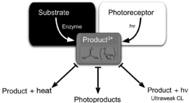
In the early seventies, Giuseppe Cilento (São Paulo University), Emil White (Johns Hopkins University) and Angelo Lamola (AT&T Bell Laboratories) postulated that typical photochemical reactions could occur in dark parts of living organisms if coupled to enzymatic sources of electronically excited products. Their paradoxical hypothesis of “photochemistry without light” was chemically anchored on the synthesis and weak chemiluminescence of several 1,2-dioxetanes, unstable cyclic peroxides whose thermal cleavage produces long-lived and reactive triplet carbonyls. Collisional reactions or energy transfer of triplet species to cellular targets could eventually result in “photo” products that potentially trigger normal or pathological responses. These ideas lourished in the labs of various researchers who attempted to explain the presence and biological roles of “dark” secondary metabolites, including plant hormones, pyrimidine dimers, alkaloid lumi-isomers, protein adducts, and mitochondrial permeators, thereby broadening the ield of photobiology.
Keywords: photochemistry in the dark, peroxidase, 1,2-dioxetanes, triplet carbonyl, chemiluminescence
1. Chemiluminescence and Bioluminescence
Chemiluminescence (CL)1 and bioluminescence (BL)2
are cold and visible light emissions from chemical reactions in the absence and in the presence of enzymes, respectively. These phenomena are the opposite of photochemical reactions, whose chemical transformations are initiated by light. In the former case, the energy of chemical bonds is converted into electronic excitation energy, whereas in photochemical processes the energy of the electromagnetic radiation is utilized to drive chemical transformations. Light emission in BL and CL can be intense, as in the case of irely BL or the peroxyoxalate CL; moderate, as in the case of luminol oxidation; weak, as the direct emission observed during 1,2-dioxetane decomposition or in fungal BL; or ultraweak, like that accompanying lipid peroxidation or peroxidase catalyzed aldehyde oxidation. In each case,
light can be considered to be one of the reaction products.3
In the last few decades, many chemiluminescent substrates have been discovered and utilized for the
development of a wide variety of analytical assays of environmental, clinical, biological and forensic
samples.3 One of the most important and well-known
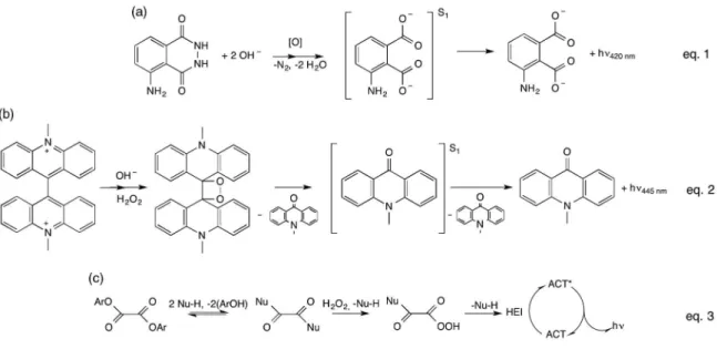
CL transformations is the oxidation of luminol (5-aminophthalhydrazide) catalyzed by many transition metals (Figure 1a) and widely employed in the detection of hydrogen peroxide and a vast number of transition metal ions. It is used, for example, in the characterization of redox imbalance in cells and biological tissues, as a sensitive detection system in immunoassays or in an antioxidant capacity assay. Noteworthy is its use to reveal traces of
blood in forensic chemistry.3
Other classical CL processes with wide analytical
application potential are (i) the transition metal-catalyzed
reaction of lucigenin (10,10'-dimethyl-9,9'-biacridylium salt) with hydrogen peroxide (Figure 1b) used mainly for transition metal quantiication, but also as a detection
system for oxidative metabolism, and (ii) the base-catalyzed
Emil White contributed to the development of this area by describing the synthesis and properties of luminol and the irely luciferin, two of the luminescent systems most exhaustively studied and widely used in analytical kits for
pure and applied chemistry.1,2
1.1. Peroxide intermediates in chemiluminescence: 1,2-dioxetanes, 1,2-dioxetanones, and 1,2-dioxetanedione
T h e d e p e n d e n c e o f c h e m i l u m i n e s c e n t a n d bioluminescent reactions on molecular oxygen or hydrogen peroxide led to the proposal that unstable four-membered ring peroxides, called 1,2-dioxetanes and 1,2-dioxetanones, are the “energy-rich” intermediates responsible for the
creation of excited products upon thermal cleavage.5 A
signiicant advance in the elucidation of chemiexcitation mechanisms of diverse substrates was achieved with the
synthesis of these peroxides in the 1960s and 1970s.5
Although the inal CL and BL products were indeed those expected from the cleavage of these cyclic peroxide intermediates, it was believed that their synthesis would be an arduous task, given the high steric strain of their 1,2-dioxacyclobutane structures. Moreover, their weak
O−O bond (ca. 140 kJ mol-1) and the strong thermodynamic
driving force towards their conversion into extremely stable carbonyl products (Figure 2) would contribute to their decomposition. 1,2-Dioxetanones should be even less stable
owing to the presence of an sp2 carbonyl carbon atom in
the four-membered ring. Therefore, it was expected that 1,2-dioxetanes would be too unstable to be isolated and could only exist as highly reactive intermediates, prone to cleave and release their intrinsic chemical energy in the
form of electronic excited products, which either emit light or undergo photochemical changes.
Despite the above-mentioned constraints, in 1969,
Kopecky and Mumford6 (University of Alberta, Canada)
reported the first synthesis of a 1,2-dioxetane at low temperature, 3,3,4-trimethyl-1,2-dioxetane, whose decomposition upon heating generated the expected decomposition products, acetone and acetaldehyde, and a bluish light emission. Soon thereafter, in 1972, Adam and
Liu7 (University of Puerto Rico, USA) reported the irst
synthesis of a 1,2-dioxetanone (α-peroxylactone), namely
the 3-tert-butyl-1,2-dioxetanone.
The presence of a carbonyl group in the peroxidic ring
makes it much less stable (Ea ca. 80 kJ mol-1, where Ea is
the thermolysis activation energy) than
3,3,4-trimethyl-1,2-dioxetane (Ea ca. 100 kJ mol-1).8
The unimolecular decomposition of 1,2-dioxetanes leads to the preferential formation of triplet-excited carbonyl compounds (Figure 2); the stability and quantum
Figure 1. Classical chemiluminescent reactions: (a) luminol; (b) lucigenin; (c) peroxyoxalate (adapted from reference 4).
Figure 3. Chemically initiated electron exchange luminescence (CIEEL) mechanism proposed for the decomposition of a 1,2-dioxetanone catalyzed by an activator (ACT) with low oxidation potential (adapted from reference 4).
yields of excited products are crucially dependent on the number and the nature, mainly the size, of the substituents in the peroxidic ring; and the stability of disubstituted 1,2-dioxetanes proved to be similar to that of
1,2-dioxetanone derivatives.9-12 The preferential formation
of triplet excited states (up to 60%) and the low quantum yields for singlet excited state formation (< 1%) imply a low CL emission quantum yield in the unimolecular decomposition of 1,2-dioxetanes. Therefore, this system is not a suitable model for eficient BL transformations,
contrary to the initial prediction.1,2 On the other hand, as
will be discussed later herein, triplet carbonyls have long lifetimes (> μs) and behave similarly to oxyl radicals, which gives these excited molecules the ability to promote radical chain reactions, ultimately leading to a plethora of photoproducts originating from isomerization, cyclization, cleavage, substitution, and hydrogen abstraction reactions.
The thermal cleavage of 1,2-dioxetanones shows characteristics similar to those presented by 1,2-dioxetane decomposition, with dominant triplet excited state formation and very low singlet excitation yields, and consequently low CL emission quantum yields (Figure 2). Interestingly, the chemiexcitation quantum yields of the thermolysis of 1,2-dioxetanones are lower than those of corresponding 1,2-dioxetanes, although the former possess higher energy
content.5 Nonetheless, studies conducted independently
by Schuster, Adam, Turro, and Wilson3,4 showed that
dioxetanones, speciically 3,3-dimethyl-1,2-dioxetanone,
the only α-peroxylactone derivative whose CL properties
have been thoroughly investigated, decompose faster in the presence of luorescent aromatic hydrocarbons, yielding the aromatic compound in its singlet excited state. The decomposition rate and eficiency of excited state formation were shown to depend on the concentration and oxidation potential of the aromatic hydrocarbon, called an activator (ACT), because these compounds “activate” peroxide decomposition. These experimental observations led to the formulation of the “chemically initiated electron exchange luminescence” (CIEEL) mechanism, which consists of an initial electron transfer from the ACT to the cyclic peroxide and concomitant O−O bond cleavage. The electron back-transfer from a carbonyl radical anion, formed by cleavage of the central C−C bond to the ACT radical cation, is responsible for the ACT’s excited state formation and
subsequent luorescence emission (Figure 3).13-16
The CIEEL mechanism was greeted with enthusiasm by the research groups of this area and frequently utilized to rationalize excited state formation in numerous CL
transformations,16 and frequently cited to explain the
chemiexcitation step of irely BL.17
The quantum yields initially determined for the catalyzed decomposition of 3,3-dimethyl-1,2-dioxetanone by various research groups (ca. 10%) indicated a reasonably eficient process, in agreement with the high emission quantum yields generally observed in BL transformations, thereby justifying the adoption of the CIEEL mechanism as a model for the bioluminescence of a number of luminescent organisms. However, recent redeterminations of the quantum yields obtained in the catalyzed decomposition of 3,3-dimethyl-1,2-dioxetanone and two other more stable 1,2-dioxetanone derivatives indicated that the quantum yields for these transformations are actually at least two
orders of magnitude lower than that initially reported.18
Although these observations might lead one to question the validity of the CIEEL hypothesis and its application to eficient BL transformations, recent experimental evidence has conirmed the occurrence of electron or charge transfer processes in these transformations. In addition, their low chemiexcitation eficiency has been associated with steric effects on complex formation between the peroxide and the
activator, using the supermolecule approach.19
Moreover, as early as the 1980s, it had been observed that the decomposition of certain 1,2-dioxetanes containing electron donor substituents occurs with the efficient
formation of singlet-excited states.20 The decomposition of
1,2-dioxetane derivatives, whose electron donor moiety is protected, can be induced by suitable deprotection agents (“induced 1,2-dioxetane decomposition”), namely chemical
reagents or enzymes.21 In the latter case, enzyme-induced
decomposition is the chemical basis of the detection system
of numerous immunoassays used in clinical assays.22 The
corresponding reaction mechanism involves, after chemical or enzymatic deprotection, an intramolecular electron transfer from the electron-rich substituent, generally a phenolate oxygen atom, to the cyclic peroxide unit, accompanied by subsequent O−O and C−C bond cleavage and a inal electron back-transfer, which may occur in either an inter- or intramolecular fashion and can lead to eficient singlet-excited state formation (Figure 4: path A,
mechanism of the induced 1,2-dioxetane decomposition constitutes the intramolecular version of the CIEEL mechanism.
Various research groups have shown that these 1,2-dioxetane derivatives possess high thermal stability and their induced decomposition leads to the eficient formation of singlet-excited states with excitation quantum yields of up
to 100%.23-25 The occurrence of an intramolecular electron
transfer from the electron donor substituent to the peroxidic ring has been demonstrated experimentally in a Hammett substituent study on a series of acridinium-substituted
1,2-dioxetanes.26,27 Additionally, it has been shown that
the formerly observed solvent-cage effect on the quantum
yields in the induced 1,2-dioxetane decomposition28-30 can
still be in agreement with an intramolecular electron back-transfer, indicating that this highly eficient process occurs
in an entirely intramolecular fashion.31
The results outlined above indicate an empirical general rule that the transformations of cyclic peroxides that involve intermolecular electron transfer processes exhibit low chemiexcitation eficiency, whereas the corresponding
intramolecular processes occur with high quantum yields.32
However, there is a CL system involving an intermolecular chemiexcitation process that produces extremely high CL emission yields: the peroxyoxalate
reaction.33 This reaction was discovered by Chandross,34
who observed intense light emission during the reaction of oxalyl chloride with hydrogen peroxide in the presence of a
luorescent compound. Rauhut35 (American Cyanamid Co.)
subsequently developed commercial applications for this system in the so-called ‘light sticks’ by using several oxalate derivatives, mainly esters and amides. The base-catalyzed reaction of oxalic esters with hydrogen peroxide occurs in a series of consecutive and parallel reaction steps and results in the formation of a high-energy intermediate, which is responsible for excited state formation upon interaction
with the luorescent activator (ACT) (Figure 1a).33 The
putative intermediate is the 1,2-dioxetanedione, a carbon
dioxide dimer, as already suggested by Rauhut;35 however,
to date there is no unequivocal experimental proof of its
existence.33 Excited state formation, which is responsible
for CL emission, occurs in this reaction in a sequence of electron transfers from the ACT to the peroxidic intermediate, bond cleavages and electron back-transfer steps in a viscous solvent cage, as indicated in a series of
recent studies.36-38 As the eficiency of the transformation
is undoubtedly high,33,35,36 this reaction is the only
chemiluminescent system occurring by an intermolecular CIEEL mechanism with proven high chemiexcitation
quantum yields.32 Additionally, the peroxyoxalate reaction
has found widespread analytical application and can be useful in chemistry education through experiments that illustrate the effects of concentration, pH, temperature and catalyst on the kinetics of a chemical reaction.
The reaction kinetics can be easily monitored visually from the course of emission intensity decay, which is
suficiently high to be photographed.39 Various oxalates and
CL activators, which elicit different colors (e.g., rubrene-yellow; perylene-green; 9,10-diphenylanthracene-blue), are sold in the form of ‘light sticks’ and used as attractors for ishing, in emergency kits, and as recreational objects. Although the contents are highly cyto- and genotoxic (in particular the activators), they are labeled as safe (provided the contents are not ingested or applied on the skin) and no
instructions are given for their proper disposal after use.40
Thousands of light sticks are used to attract pelagic ish and can be found discarded on beaches in Brazil’s northeastern regions, where naive locals use the oily content of the sticks for several purposes, e.g., as sun ilters, massage, insect repellent, or as an ointment to alleviate joint pain.
2. Why “Photochemistry Without Light”?
T h e d e c o m p o s i t i o n o f 1 , 2 - d i o x e t a n e a n d 1,2-dioxetanones leads to the generation of excited carbonyl products, mainly in the triplet state, which can undergo the same photophysical and photochemical
processes as when electronically excited by irradiation.41
Excited aldehydes and ketones decay by a variety of processes from the singlet as well as the triplet manifold,
which encompass homolytic C−C bond cleavage (α- and
β-cleavage), hydrogen abstraction (photoreduction), [2 + 2]
cycloadditions (Paternò-Büchi reaction), quenching by
conjugated dienes, and others (Figure 5).42
In the early seventies, anchored on the chemistry of 1,2-dioxetanes, which tend to yield long-lived and reactive triplet carbonyls, and on the identification of typical photoproducts in tissues of plants and animals never directly exposed to light, Emil White (Johns Hopkins University), Angelo Lamola (AT&T Bell Laboratories) and Giuseppe Cilento (University of São Paulo) postulated the hypothesis of “photochemistry without light” or “photochemistry in the dark,” which seemed at irst sight to be a paradox. The idea behind their hypothesis is that “photoproducts” can be formed in living cells from electronically excited precursors, which have been formed in the dark from appropriate enzyme-catalyzed or chemical transformations, not from direct light absorption (Figure 6). Triplet carbonyl species seemed to be excellent candidates for “photochemistry in the dark,” as they are long-lived (> microseconds) and can react
like a diradical, particularly as an alkylperoxylradical.5,43
Accordingly, they are expected to: (i) abstract hydrogen
atoms from polyunsaturated fatty acids (PUFAs), initiating
their peroxidation; (ii) undergo cleavage, yielding
carbon-centered or oxygen-carbon-centered radicals; and (iii) transfer
electronic energy to several biological acceptors, followed by light emission or chemical transformations.
This hypothesis is strongly supported by the occurrence of some “dark” photoproducts in living organisms, such as cyclobutane dimers, which cannot be credited to ground state reactions because, according to the Woodward-Hoffmann rules, these [2 + 2] cycloaddition reactions are forbidden in the ground state, but allowed in the electronically excited state. According to these rules, concerted transformations such as cycloaddition, electrocyclic, sigmatropic and group transfer reactions, are “allowed” or “forbidden” in the ground state or in the excited state because of changes in the orbital symmetry of reagent and product, depending on
the electronic distribution.44
Figure 5. Photophysical and photochemical transformations of acetone upon excitation to singlet and triplet states (*); (i) thermal deactivation; (ii) luorescence and phosphorescence emission; (iii) energy transfer to an acceptor molecule (A), possibly followed by photophysical (hν, heat) or photochemical processes of A (photoproducts); (iv) energy transfer from triplet acetone to molecular oxygen, generating highly reactive singlet oxygen; (v) 1,2-cycloaddition to alkenes, yielding an oxetane (Patern® -Büchi reaction); (vi) hydrogen abstraction from suitable H-donors like alcohols and 1,4-dienes, leading to (vii) the reduction product 2-propanol and dimerization product 2,3-dihydroxy-2,3-dimethylbutane (pinacol); (viii) C−C bond homolysis (α-cleavage) to a methyl and an acetyl radical, which can undergo decarbonylation or dimerization to diacetyl40 (adapted from reference 4).
Motivated by the photochemistry of excited carbonyls, Cilento and White looked for secondary metabolites in the biochemical and biological literature, whose origin is “allowed” preferentially from the excited state, aiming to validate the hypothesis of dark photochemistry. Their search for enzymatic sources of triplet species was
founded upon: (i) the reported chemical mechanisms
of chemiluminescence and bioluminescence; (ii) the
structural similarity between luciferins and potential sources with respect to the presence of a carbonyl-activated
α-hydrogen atom, and (iii) enzymatic products identical
to those obtained from a hypothetical 1,2-dioxetane or 1,2-dioxetanone intermediate.
2.1. Emil White’s contributions to “photochemistry in the dark”
White’s work focused on the synthesis and use of 1,2-dioxetanes as clean sources of excited carbonyl species that could transfer electronic energy to classical photoreceptors, whose products are chemically similar to
those found in plants. In White et al.45 review published in
1974, it was exempliied several photochemical reactions that could take place in the dark at the cost of dioxetane
thermolysis. These reactions included: (i) isomerization
of trans-stilbene to the cis-isomer coupled with the thermolysis of 3,3,4-trimethyl-1,2-dioxetane, a reaction analogous to the isomerization of cinnamates in sweet
clover (Figure 7); (ii) [2 + 2] cycloaddition of
dioxetane-generated singlet acetone to 1,2-dicyanoethylene, yielding an oxetane somewhat similar to the dimerization of
cinnamates to truxillates in coca (Erythroxylum coca),
and triplet acetone-induced isomerization of trans
-dicyanoethylene (Figure 8); (iii) cyclic rearrangement
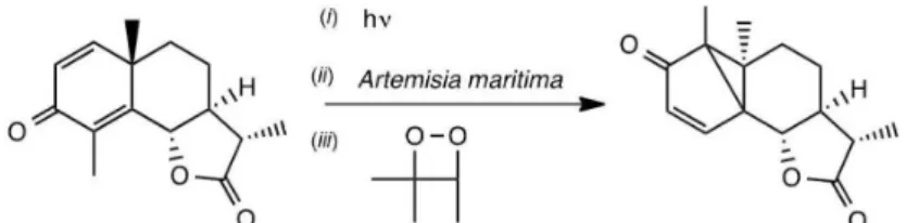
of santonin into lumisantonin, both present in absinthe (Artemisia maritima), coupled to the thermolysis of 3,3,4-trimethyl-1,2-dioxetane (Figure 9).
To the best of our knowledge, the first in vivo
demonstration of “photochemistry in the dark” was given
by Bechara and co-workers,46 in a study of the electrocyclic
ring closure of the tropolonic alkaloid colchicine into
lumicolchicines in the corms of autumn crocus (Colchicum
autumnale). This is a short-day plant used since ancient times as a source of colchicine to alleviate gout pain. In the
Figure 7. Photochemical and dioxetane-induced cis,trans-isomerization of stilbene and analogous reaction of cinnamic acid in sweet clover (Melilotus albus) (adapted from reference 4).
Figure 9. Isomerization of santonin to lumisantonin in absinthe, either photochemical or induced by chemically-generated triplet acetone (adapted from reference 4).
Figure 10. Disrotatory electrocyclic ring closure of colchicine into isomeric β- and γ-lumicolchicines, under sunlight radiation or in underground corms of autumn crocus Colchicum autumnale. Continuous exposure of colchicine to light leads to the dimerization of β-lumicolchicine to the cyclobutene derivative
α-lumicolchicine (adapted from reference 4).
winter, 14C-colchicine was infused into underground corms
of the plant, without leaves and lowers. After two days,
radiolabeled β- and γ-lumicolchicines (respectively, trans-
and cis-cyclobutene isomers) resulting from the disrotatory
electrocyclic ring closure of colchicine, reportedly formed by exposure of colchicine to light, were detected in the corm extracts (Figure 10).
Unexpectedly, in vitro experiments with colchicine
treated with 3,3,4,4-tetramethyl-1,2-dioxetane in the dark under heating for two hours were not consistent with a triplet acetone-induced process. Instead, lash photolysis studies revealed that colchicine isomerization was driven
by singlet acetone.47 The colchicine system must also be
revisited using more accurate methods, because of another intriguing finding: colchicine successfully underwent
isomerization when challenged with Fe(CO)5 (unpublished
results). Two conjugated π bonds of the colchicine
tropolone ring are expected to displace two CO molecules
of the iron complex, concomitantly strengthening the π
character of the central σ-bond, which could ultimately
facilitate the intramolecular cyclization of colchicine to the “lumi” derivatives. This observation raises the question of whether transition metal complexes or metalloenzymes
could also promote colchicine isomerization in the ground state.
2.2. Cilento’s contributions to “photochemistry in the dark”
In contrast, up to his death in 1994, Cilento, together with his students and collaborators, persisted in the search for substrates of horseradish peroxidase (HRP) and other peroxidase that might generate triplet excited carbonyl
species via 1,2-dioxetanes intermediates.48 Triplet carbonyls
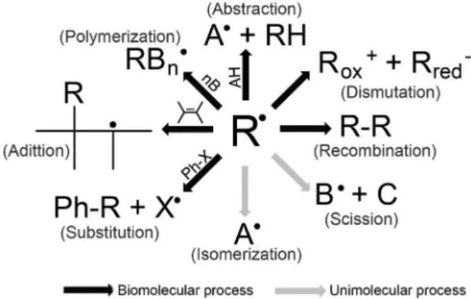
are weak emitters or non-emissive, have long lifetimes in aqueous and hydrophobic media, albeit quenchable by dissolved molecular oxygen, and react as alkoxyl radicals
that play important roles in biological peroxidation.49
Alkoxyl radicals undergo C−C cleavage and hydrogen abstraction reactions, can initiate radical polymerization, dimerize, and add to unsaturated functional groups (Figure 11), like triplet acetone illustrated in Figure 5.
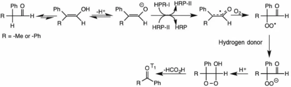
pH (7.4), an adequate model for a close approximation
to physiological conditions (Figure 12).50 Moreover,
IBAL is structurally similar to the metabolite methyl malondialdehyde, which also contains a hydrogen atom activated by the carbonyl group. The HRP enzymatic cycle
is initiated by H2O2 and involves a two-electron oxidation
of its native form [HRP-FeIII] to HRP-compound 1
[HRP-•+FeIV], a highly oxidizing species that promotes
oxidation of the substrate’s enol form. The initially formed resonance-stabilized enol radical reacts with dissolved oxygen, yielding a peroxyl radical whose reduction by sugar portions of the enzyme leads to a hydroperoxide (IBAL-OOH), which can cyclize to a 1,2-dioxetane
derivative (IBALO2), whose thermal cleavage results in the
formation of formic acid and acetone, partly in the triplet state (Figure 12).
The rationale for triplet acetone generation by IBAL/
HRP/O2 implies previous H2PO4−catalyzed enolization of
the substrate. Enolates are oxidized more easily than their
carbonyl form, thus favoring hydrogen abstraction from IBAL
by the highly oxidizing HRP-compound 1 intermediate.
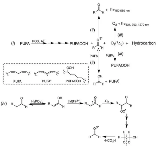
Once formed inside the enzyme active site, triplet acetone removes hydrogen atoms from the carbohydrate portion of HRP (18% carbohydrate content), leading to pinacol and 2-propanol (Figure 12). A large volume of kinetic and spectroscopic data strongly supports this mechanism. Importantly, excited carbonyls can also be formed from other sources, such as the dismutation of alkoxyl and alkylperoxy
intermediates of lipid peroxidation (Figure 13).51,52
It took only a few years for detailed mechanistic studies of the reaction to be unveiled and the formation of acetone in
the triplet state (roughly 30%) to be proven by: (i) matching
the CL emission spectrum with the phosphorescence
spectrum of acetone (λmax ca. 430 nm); (ii) eficient energy
transfer to the water-soluble
9,10-dibromoanthracene-2-sulfonate (DBAS) anion; (iii) quenching of the emission
with sorbate (2,4-hexadienoate) anion, a water soluble
conjugated diene; and (iv) detection of photoproducts
originated from triplet excited acetone, namely isopropanol
and pinacol (Figure 12).51,53
Additional studies indicated the need for H2O2 as a HRP
co-substrate and enolic IBAL as the enzyme substrate. IBAL is oxidized by peroxidase, which acts as an oxidase in a
typical enzymatic cycle involving peroxidase compounds 1
and 2, as mentioned earlier.54,55 The involvement of the
enolic form of IBAL was deinitively proven by the use of the corresponding IBAL silyl enol ether. The silyl IBAL derivative resulted in higher reaction rate constants, and
Figure 11. Typical reactions of free radicals centered on carbon, oxygen or other atoms (adapted from reference 4).
Figure 12. Generation of triplet acetone by the horseradish peroxidase (HRP)-catalyzed oxidation of isobutanal (IBAL). The substrate evokes irely luciferin with respect to the presence of a carbonyl-activated hydrogen atom, insertion of oxygen in the α-carbon yielding an α-hydroperoxide (IBAL-OOH), cyclization to a hypothetical 1,2-dioxetane (IBALO2), whose cleavage yields triplet acetone, which decays by light emission and reduction to isopropanol and pinacol.51 Reproduced from reference 4, by permission of Rev. Virtual de Química.
Figure 14. HRP-catalyzed or peroxynitrite (ONOO−)-initiated aerobic oxidation of methyl acetoacetone (MAA) to acetate and triplet excited diacetyl
(adapted from reference 4).
signiicantly increased emission intensities and quantum
yields (Figure 12).56 Under these experimental conditions,
acetone phosphorescence is enhanced to the point that it can be easily seen by eyes adapted to the dark. In the presence of the triplet energy acceptor DBAS, the chemiluminescence
of IBAL/HRP could even be photographed.53,57 Using the
enolic substrate, it was also possible to show that triplet acetone is generated inside the chiral environment of the active site, as indicated by observed differential emission
quenching by D- and L-tryptophan.56
Unlike aldehydes, the corresponding carboxylic acid derivatives are not peroxidase substrates in analogous experimental conditions, probably due to their much lower enol content. However, the utilization of protected enol equivalents of carboxylic acid derivatives containing
active α-hydrogen atoms results in substrate oxidation
accompanied by light emission. This indicates the production of excited species by a mechanism similar to
the aldehyde reaction.58
In parallel, another interesting HRP substrate named methyl acetoacetone (MAA, 3-methylpentane-2,4-dione) was studied as a putative source of excited diacetyl. MAA was chosen as a model for methyl acetoacetate, a ketone body accumulated in diabetes and isoleucinemia patients.
MAA is a β-diketone long known to enolize in aqueous
medium. Indeed, the MAA/HRP system was found to
generate diacetyl in the triplet state (τ ca. 20 μs), which
undergoes quenching by sorbate and shows a CL emission spectrum identical to the phosphorescence spectrum of
diacetyl (λmax ca. 520 nm, shoulder at 550 nm) (Figure 14).59
The mechanism of the MAA oxidation reaction was corroborated by product analysis (acetate and diacetyl), oxygen and peroxynitrite consumption, detection of
MAA• and acetyl radical adducts by electron paramagnetic
resonance (EPR) spin trapping with methylnitrosopropane
(MNP) (aN = 1.52 and 0.82 mT), and the spectral
coincidence between CL and phosphorescence of diacetyl. Moreover, the substrates 2-phenylpropanal and diphenylacetaldehyde were oxidized by dissolved oxygen in the presence of HRP by a mechanism analogous to that of IBAL to acetophenone and benzophenone, respectively,
in their triplet excited states (Figure 15).60 In the latter case,
the observed red light emission was assigned to singlet oxygen derived from excited benzophenone energy transfer to ground state oxygen. Additionally, mitochondria isolated from mouse liver challenged with diphenylacetaldehyde led to oxidative damage to their proteins, lipids and DNA,
which was attributed to triplet benzophenone (τ ca. 100 μs)
formed by the aerobic oxidation of the substrate catalyzed by cytochrome c present in the inner mitochondrial
membrane.61 In this regard, various reports have revealed
that different hemoproteins, acting as peroxidases (e.g., HRP, myoglobin, cytochrome c, lipoxygenase), catalyze ultraweak chemiluminescent reactions, thus possessing the potential to cause deleterious effects induced by excited species.
Other peroxidase substrates of biological interest are the plant growth hormones phenylacetaldehyde and indole acetaldehyde, generators of formate and benzaldehyde
or indole aldehyde, respectively.62 Potentially important
in plant biochemistry is the HRP catalyzed oxidation of
n-pentanal yielding formic acid and triplet n-butanal, whose
intramolecular γ-hydrogen abstraction and subsequent
β-cleavage (Norrish type II photochemical reaction) yield
acetaldehyde (ethanal) and ethylene (ethene), another plant
growth hormone.63
Using the IBAL/HRP system as a triplet acetone source,
Cilento et al.48 successfully excited and/or chemically
for singlet oxygen formation); red- and infrared-sensitive phytochromes (day-period mediators in phototropism and photoperiodism); chlorophyll (involved in photosynthesis); diethylstilbestrol (an estrogen with tumorigenic properties) and tetracyclines (antibiotics with bactericidal activity) (Figure 16).
For many years, G. Cilento collaborated closely with W. Adam at the Universität Würzburg, Germany. One of the most outstanding works resulting from this collaboration in dark-photochemistry appeared in 1992, describing how various 1,2-dioxetane derivatives are able to induce chemical modiications of DNA, mainly the [2 + 2] cycloaddition (Paternò-Büchi) reaction of adjacent DNA pyrimidines to cyclobutane dimers (CPDs, “cyclobutane pyrimidine dimers”) and the oxidation of
guanosine to 8-hydroxy-2’-deoxyguanosine.64 The CPDs
generated by energy transfer from a triplet carbonyl to the ground state of guanine were detected with a UV speciic endonuclease. According to the authors, oxidized guanine might be formed by energy transfer from a triplet carbonyl compound, followed by reaction with dissolved molecular oxygen, or by direct reaction with residual 1,2-dioxetane.
Noteworthy in this respect is Lamola’s report43 twenty
years earlier that the incubation of
3,3,4-trimethyl-1,2-dioxetane with isolated 14C-labeled Escherichia coli
DNA in nitrogen-purged phosphate buffer at 70 ºC produces a major compound detected by descending paper chromatography, attributed to thymine dimers (TT, 6.5%). Minor amounts of UT dimer (0.8%) derived from CT were also identiied (Figure 17). Accordingly, irradiation of the TT-containing fraction with 254 nm light re-formed thymine, thereby confirming triplet acetone-induced thymine dimerization.
It is important to emphasize that the development of the ields of chemiluminescence and bioluminescence was signiicantly advanced by the Workshop Brazil-United
States on Chemiluminescence and Bioluminescenceheld
at the Chemistry Institute of the University of São Paulo (USP), and by the International Conference on Chemi- and Bioenergized Processes, organized by Giuseppe Cilento and Waldemar Adam in 1978 in the municipality of Guarujá, SP, Brazil. These meetings were attended by prominent scientists who established the fundamentals for the identiication of the sources, targets, mechanisms and biological responses of excited states in CL, BL and photo(bio)chemistry in the dark (Figure 18).
3. Recent Advances in Photochemistry in the Dark
New advances and inspiring insights into “dark” photobiochemistry have been triggered by modern
Figure 15. HRP-catalyzed oxidation of 2-phenylpropanal (R = Me) and diphenylacetaldehyde (R = Ph) to acetophenone and benzophenone, respectively, in the triplet state (adapted from reference 4).
Figure 16. Target molecules for triplet acetone generated by isobutanal/ HRP. Pr and Pfr, red-absorbing and far-red absorbing phytochromes; 9,10-dibromoanthracene (DBA); xanthene dyes: luorescein, eosin and rose bengal; indoles, tryptophan and plant hormones (adapted from reference 4).
Figure18. Attendees of the International Conference on Chemi- and Bioenergized Processes, held in 1978 in Guarujá (SP, Brazil). From left to right: 1st row (seated): Edy Rivas, Carmem Vidigal, Michael Kasha, John Woodland (“Woody”) Hastings, Eduardo Lissi, Etelvino Bechara; 2nd row: Christopher Foote, Giuseppe Cilento, Waldemar Adam, Frank M. Thérèse Wilson; 3rd row: William Richardson, Adelaide Faljoni-Alário, Ohara Augusto, Rex Tyrrell, Roberto C. de Baptista, Paul Schaap, Nelson Duran, Marcela Haún; 4th row: Gary B. Schuster, Norman Krinsky, Pill-Soon Song, Alfons Baumstark, K. Zaklika, Yoshitaki Shimizu, Rogerio Meneghini; 5th row: R. Srinivasan, Karl Kopecky, Klaus Zinner, Frank Quina, Bechara Kachar.
methodologies and technology. For instance, (i)
diffusion-controlled quenching of triplet acetone by 2,4-hexadienoates
(kq, rate constant ≥ 109 mol L-1 s-1 in aqueous media at room
temperature), commonly known as sorbates, yielding the
cis,trans-isomers of the diene, has been veriied, as well
as (ii) the ability of triplet species to abstract the
double-allylic hydrogen atoms from linoleic and arachidonic acids,
triggering peroxidation.51,65 Furthermore, the role of triplet
carbonyls in mitochondrial swelling, accompanied by lipid, protein and DNA damage, has been clearly demonstrated. For many decades, the impairment of mitochondria properties by phosphate buffer was not fully understood, making it imperative to isolate these organelles in amino-alcohol buffers, mainly Tris [tris(hydroxymethyl) aminomethane] and HEPES [4-(2-hydroxyethyl)-1-piperazineethanesulfonic acid] buffers. In phosphate buffers, isolated mitochondria rapidly undergo perforation with consequent swelling, collapse of the transmembrane potential, loss of respiratory control, accumulation of calcium, and decrease of ATP synthesis. In 1996, the hypothesis was put forward that phosphate could be responsible for amplifying the peroxidation chains of the mitochondrial membrane lipids, because phosphate reportedly catalyzes the enolization of aldehydes resulting from spontaneous lipid peroxidation, which is followed by cytochrome c catalyzed oxidations, yielding triplet carbonyls (Figure 19). This was shown to be accompanied by the formation of mitochondrial permeability transition pores (MPTs), leading to organelle deterioration and
death (Figure 19).66 This proposition was endorsed by the
inhibitory effect of the phosphate-promoted mitochondrial
swelling by both the antioxidant 2,6-di-tert
-butyl-4-methylphenol (BHT, “butylated hydroxyltoluene”) and by cyclosporin A, which prevents MPTs from opening, thus inhibiting cytochrome c release, a potent apoptotic stimulation factor. Both compounds reportedly block mitochondrial peroxidation and, accordingly, were found to prevent formation of MPTs upon the addition of sorbate, a potent quencher of triplet carbonyls.
Also notable was the demonstration that myoglobin catalyzes the aerobic oxidation of acetoacetate and 2-methylacetoacetate to formate plus methylglyoxal or biacetyl, respectively, accompanied by ultraweak light
emission.67 Like the HRP-catalyzed oxidation of IBAL,
the β-ketoacid oxidation by myoglobin was envisaged as
involving the following steps: oxygen insertion into the
α-carbon of the substrate, cyclization to a dioxetane, and
cleavage to triplet dicarbonyls. Using EPR spin trapping with MNP, acetyl radicals were detected in the reaction mixtures, probably resulting from the cleavage of excited dicarbonyl
products. These two β-ketoacids are included as the “ketone
bodies” that accumulate at millimolar concentrations in the blood of diabetics and individuals under ketogenic diet and may be involved in rhabdomyolysis.
3.1. Light, oxygen, and melanin: a dangerous combination
According to the World Health Organization, two to three million individuals acquire skin cancer annually, of which about 130 thousand cases were diagnosed as melanoma, the most lethal kind of cancer. Among genetic and environmental factors triggering carcinogenesis, UV exposure appears as the main cause, and has been imputed to atmospheric ozone depletion. The skin pigment, melanin, predominantly black (eumelanin) in dark individuals, brown (pheomelanin) in blondes and redheads and almost absent in albinos, absorbs the sunlight and dissipates the energy as heat, thereby protecting the skin against photochemical lesions in DNA, which may induce mutagenesis and carcinogenesis. Thus, overexposure to sunlight may trigger skin burns, mutations and cancer. Most skin cancers have been attributed to the photochemical [2 + 2] dimerization of adjacent DNA pyrimidine bases, mainly thymine (T) and
cytosine (C) when they absorb UVB light (290-320 nm).68
The dimers are commonly referred as CPDs, i.e., “cyclobutane pyrimidine dimers,” which reportedly lead
to mutagenic transitions C→T and CC→TT. Melanoma
has been increasingly related to C→T mutations induced
Surprisingly, experiments carried out at Yale University
by Brash and co-workers69 revealed continuing CPDs
formation 3-4 hours after UVA and UVB illumination of mouse melanocytes, hence, in the dark. As expected, direct UV exposure of ibroblasts, brown and albino melanocytes generates CPDs within one picosecond. The CPD peak then slowly decays to the baseline due to the DNA repair systems in action. However, long after UV irradiation of dark melanocytes, but not fibroblasts and albino melanocytes, CPDs persistently formed. The addition of kojic acid, an inhibitor of melanin synthesis, inhibited the formation of CPDs, thus conirming that the observed DNA damage is melanin dependent. Also, as expected, production of CPDs signiicantly decreased in response to the addition of inhibitors of the nuclear DNA repair systems. Special attention was given to thymine-cytosine dimers, which prevailed among the detected CPDs, and are the UV-signature for C- > T mutations, the putative cause of melanoma.
These results were later interpreted as an outstanding case of “photochemistry in the dark” on the basis of evidence unveiled by classical quenching tests of triplet carbonyls. “Dark” CPDs signiicantly decreased upon the addition of sorbate to melanocyte cultures, and the DBAS-enhanced chemiluminescence of the cell cultures also hindered the formation of “dark” CPDs. These data provided clues to design additional experimental strategies to postulate a reaction mechanism, which is sketched in Figure 20.
In summary, UVA absorbed by melanin results in the latter’s fragmentation and activation of nitric oxide synthase (iNOS) and NADPH-dependent oxidase (NOX), respectively, sources of NO and superoxide anion-radical, whose bimolecular reaction rapidly yields highly oxidizing peroxynitrite. Gradually, melanin fragments and precursors as well as peroxynitrite diffuse to the nucleus, where they form melanin-derived radicals. Melanin radical fragments then add molecular oxygen to ultimately produce a
Figure 20. “Dark” generation of cyclobutane pyrimidine dimers (CPDs) several hours after melanocyte exposure to UVA,B light. Red arrows: solar UVA and UVB light is absorbed by the DNA thymine and cytosine bases of skin melanocytes, yielding their electronically excited states, which undergo [2 + 2] cycloaddition within picoseconds to the mutagenic pyrimidine dimers: C=C, C=T, and predominantly T=T. Grey arrows: concomitantly, UV light induces melanin synthesis, the natural skin solar protection pigment, and activation of nitric oxide synthase (NOS) and NADPH oxidase, respectively, sources of superoxide radical (O2•−) and nitric oxide (NO•) whose diffusion-controlled reaction produces peroxynitrite (ONOO−). These reactive oxygen species (ROS) continuously attack melanin, leading to its fragmentation. The melanin derivatives migrate to the nuclear space where they are oxidized by peroxynitrite to a hypothetical dioxetane indole intermediate, whose cleavage yields a kynurenine analogue excited to triplet state. Exothermic energy transfer from the triplet carbonyl product to adjacent T and C residues produces a blend of T=T, C=C, and predominantly T=C, which is a pre-mutagenic C→T transition that is reportedly a melanoma signature. Parallel oxidative damage to guanosine also reportedly promotes G→T mutations that may lead to apoptosis (adapted from reference 70).
hypothetical indole dioxetane, whose thermal cleavage yields a triplet kynurenine analogue. Energy transfer from the excited product to adjacent DNA pyrimidines then sensitizes dimerization and CPDs formation. Accordingly, iNOS and NOX inhibitors hampered “dark” CPDs formation; nitrotyrosine-containing nuclear proteins were detected by immunoluorescent techniques; melanin fragments were found surrounding the nuclei before UVA irradiation and inside the nuclear volume during “dark” CPD formation; sorbate and DBAS were effective as triplet interceptors and “dark” CPD blockers, and the use of silencing genes of DNA repair systems maintained the levels of CPDs for much longer. Last but not least, the triplet-triplet energy transfer from excited melanine products (3.8 eV) to pyrimidines (3.0 eV), which leads to CPDs, is exothermically favored.
Numerous questions remain to be answered, particularly about the reaction mechanisms and carcinogenesis. These indings reinforce the need for extra care against excessive exposure to sunlight between 9:00 a.m. and 4:00 p.m., and the recommendation to the cosmetic industry to add triplet quenchers to its formulations of sun protection creams, lotions, sunscreens and antioxidants, in order to prevent “dark” CPDs and carcinogenesis. Triplet carbonyls have been neglected as reactive oxygen species in biomedicine, although they react like alkoxyl radicals and are produced by dioxetane thermolysis and in peroxidation chains by dismutation of oxyl and peroxyl radicals. More investment in research on the pathophysiological roles of triplet species
would beneit our understanding of the molecular aspects of health maladies.
3.2. Generation of singlet oxygen in the dark
The spin prohibition for ground state molecular oxygen (3Σ
g−) to directly react with diamagnetic molecules is known
to be circumvented by its photo- or chemiexcitation to its
singlet state (1∆
g).71,72 The discovery of singlet oxygen in the
1930s by Kautsky using very simple dye-photosensitization of molecular oxygen, and its “rediscovery” by Seliger in
1960 by reacting hypochlorite with H2O2, was followed
in the early sixties by the spectroscopic identiication of its dimol and monomol emission bands, respectively, in the red (634, 703, and 762 nm) and infrared (1270 nm) spectral regions by Kasha, Khan and Ogryslo. Given its high electrophilicity, the ability of singlet oxygen to react with unsaturated compounds (1,2-, 1,3-, and 1,4-cycloadditions) and sulides leading to peroxides and
sulfoxides, respectively, was soon characterized.72
Quenching by azide, tertiary amines, histidine, tocopherol, carotenoids, among others, was also introduced as a simple pretest to conirm the presence of singlet
oxygen.73 However, unequivocal identiication of singlet
oxygen in a given in vitro or in vivo system is currently
considered to be as the detection of its monomol emission at 1270 nm and/or trapping with anthracene derivatives
as the corresponding 9,10-endoperoxides, using 18O
2
as compared to 16O
Figure 21 illustrates some photochemical, chemical and enzymatic sources of singlet oxygen and several biological
targets and responses reported in the literature.74
Recently, the triplet-triplet energy transfer from acetone generated from either 1,2-dioxetane thermolysis or the IBAL/HRP system to ground state oxygen yielding the
molecular oxygen excited singlet state (1∆
g) was achieved
and unequivocally demonstrated (Figure 22).71 First,
concomitant emission of triplet acetone (λmax ca. 430 nm)
and singlet oxygen (λmax ca. 1270 nm) was measured
during the course of the reaction. Then, after purging the
dioxetane or IBAL/HRP reaction mixture with 18O
2 in
the presence of the singlet oxygen water-soluble probe 9,10-diethylanthracene sulfonate, the corresponding
18O-incorporated 9,10-endoperoxide. These data reinforce
the hypothesis that singlet oxygen can potentially be generated and play normal or pathogenic roles in the absence of light when sensitized by triplet carbonyls.
Finally, singlet oxygen was also detected as a by-product of the reaction of glyoxal with peroxynitrite
in aerated buffer.75 Glyoxal, methylglyoxal and diacetyl
are endogenous toxicants overproduced in tissues through the peroxidation of carbohydrates, lipids, and proteins.
The former two α-dicarbonyls have been detected in
diabetes, and diacetyl is well-known as flavorant in buttered foods such as popcorn and cookies, although
it causes bronchiolitis.76 In cells, dicarbonyls have been
shown to attach to proteins through Schiff reactions, leading to protein cross-linking, precipitation, and loss of biological functions. In addition, they were found to undergo phosphate-catalyzed nucleophilic addition of peroxynitrite, causing carbonyl-carbonyl cleavage to carboxylic acids via acyl radicals: acetyl radical from diacetyl and methylglyoxal, and formyl radical from
glyoxal.77,78
Acetyl radical was able in vitro to acetylate amino acids,
synthetic peptides, albumin, and 2’-deoxyguanosine, which raises the hypothesis that these reactions may be involved in post-translational modiications of proteins (epigenetics) and mutagenesis. In turn, formyl radical added molecular oxygen, yielding a formyl peroxyl radical whose geminal hydrogen atom makes it prone to undergo the Russell
annihilation reaction, yielding singlet oxygen.75 Thus, the
glyoxal/peroxynitrite system constitutes another interesting potential route to generate deleterious singlet oxygen in cells not exposed to light (Figure 23).
Figure 21. Sources, targets and biological response of singlet oxygen (adapted from reference 4).
Figure 22. Generation of singlet oxygen (1∆
Figure 23. Reaction mechanism of peroxynitrite addition to diacetyl, methylglyoxal, and glyoxal, ultimately leading to acetic acid, acetic acid plus formic acid, and formic acid. The acetyl radical intermediate attaches to added amino acids and nucleobases, whereas formyl radical inserts ground state oxygen (3Σ
g−) to form formyl peroxyl radical, whose annihilation yields highly reactive singlet oxygen (1∆g). Singlet oxygen was scavenged by a water-soluble anthracene derivative (AVS) and the corresponding 9,10-endoperoxide (AVSO2) adduct was identiied by HPLC-MS.
4. Conclusions
This review updates the advances that further corroborate Cilento-Lamola-White hypothesis of “photo(bio)chemistry without light.” It emphasizes not only that photoexcited biomolecules play crucial roles in living organisms, e.g., chlorophyll in photosynthesis, rhodopsin in vision, and phytochrome in phototropism, but also that chemically and enzymatically generated excited products - triplet carbonyls and singlet oxygen - may trigger important biological events in tissues never exposed to light. The hypothesis of “photochemistry in the dark” is illustrated here with examples of isomerizations and cycloadditions of natural products in plants, phosphate-induced permeabilization and inactivation of isolated mitochondria, production of plant hormones (ethylene and phenylacetaldehyde), mutagenesis associated with pyrimidine dimerization, endogenous and xenobiotic toxicants, and singlet oxygen generation, among others. The substrates, mainly luciferin-like compounds,
that possess an abstractable α-hydrogen atom vicinal to
a carbonyl group, are prone to form a 1,2-dioxetane after oxygen insertion, and produce cleavage products in the electronically excited state. Highly emissive singlet excited states are produced in bioluminescence, whereas non-emissive but extremely reactive triplet states are involved in “dark” photobiochemistry. Their potential biological
targets are the same as those attacked by radicals and other strong oxidants such as oxygen and carbonate radicals,
hypochlorite, peroxynitrite, and peroxidase/H2O2, which
are recognized participants in so-called oxidative stress or
redox imbalance.79
Investigating the nature, source and role of excited species in dark processes is not an easy task, although remarkable success has been achieved in studies of the biochemistry and biomedicine of radicals, which can sometimes be short-lived as triplet carbonyls and present in comparably low concentrations. All too often we make frustrating attempts to determine concentrations and luxes of reactive species in cell cultures and tissues, to synthesize speciic probes for reliable establishment of mechanistic routes, and to ind selective biomarkers for the diagnosis of inherited and acquired maladies. The astounding progress that has been made in the development of new analytical separation and spectroscopic techniques in recent years has paved the way for clarifying and resolving many of these technical problems.
Acknowledgments
C. V. S. 2013/16885-1), the INCT Processos Redox em Biomedicina (FAPESP/CNPq/CAPES 573530/2008-4) and the Conselho Nacional de Desenvolvimento Cientíico e Tecnológico (CNPq: E. J. H. B. 302326/2011) for their inancial support. We also thank Fernanda F. Ventura for her assistance in adapting the manuscript to the journal guidelines.
Wilhelm J. Baader was born in 1954 in Spalt (Germany), and studied Chemistry at the University of Würzburg, where he got his PhD in 1983 under the supervision of Prof Waldemar Adam, working on the mechanism of 1,2-dioxetane decomposition. After post-doctoral studies at the Universities of São Paulo (Brazil) and Konstanz (Germany), he became a Professor at the University of São Paulo in 1989, and since then he has been working in the field of mechanistic and applied organic chemiluminescence.
Cassius V. Stevani was born in São Paulo, SP. He received his BSc in Chemistry in 1992 from the Chemistry Institute of University of São Paulo (IQ-USP). He completed his PhD in Organic Chemistry in 1997 under supervision of Prof J. W. Baader and was a post-doctoral fellow working with Prof E. J. H. Bechara (1998-2000) both at IQ-USP. Currently, he lectures in Organic and Environmental Organic Chemistry as Associate Professor at IQ-USP, where his group studies the mechanism and environmental application of bioluminescent fungi.
E t e l v i n o J . H . B e c h a r a re c e i v e d h i s B S c d eg re e i n C h e m i s t r y ( 1 9 6 8 ) a n d P h D degree in Biochemistry (1972) from the Chemistry Institute/São Paulo University (IQUSP) under direction of Prof Giuseppe Cilento. He was appointed Assistant (1972), Associate (1979), and Professor of Biochemistry at the IQUSP, where he is now a Senior Collaborator. He did his postdoctoral work with Prof Emil H. White (Johns Hopkins University; 1974-5) and Prof Thérèse Wilson (Harvard University; 1975-6). Research interests and publications encompass peroxide chemiluminescence, triplet carbonyls (“photochemistry in the dark”), singlet oxygen, Coleoptera bioluminescence,
free radicals in biomedicine, lead poisoning, and alpha-dicarbonyls.
References
1. Campbell, A. K.; Chemiluminescence-Principles and Applications in Biology and Medicine; Ellis Horwood: Cambridge, 1988.
2. Wilson, T.; Hastings, J. W.; Bioluminescence-Living Lights, Lights for Living; Harvard University Press: Cambridge, 2013. 3. Bartoloni, F. H.; Ciscato, L. F. M. L.; Peixoto, M. M. M.; Santos, A. P. F.; Santos, C. S.; Oliveira, S.; Augusto, F. A.; Pagano, A. P. E.; Baader, W. J.; Quim. Nova 2011, 34, 544.
4. Baader, W. J.; Stevani, C. V.; Bechara, E. J. H.; Rev. Virtual Quim.2015, 7, 74.
5. Adam, W.; Cilento, G.; Angew. Chem., Int. Ed. 1983, 22, 529. 6. Kopecky, K. R.; Mumford, C.; Can. J. Chem. 1969, 47, 709. 7. Adam, W.; Liu, J. C.; J. Am. Chem. Soc. 1972, 94, 2894. 8. Nery, A. L. P.; Baader, W. J.; Quim. Nova 2001, 24, 626.
9. Adam, W.; Baader, W. J.; Angew. Chem., Int. Ed. 1984, 23, 166. 10. Adam, W.; Baader, W. J.; J. Am. Chem. Soc. 1985, 107, 410. 11. Bechara, E. J. H.; Wilson, T.; J. Org. Chem. 1980, 45, 5261. 12. Bastos, E. L.; Baader, W. J.; ARKIVOC 2007, 8, 257. 13. Schuster, G. B.; Acc. Chem. Res. 1979, 12, 366.
14. Turro, N. J.; Chow, M.-F.; J. Am. Chem. Soc. 1980, 102, 5058. 15. Adam, W.; Cueto, O.; J. Am. Chem. Soc. 1979, 101, 6511. 16. Wilson, T.; Photochem. Photobiol. 1995, 62, 601.
17. Koo, J.-Y.; Schmidt, S. P.; Schuster, G. B.; Proc. Natl. Acad. Sci. USA 1978, 75, 30.
18. Oliveira, M. A.; Bartoloni, F. H.; Augusto, F. A.; Ciscato, L. F. M. L.; Bastos, E. L.; Baader, W. J.; J. Org. Chem. 2012, 77, 10537.
19. Bartoloni, F. H.; Oliveira M. A.; Ciscato, L. F. M. L.; Augusto, F. A.; Bastos, E. L.; Baader, W. J.; J. Org. Chem. 2015, 80, 3745. 20. Schaap, A. P.; Gagnon, S. D.; J. Am. Chem. Soc. 1982, 104,
3504.
21. Schaap, A. P.; Sandison, M. D.; Handley, R. S.; Tetrahedron Lett. 1987, 28, 1159.
22. Beck, S.; Köster, H.; Anal. Chem. 1990, 62, 2258.
23. Schaap, A. P.; Chen, T.-S.; Handley, R. S.; de Silva, R.; Giri, B. P.; Tetrahedron Lett. 1987, 28, 1155.
24. Nery, A. L. P.; Weiss, D.; Catalani, L. H.; Baader, W. J.; Tetrahedron 2000, 56, 5317.
25. Nery, A. L. P.; Ropke, S.; Catalani, L. H.; Baader, W. J.; Tetrahedron Lett. 1999, 40, 2443.
26. Ciscato, L. F. M. L.; Weiss, D.; Beckert, R.; Bastos, E. L.; Bartoloni, F. H.; Baader, W. J.; New J. Chem. 2011, 35, 773. 27. Ciscato, L. F. M. L.; Bartoloni, F. H.; Weiss, D.; Beckert, R.;
Baader, W. J.; J. Org. Chem. 2010, 75, 6574.
29. Adam, W.; Troimov, A. V.; J. Org. Chem. 2000, 65, 6474. 30. Adam, W.; Matsumoto, M.; Troimov, A. V.; J. Am. Chem. Soc.
2000, 122, 8631.
31. Bastos, E. L.; da Silva, S. M.; Baader, W. J.; J. Org. Chem. 2013, 78, 4432.
32. Augusto, F. A.; Souza, G. A.; Souza Jr., S. P.; Khalid, M.; Baader, W. J.; Photochem. Photobiol. 2013, 89, 1299. 33. Ciscato, L. F. M. L.; Augusto, F. A.; Weiss, D.; Bartoloni,
F. H.; Albrecht, S.; Brandl, H.; Zimmermann, T.; Baader, W. J.; ARKIVOC 2012, 3, 391.
34. Chandross, E. A.; Tetrahedron Lett. 1963, 4, 761. 35. Rauhut, M. M.; Acc. Chem. Res. 1969, 2, 80.
36. Stevani, C. V.; Silva, S. M.; Baader, W. J.; Eur. J. Org. Chem. 2000, 24, 4037.
37. Ciscato, L. F. M. L.; Bartoloni, F. H.; Bastos, E. L.; Baader, W. J.; J. Org. Chem. 2009, 74, 8974.
38. Bartoloni, F. H.; Ciscato, L. F. M. L.; Augusto, F. A.; Baader, W. J.; Quim. Nova2010, 33, 2055.
39. Albertin, R.; Arribas, M. A. G.; Bastos, E. L.; Röpke, S.; Sakai, P. N.; Sanches, A. M. M.; Stevani, C. V.; Umezu, J. I.; Yu, S.; Baader, W. J.; Quim. Nova 1998, 21, 772.
40. Oliveira, T. F.; Silva, A. L. M.; Moura, R. A.; Bagattini, R.; Oliveira, A. A. F.; Medeiros, M. H. G.; di Mascio, P.; Campos, I. P. A.; Barreto, F. P.; Bechara, E. J. H.; Loureiro, A. P. M.; Sci. Rep. 2014, 4, 5359.
41. Adam, W.; Baader, W. J.; Babatsikos, C.; Schmidt, E.; Bull. Soc. Chim. Belg. 1984, 93, 605.
42. Turro, N. J.; Ramamurthy, V.; Scaiano, J. C.; Modern Molecular Photochemistry of Organic Molecules;University Science Books: Sausalito, CA, USA, 2010.
43. Lamola, A. A.; Biochem. Biophys. Res. Commun. 1971, 43, 893.
44. Woodward, R. B.; Hoffmann, R.; Angew. Chem., Int. Ed. 1969, 8, 781.
45. White, E. H.; Miano, J. D.; Watkins, C. J.; Breaux, E. J.; Angew. Chem., Int. Ed. 1974, 13, 229.
46. Brunetti, I. L.; Bechara, E. J. H.; Cilento, G.; White, E. H.; Photochem. Photobiol. 1982, 36, 245.
47. Nery, A. L. P.; Quina, F. H.; Moreira, P. F.; Medeiros, C. E. R.; Baader, W. J.; Shimizu, K.; Catalani, L. H.; Bechara, E. J. H.; Photochem. Photobiol. 2001, 73, 213.
48. Cilento, G.; J. Theor. Biol. 1975, 55, 471; Cilento, G.; Acc. Chem. Res. 1980, 13, 225; Cilento, G.; Zinner, K.; Bechara, E. J. H.; Durán, N.; Baptista, R. C.; Shimizu, Y.; Augusto, O.; Faljoni-Alário, A.; Vidigal, C. C. C.; Oliveira, O. M. F.; Haun, M.; Ciên. Cult. 1979, 31, 290.
49. Cilento, G.; Adam, W.; Free Radical Biol. Med. 1995, 19, 103. 50. Bechara, E. J. H.; Oliveira, O. M. M. F.; Duran, N.; Baptista,
R. C.; Cilento, G.; Photochem. Photobiol. 1979, 30, 101. 51. Velosa, A. C.; Baader, W. J.; Stevani, C. V.; Mano, C. M.;
Bechara, E. J. H.; Chem. Res. Toxicol. 2007, 20, 1162.
52. di Mascio, P.; Catalani, L. H.; Bechara, E. J. H.; Free Radical Biol. Med. 1992, 12, 471.
53. Catalani, L. H.; Wilson, T.; Bechara, E. J. H.; Photochem. Photobiol. 1987, 45, 273.
54. Dunford, H. B.; Baader, W. J.; Bohne, C.; Cilento, G.; Biochem. Biophys. Res. Commun. 1984, 122, 28.
55. Baader, W. J.; Bohne, C.; Cilento, G.; Dunford, H. B.; J. Biol. Chem. 1985, 260, 10217.
56. Adam, W.; Baader, W. J.; Cilento, G.; Biochim. Biophys. Acta 1986, 881, 330.
57. Baader, W. J.; Bohne, C.; Cilento, G.; Nassi, L.; Biochem. Educ. 1986, 14, 190.
58. Baader, W. J.; Quim. Nova 1989, 12, 325.
59. Soares, C. H. L.; Bechara, E. J. H.; Photochem. Photobiol. 1982, 36, 117.
60. Nantes, I. L.; Bechara, E. J. H.; Photochem. Photobiol. 1996, 63, 702.
61. Almeida, A. M.; Bechara, E. J. H.; Vercesi, A. E.; Nantes, I. L.; Free Radical Biol. Med. 1999, 27, 744.
62. Escobar, J. A.; Vazquez-Vivar, J.; Cilento, G.; Photochem. Photobiol. 1992, 55, 895.
63. Knudsen, F. D.; Campa, A.; Stefani, H. A.; Cilento, G.; Proc. Natl. Acad. Sci. USA 1994, 91, 410.
64. Müller, E. B.; Adam, W.; Saha-Möller, C. R.; Chem. Biol. Interact. 1992, 85, 265.
65. Indig, G.; Campa, A.; Bechara, E. J. H.; Cilento, G.; Photochem. Photobiol. 1988, 48, 719.
66. Kowaltowski, A. J.; Castilho, R. F.; Grijalba, M. T.; Bechara, E. J. H.; Vercesi, A. E.; J. Biol. Sci. 1996, 271, 2929.
67. Ganini, D.; Christoff, M.; Ehrenshaft, M.; Kadiiska, M. B.; Mason, R. P.; Bechara, E. J. H.; Free Radical Biol. Med. 2011, 51, 733.
68. Kappes, U. P.; Luo, D.; Potter, M.; Schulmeister, K.; Rünger, T. M.; J. Invest. Dermatol. 2006, 126, 667.
69. Premi, S.; Wallisch, S.; Mano, C. M.; Weiner, A. B.; Bacchiocchi, A.; Wakamatsu, K.; Bechara, E. J. H.; Halaban, R.; Douki, T.; Brash, D. E.; Science 2015, 347, 842.
70. Taylor, J.-S.; Science2015,824,347.
71. Mano, C. M.; Prado, F. M.; Massari, J.; Ronsein, G. E.; Martinez, G. R.; Miyamoto, S.; Cadet, J.; Sies, H.; Medeiros, M. H.; Bechara, E. J. H.; di Mascio, P.; Sci. Rep. 2014, 4, 5938. 72. Foote, C. S.; Acc. Chem. Res. 1968, 1, 104.
73. Krinsky, N. I.; Trends Biochem. Res. 1977, 2, 35.
74. Miyamoto, S.; Martinez, G. R.; Medeiros, M. H. G.; di Mascio, P.; J. Photochem. Photobiol. B2014, 139, 24. 75. Massari, J.; Tokikawa, R.; Medinas, D. B.; Angeli, J. P. F.; di
Mascio, P.; Assunção, N. A.; Bechara, E. J. H.; J. Am. Chem. Soc. 2011, 133, 20761.
76. Kreiss, K.; Goma, A.; Kullman, G.; Fedan, K.; Simões, E. J.; Enright, P. L.; N. Engl. J. Med. 2002, 347, 330.
Micke, G. A.; Tavares, M. F. M.; Tokikawa, R.; Assunção, N. A.; Bechara, E. J. H.; Chem. Res. Toxicol. 2008, 21, 879. 78. Massari, J.; Tokikawa, R.; Zanolli, L.; Tavares, M. F. M.;
Assunção, N. A.; Bechara, E. J. H.; Chem. Res. Toxicol. 2010, 23, 1762.
79. Toledo Jr., J. C.; Augusto, O.; Chem. Res. Toxicol. 2012, 25, 975.
Submitted:July 6, 2015
Published online: October 2, 2015

![Figure 8. [2 + 2] Cycloaddition and cis,trans-isomerization driven by excited acetone in a model system with 1,2-dicyanoethylene and hypothetical “dark”](https://thumb-eu.123doks.com/thumbv2/123dok_br/18998713.463021/6.892.223.706.857.1061/figure-cycloaddition-isomerization-driven-excited-acetone-dicyanoethylene-hypothetical.webp)