Faculdade de
Cienctes
e
Tecnologia
THERMODYNAMIC AND KINETIC MODELLING OF
THE REDOX PROPERTIES OF TETRAHAEM
CYTOCHROMES
C3
MARlA
TERESA NUNES
MANGAS
CATARINO
Dissertacao apresentada para obtencao do grau de Doutor em Bioqufmica, especialidade Bioqufmica-Ffsica, pela Universidade Nova de Lisboa, Faculdade de Ciencias e Tecnologia.
Agradecimentos
Agradecimentos
Em primeiro lugar quero agradecer ao Pro! Antonio Xavier, meu orientador ao longo destes anos, pelo interesse e empenhamento sempre demonstrados em relaciio ao meu trabalho. Admiro particularmente a sua intuiciio cientifica e a disponibilidade que consegue encontrar, apesar da grande responsabilidade que a direcciio do Instituto acarreta. As discussoes que tivemos, por vezes acaloradas, serviram sempre para levantar questiies pertinentes ou esclarecer duvidas. A sua capacidade para encontrar alternativas inteligentes, mesmo nas situaciies em que parecia niio existir altemativa possivel, foi fundamental para0 desenvolvimento e conclusiio deste trabalho.
Ao Pro! Massimo Coletta devo a minha 'iniciaciio' no campo da cinetica. Durante os estdgios realizados no seu laboratorio em Roma tive os primeiros contactos com a tecnica de 'stopped-flow', que permitiram 0 posterior desenvolvimento desta tecnica no nosso laboratorio. As experiencias que realizamos em conjunto constituem grande parte do trabalho experimental apresentado nesta tese. A sua colaboraciio foi fundamental para 0 desenvolvimento do modelo termodiniimico AlB, bem C011l0 do modelo cinetico. Agradeco muito especialmente a sua amizade e a forma como a Carmen, 0 Andrea, 0 Matteo eo Jacopo me tem sempre recebido ao longo destes anos, quer em Roma quer em Panicale.
Ao Pro! David Turner agradeco0 desenvolvimento do modelo termodindmico dos cinco centros aqui apresentado. Os conhecimentos que me tem transmitido contribuiram substancialmente para uma melhor compreensiio dos problemas associados
a
modelizaciio termodintimica e cinetica do citocromofJ. Agradeco a sua sugestiio para a aplicaciio da teoria de Marcus aos factores sigma do modelo cinetico. 0 apoio fornecido durante a construciio dos programas em FORTRAN foi tambem fundamental. Devo-lhe ainda, a revisiio do manuscrito em lingua inglesa. Esse processo, no inicio certamente moroso, acabou por ser classificado como 'moving commas around', pelo que Ihe devo tambem os progressos que
fi:
no dominio lingua inglesa escrita. Por ultimo, agradeco todas as discussoes que tivemos durante os jantares no 'Restaurant at the End ofthe Universe'.Ao Pro! Jean LeGall agradeco os conselhos relativos a aspectos da microbiologia das bacterias sulfato-redutoras e ainda os presentes de biomassa ou de proteina purificada.
A
/sabel Pacheco agradeco a purificaciio das proteinas utilizadas neste trabalho. A sua capacidade de trabalho e eficiencia, tanto no dominio das tecnicas de purificaciio como na gestiio do laboratorio, contribuem em larga escala para que0 trabalho de todo0 grupo decorra da melhor forma. Agradeco ainda a amizade resultante de longos anos de convivio e defestas de aniversdrio comuns.Ao Eng. Pedro Femandes, representante da Hi-Tech Scientific em Portugal, agradeco todo 0 empenho demonstrado na resoluciio do problema da anaerobicidade do aparelho de 'stopped-flow'. Durante quase dois anos 0 Pedro foi uma presence constante no nosso laboratorio. Juntos, de chave de fendas em punho, desmontdmos e montdmos 0 aparelho inumeras vezes, testando diferentes tipos de vedantes, ora vermelhos, ora pretos... Quando por fim tomamos a decisiio de colocar0 aparelho dentro de uma ctimara anaerobica, as solucoes encontradas e parte das pecas necessdrias
a
instalaciio, foram resultado do nosso esforco conjunto.eisss
seguidas por NMR Cl temperatura ambiente. De um modo especial gostaria de salientar a disponibilidade e a paciencia com que0 Ricardo me tem ouvido ao longo destes anos de trabalho em conjunto. A ajuda prestada durante a realizaciio de experiencias de 'stopped-flow' na ctimara anaerobica, a pesquisa bibliogrdfica, os 'Services Informdticos ON-LINE' e, acima de tudo, as numerosas discussiies acerca dos modelos, contribuiram decisivamente para0 hito deste trabalho. 0 convivio diario resultou numa amizade solida que tem sido0 suporte do bom ambiente que se vive no grupo.Ao Carlos Salgueiro agradeco a utilizaciio dos resultados de titulaciies redox do citocromo f.3de D. vulgaris seguidas por NMR. Agradeco de uma forma especial as 'discussiies inesperadas'. Quando tudo parecia obvio, 0 Carlos, quase pedindo desculpa, soube sempre levantar a questiio certa no
momento exacto. As discussiies assim despoletadas serviram invariavelmente para atingir um melhor conhecimento do sistema. A amizade e 0 espirito de entre-ajuda existentes entre n6s foram muito importantes durante a fase de redaccdo da tese.
Ao Prof. P. L.Dutton agradeco a prontidiio com que respondeu aos meus pedidos de esclarecimento. Agradeco tambem 0 interesse com que tem seguido 0 meu trabalho, desde que, ha alguns anos atrds,
visitou0 nosso laboratorio. Nessa altura, 0 envio de um 'pre-print'<do seu artigo sobre transferencia
electr6nica em sistemas biologicos, publicado na revista Nature, deixou-me muito sensibilizada. Ao meu irmiio agradeco0 arranjo grafico da capa.
Aos colegas da secciio de Bioquimica da FCTIUNL, Prof. lsabel Moura, Prof. Teresa Moura, Prof. Helena Santos, Prof. lsabel Coutinho e Dr. Carlos Salgueiro, agradeco 0 trabalho suplementar que tiveram durante a minha dispensa de service docente.
A
Maria Manuel e Cl Maria Jose, secretdrias do Departamento de Quimicada FCT, agradeco a forma como, com eficiencia e amizade, tem tratado dos assuntos relativos ao Departamento e ao service docente ao longo destes anos. A Ermelinda agradeco todo 0 apoio as aulas de laboratorio. Todoscontribuiram para uma maior dedicaciio
a
investigaciio nos intervalos dos periodos lectivos.Aos colegas do ITQB agradeco a amizade, 0 espirito de entre-ajuda e todas as discussiies que tivemos
sobre os temas mais diversos.
Aos funcionarios das oficinas, services administrativos, centro de apoio informatico e secretdrias do ITQB agradeco todo0 apoio prestado.
As colegas e amigas, lsabel Coutinho, Teresa Moura, Anjos Macedo e Margarida Martinez; agradeco o incentivo e apoio psico16gico que foi fundamental nos periodos mais criticos.
Aos meus pais, ao Sergio, e Cl Cristina, agradeco a amizade, 0 interesse e0 respeito com que sempre
encararam 0 meu trabalho. Agradeco de um modo especial todo 0 apoio que, nesta ultima fase,
permitiu a conclusiio da escrita dentro do prazo. 0 facto de nunca terem deixado de acreditar nas minhas capacidades foi muito importante nos momentos de maior des animo.
Resumo
RESUMO
o
trabalho apresentado nesta dissertacao descreve e modeliza as propriedades termodinamicase cineticas de citocromos C3tetrahernicos, isolados a partir de bacterias pertencentes ao genero
Desulfovibrio.
o
capftulo I consta de uma breve introducao geral na qual sao referidas algumas caracterfsticas das bacterias sulfato-redutoras, dando especial enfase aos aspectosrelacionados corn 0 metabolismo bioenergetico, Neste capftulo, sao ainda abordados aspectos
gerais do papel fisiol6gico do citocromo C3, assim coma da sua estrutura e das suas
propriedades termodinamicas e cineticas. Este capftulo termina corn a descricao da
organizacao e conteudo da dissertacao.
Os capftulos 11 e III versam as propriedades termodinamicas e cineticas do citocromo C3,
respectivamente. Estes capftulos SaD auto-suficientes, cada urn incluindo uma introducao onde
se definem os objectivos do presente estudo e se faz uma revisao da literatura sobre 0 assunto,
bem coma uma seccao de materiais e metodos, contendo a descricao das tecnicas utilizadas no
desenvolvimento do trabalho experimental. Nestes capitulos SaD apresentados e discutidos os
modelos matematicos utilizados na simulacao dos resultados experimentais, assim coma os
valores dos parametros que melhor descrevem as propriedades do sistema em cada caso. No
final de cada capitulo sao apresentadas perspectivas futuras, quer ao nfveldo desenvolvimento
experimental quer ao nfvel da modelizacao,
No capftulo IV encontram-se resumidas as principais conclusoes retiradas deste trabalho. Na
seccao relativa aos estudos cineticos foi ainda incluida uma discussao acerca do tipo de
informacao que se pode retirar de experiencias de fluxo interrompido semelhantes as
realizadas, consoante as propriedades do sisterna. Sao ainda discutidos alguns resultados da
literatura a luz do modelo cinetico desenvolvido nesta tese. Este capitulo termina corn a
integracao das propriedades termodinamicas e cineticas ao nivel de urn modelo proposto para
o envolvimento do citocromo C3 nos processos de transducao de energia que ocorrem nas
Os citocromos C3 tetrahamicos sao protefnas relativamente pequenas, corn cerea de 120
resfduos de aminoacidos e massa molecular entre 13 e 15 kD. Esta protefna e produzida em
grandes quantidades pelas bacterias redutoras de sulfato, encontrando-se localizada no espaco
periplasmatico da celula, onde se pensa que funcione coma cofactor da enzima hidrogenase,
participando no metabolismo bioenergetico. Os quatro hemos encontram-se covalentemente
ligados
a
protefna atraves de ligacoes tioeter corn resfduos de cistefna, sendo a coordenacaoaxial do ferro feita sempre por duas histidinas. Apesar do pequeno grau de homologia
encontrado para as sequencias de aminoacidos dos citocromos C3 isolados a partir de
diferentes estirpes, 0 arranjo espacial dos quatro hemos
e
conservado. Os quatro hemosapresentam potenciais redox distintos e bastante negativos (entre -100 e -500 mY), tendo sido
demonstrada a existencia de potenciais de interaccao entre eles. Alem disso, os valores dos
potenciais redox dependem do pH, apresentando0 chamado efeito 'redox-Bohr'. Do ponto de
vista cinetico ha que referir que os processos de transferencia electronica intramolecular sao
mais rapidos do que os processos de transferencia electronica intermolecular, quer entre
diferentes moleculas de citocromo C3, quer envolvendo dadores ou aceitadores exogenos de
electroes. Desta forma, independentemente do hemo atraves do qual se da a entrada dos
electroes na protefna, a distribuicao electronica dentro da molecula e feita de acordo corn os
potenciais relativos dos varies centros. Por esta razao, 0 citocromo C3 e urn sistema
termodinamicamente controlado. A reducao do citocromo C3 por agentes redutores exogenos,
ou por outras proteinas envolvidas em processos de transferencia electronica, apresenta
caracterfsticas bifasicas na maior parte dos casos, tendo as constantes de velocidade de
segunda ordem valores compreendidos entre 106 - 107 M-Is-I. Os processos de oxidacao
apresentam constantes de velocidade inferiores, corn valores da ordem de 104- 105M-Is-I.
Propriedadestermodinamicas
Apresentam-se os modelos termodinamicos desenvolvidos ate ao presente para a descricao
dos processos de oxidacao-reducao em que 0 citocromoC3 se encontra envolvido. Os modelos
mais simples consideram 0 processo de reducao global coma 0 resultado dareducao de quatro
centros redox independentes ('modelo individual'), ou coma quatro passos consecutivos de
urn electrao ('modelo sequencial'). 0 significado ffsico dos parametros obtidos em cada caso
Resumo
o
primeiro modelo que inclui interaccoes entre os hemos foi proposto em 1984 (Santos, H.,Moura, J.1.G., Moura,1.,LeGall, J. and Xavier, AV. (1984), Eur. J. Biochem. 141,283-296).
De acordo corn este modelo a distribuicao dos electroes entre os quatro centros redox da
origem a 16 microestados que se relacionam entre si atraves de 32 equacoes de Nemst.
Contudo, ficou demonstrado nesse trabalho, que
e
possfvel descrever as propriedades do sistema, nomeadamente os desvios qufmicos dos grupos metilo dos hemos em diferentesestados de oxidacao, utilizando apenas dez parametros: os potenciais redox dos quatro centros
definidos para a forma reduzida e seis potenciais de interaccao definidos entre os hemos dois
a dois. A determinacao destes parametros para dois valores de pH mostrou que os potenciais
microscopicos dos hemos dependem do pH e que 0 mesmo parecia acontecer corn os valores
das interaccoes hemo-hemo, ficando demonstrada a existencia de interaccoes herno-protao.
A este modelo seguiu-se 0 'modelo NB' que, para explicar a dependencia dos potenciais e
das interaccoes corn 0 pH, propoe a existencia de uma alteracao conformacional da protefna
associada ao processo de protonacao (Coletta, M., Catarino, T., LeGall, J. and Xavier, AV.
(1991), Eur. J. Bioclzem. 202, 1101-1106).0 modelo acido/base (A/B) foi 0 primeiro modelo
aincluir simultaneamente 0 pH e 0 potencial da solucao coma variaveis do sistema. Devido
a
introducao doequilfbrio acido/base, 0 mimero total de microestados aumenta para 32, pois
cada urn dos 16 microestados anteriores se pode encontrar protonado ou desprotonado.
Segundo 0 modelo A/B, cada conformacao
e
caracterizada pelos quatro potenciaisrnicroscopicos dos hemos e por seis potenciais de interaccao hemo-hemo, corn a constante de
equilfbrio acido/base definida para 0 estado de referencia, 0 mirnero total de parametros
necessaries para descrever 0 sistema eleva-se para 21. Atraves da comparacao entre os
resultados experimentais anteriormente obtidos (Santos, H., Moura, J.1.G., Moura,1.,LeGall,
J. and Xavier, A V. (1984),Eur. J.Biochem. 141, 283-296) e curvas simuladas de acordo corn
o formalismo matematico do modelo NB, deterrninaram-se os parametros por aproxirnacoes
sucessivas, tendo coma ponto de partida os valores dos parametros anteriormente
determinados. Foram ainda utilizadas titulacoes espectrofotometricas para definir a escala
absoluta de potencial, impossfvel de obter apartir dos dados de ressonancia rnagnetica nuclear.
Como resultado da aplicacao do modelo A/B obtiveram-se potenciais e interaccoes
hemo-hemo diferentes para as conformacoes protonada e desprotonada. Curiosamente, apenas as
interaccoes envolvendo 0 hemo IV (definido de acordo corn a ordem de ligacao
a
cadeiapolipeptfdica) pareciam ser dependentes do pH, 0 que foi interpretado coma sendo0 resultado
de uma alteracao conformacional significativa envolvendo as relacoes estruturais entre 0
as interaccoes hemo-hemo englobam as interaccoes hemo-protao. Este problema, e 0 facto de
o potencial do hemo IV se encontrar relativamente separado dos potenciais dos outros hemos,
aumenta a imprecisao dos parametros determinados para 0 citocromo C3 de Desulfovibrio
gigascorn0 modelo A/B. No entanto,
e
importante referir que, apesar de envolver urn mimerode parametros superior ao necessario, 0 modelo A/B permitiu antever0 acoplamento entre 0
processo de reducao e a protonacao da moleculaque ocorre a pH fisio16gico.
Posteriormente, corn 0 desenvolvimento do modelo dos cinco centros (Turner, D.L.,
Salgueiro,
c.x.,
Catarino, T., LeGall, J. and Xavier, A.V. (1996), Eur. J. Biochem.241,723-731), demonstrou-se que era possfvel simular todo 0 conjunto de resultados experimentais
corn potenciais de interaccao herno-hemo independentes do pH. 0 novo modelo parte de urn
pressuposto diferente pois, em vez assumira prioria existencia de alteracoes conformacionais
da protefna, considera apenas a existencia de cinco centros, quatro centros redox e urn centro
acido/base, que, interactuando entre si, condicionam mutuamente as respectivas propriedades.
Os parametros deste modelo sao os potenciais redox dos quatro hemos, 0 pKa do centro
acido/base para 0 estado de referencia e, ainda, as interaccoes dos centros dois a dois. Mais
concretamente: seis interaccoes hemo-hemo e quatro interaccoes hemo-protao. Apesar do
numero de microestados envolvidos nos equilfbrios redox e acido/base continuar a ser32,este
modelo necessita de apenas 15 parametros para descrever 0 sisterna. Os parametros que
caracterizam as propriedades termodinamicas dos citocromos C3isolados a partir deD. gigas e
de D. vulgaris foram determinados a partir do ajuste de curvas sirnuladas de acordo corn 0
modelo dos cinco centros aos resultados experimentais obtidos em titulacoes redox seguidas
quer por ressonancia magnetica nuclear quer por espectroscopia de visfvel, utilizando 0
metodo de Marquardt. Demonstra-se que 0 modelo dos cinco centros consegue simular as
propriedades termodinamicas do citocromoC3, incluindo a dependencia corn 0 pH dos desvios
qufmicos dos grupos metilo dos hemos, corn menor numero de parametros. Por essa razao, os
valores dos parametros encontram-se mais bem definidos do que no rnodelo anterior e 0 seu
significado ffsico
e
mais evidente. Como nao foi necessario introduzir interaccoes dos centros tres a tres para explicar os resultados experimentais, nao existe evidencia para alteracoesconformacionais significativas associadas quer ao processo de reducao quer ao processo de
protonacao. No entanto, 0 aparecimento de uma cooperatividade positiva entre dois centros
corn carga do mesmo tipo (os hemos I e Il no citocromo C3de D. vulgaris e os hemos Il e III
Resumo
propondo-se que resulte de pequenos movimentos de resfduos carregados, associados ao
processo de oxidacao-reducao,
A aplicacao do modelo dos cinco centros a citocromos C3 isolados a partir de organismos
diferentes mostra que, apesar de apresentarem parametros termodinamicos diferentes, em
ambos se observa uma cooperatividade positiva entre dois dos hemos e cooperatividades
positivas entre os hemos e urn centro ionisavel. No entanto, e apesar da conservacao do
arranjo espacial dos hemos, a cooperatividade positiva nao se estabelece sempre entre 0
mesmo par de hemos. Assim, parece nao existir uma correlacao 6bvia entre a arquitectura dos
centros e a rede de cooperatividades estabelecida entre eles. Contudo, qualquer destas
protefnas tern a capacidade de acoplar a transferencia de electroes corn a transferencia de
protoes, caracterfstica que lhes confere as propriedades termodinamicas necessarias para
efectuar transducao de energia, se 0 controlo cinetico dos varies passos do ciclo energetico 0
permitir.
Propriedadescineticas
Utilizando a tecnica de 'fluxo interrompido' acoplada
a
espectroscopia de visfvel, estudou-se a cinetica de reaccoes de transferencia electr6nica envolvendo 0 citocromo C3isolado a partirde D. gigas e diferentes dadores e aceitadores de electroes. A analise dos resultados obtidos
foi feita corn urn modelo cinetico especificamente desenvolvido para este sisterna.
A primeira hip6tese restritiva introduzida na modelizacao esta relacionada corn a utilizacao de
urn mecanismo cinetico puramente colisional, ou seja, no qual sao ignorados os passos de
formacao e dissociacao do complexo entre os parceiros redox, por nao serem passos
limitantes. Mesmo assumindo urn mecanismo colisional 0 esquema cinetico
e
muitocomplexo, pois envolve todas as reaccoes de transferencia electr6nica possfveis entre 0
dador/aceitador de electroes e os 32 microestados do citocromo C3. No entanto, coma a
transferencia electr6nica intramolecular
e
mais rapida do que a transferencia electr6nica intermolecular, 0 equilfbrio entre os microestados que pertencem ao mesmo estado redoxe
muito rapido e 0 esquema cinetico reduz-se a quatro passos consecutivos de urn electrao. Se,ao longo do processo redox, as constantes de velocidade dos hemos individuais forem
afectadas apenas pela variacao da energia livre padrao (~Go) para 0 processo de transferencia electr6nica, demonstra-se que as constantes de velocidade macrosc6picas que caracterizam as
Marcus, se a energia de reorganizacao (A) for muito superior ao valor do LlGo para a
transferencia electronica, as constantes de velocidade dos hemos ao longo do processo redox
podem ser relacionadas entre si atraves de factores (factores (Jij) que dependem unicamente
dos potenciais de interaccao hemo-hemo. De modo analogo, as constantes de velocidade da
forma desprotonada podem ser relacionadas corn as constantes de velocidade da forma
protonada atraves de factores (JiH que dependem exclusivamente das interaccoes
'redox-Bohr'. A abordagem colisional em conjunto corn a aproximacao dos factores (J permitiu
reduzir0 mimero de parametros do modelo para apenas quatro constantes de velocidade, uma
para cada hemo, no estado de referencia,
A aplicacao do modelo cinetico, aos resultados obtidos nas experiencias de reducao do
citocromo C3corn ditionito de sodio, mostrou que
e
impossfvel definir as quatro constantes develocidade porque alguns hemos se encontram termodinamicamente acoplados.
E
importantesublinhar que esta limitacao nao e consequencia da fraca resolucao da espectroscopia de
visfvel nem do modelo cinetico utilizado, mas sim das propriedades intnnsecas do sistema,
nomeadamente da existencia de rapido equilfbrio intramolecular. Os resultados experimentais
foram por isso simulados utilizado urn modelo corn apenas dois parametros: a constante de
velocidade do hemo IV, que se encontra bem definida porque este hemo tern urn potencial
redox relativamente separado dos potenciais redox dos outros hemos, e constantes de
velocidade iguais para os restantes hemos. A aplicacao do modelo cinetico a experiencias corn
ditionito de sodio, realizadas a diferentes valores de pH, demonstram a validade da utilizacao
dos factores (J relativos
a
protonacao, pois a dependencia da velocidade corn 0 pH pode serexplicada pelo efeito das interaccoes hemo-protao nas constantes de velocidade dos hemos
individuais. Estas interaccoes determinam que as constantes de velocidade sejam superiores
na forma protonada e, ainda, que a constante de velocidade do hemo I seja a mais afectada,
porque este hemo tern 0 maior potencial de interaccao corn 0 protao. Por outro lado, 0 hemo
IV apresenta 0 menor potencial de interaccao corn 0 protao, sendo a sua con stante de
velocidade a menos afectada pelo pH. A pH fisiologico, este efeito da origem a uma
aceleracao do processo global de reducao, pois est a inicia-se pelo hemo IV na forma
desprotonada, terminando corn a reducao do hemo I na forma protonada, devido
a
alteracao dos valores de pKa ao longo do processo. Desta forma, as cooperatividades termodinamicasresponsaveis pela transferencia concertada de dois electroes sao assistidas cineticamente pelo
Resumo
processo de reducao, acoplada a protonacao da molecula, possa contribuir para 0 controlo
cinetico do ciclo de transducao de energia.
Apresentam-se tambern os resultados de estudos cineticos envolvendo 0 citocromo C3 de
D.gigas e flavodoxina isolada a partir do mesmo organismo. A flavodoxina
e
uma proteina corn 16 kD que contern urn cofactor flavina mononucleotfdeo (FMN). Devido a sualocalizacao citoplasmatica nao se espera que ocorra transferencia electr6nica entre a
flavodoxina e0 citocromo C3 in vivo. No entanto, a homologia recentemente encontrada entre
o domfnio N-terminal da subunidade 28 kD da hidrogenase periplasmica e a flavodoxina,
alem da homologia existente entre os quatro domfnios do citocromo hexadecahemico
membranar e 0 citocromo C3, conferem relevancia fisiol6gica a destes estudos. Corn 0
objectivo de comparar0 comportamento do grupo prostetico da flavodoxina quando isolado e
quando inserido na protefna, realizaram-se tambem estudos de transferencia electr6nica entre
o citocromo C3e FMN.
A analise qualitativa dos resultados obtidos foi feita utilizando tambem urn modelo cinetico
corn apenas dois parametros: a constante de velocidade para 0 hemo IV e a constante de
velocidade para 0 hemo I. Tal coma tinha sido anteriormente observado para a reaccao corn
ditionito de s6dio, a constante de velocidade do hemo IV e sempre superior as constantes de
velocidade dos restantes hemos. Concluiu-se que do ponto de vista cinetico 0 hemo IV tern
urn comportamento distinto dos outros hemos, sendo responsavel pela fase rapida observada
nas curvas da variacao da fraccao de reducao corn 0 tempo. A simulacao por computador da
evolucao temporal das fraccoes de reducao dos hemos individuais mostra ainda que, devido as
cooperatividades do sistema e a rapida permuta intramolecular, 0 hemo IV contribui para a
reducao dos outros hemos, funcionando coma a principal porta de entrada de electroes na
molecula do citocromo C3.
E
interessante referir que este hemo fica rodeado por uma serie deABSTRACT
The work presented in this thesis concerns the description and modelling of the
thermodynamic and kinetic properties of tetrahaem cytochromes C3 isolated from different
Desulfovibrio spp.. Sulfate-reducing bacteria produce tetrahaem cytochrome C3 in large
quantities. This small (13 - 15 kD) protein is located in the periplasmic space of the cells,
where it has been proposed to work as a cofactor of the enzyme hydrogenase. The four haems
in cytochromeC3are covalently bound to the protein through thioether linkages with cysteinyl
residues and all display bis-histidinyl axial coordination. Although the degree of homology
between the amino acid sequence of cytochromes C3isolated from different organisms is low,
the architecture of the haem core, is strictly conserved.
The modelling of the thermodynamic properties of this protein has to take into account the
fact that the four haems display different, interacting, and pH dependent redox potentials.
Various approaches are described and discussed in this thesis, giving special emphasis to
models which include both haem-haem and haem-proton interactions. To explain the pH
dependence of the NMR resonances, the acid/base (NB) model was developed under the
assumption that the protein could undergo a proton-linked conformational change, each
conformation being characterised by a different set of microscopic redox potentials and
haem-haem interacting potentials. The model of five interacting centres, developed later,
demonstrated that it is not necessary to make any structural assumptions and that the
experimental data can be simulated using fewer parameters, which are therefore better
defined. According to this model, the thermodynamic properties of cytochrome C3 can be
described by the intrinsic microscopic parameters of one acid/base and four redox centres plus
the two-site interactions established between the five centres, namely, six haem-haem
interactions plus four haem-proton interactions. Application of this model to NMR and visible
redox titrations enabled the accurate determination of the complete set of microscopic
parameters that characterise the thermodynamic behaviour of cytochromes C3 isolated from
D. gigas andD. vulgaris.
It is apparent from the values of the thermodynamic parameters that these cytochromes are
able to perform a concerted two-electron step, which is coupled to the simultaneous uptake or
release of protons, depending on the direction of the electron flow. These properties allow
cytochromeC3to work as an energy transducing system. However, since kinetic control of the
properties of cytochrome C3 isolated from D. gigas are described and analysed in the second
half of this thesis, making use of a specially developed kinetic model.
Due to the existence of fast intramolecular electron equilibration within the cytochrome C3
molecule, the very complex kinetic scheme can be reduced to four consecutive steps of one
electron. It is shown that, under certain circumstances, it is possible to relate the macroscopic
rate constants of these steps to the electron transfer rate constants of the individual haems,
through the complete set of microscopic thermodynamic parameters. It is stressed that it is
impossible to define the rate constants of thermodynamically coupled haems and that this
limitation is not a consequence of the low resolution of the visible spectrum nor of the kinetic
model used, but derives from the intrinsic properties of the system, particularly from the fast
intramolecular electron exchange.
The kinetics of reduction of cytochromeC3by sodium dithionite were studied by stopped-flow
techniques coupled to visible spectroscopy at different pH values. In this work, it is
demonstrated that the pH dependence of the rate constants can be explained by the change in
the driving force induced by the existence of haem-proton interacting potentials (redox-Bohr
interactions), according to Marcus theory. The main conclusion of the pH dependence studies
is that, because of the transition from the deprotonated to the protonated form that is
associated with the reduction process at physiological pH, there is an acceleration of the
electron transfer which may provide the means for the kinetic control of the energy
transducing cycle.
Stopped-flow kinetic studies involving cytochrome C3 and flavodoxin isolated from D. gigas,
or cytochrome C3 and FMN, both in the presence and in the absence of sodium dithionite, are
also presented.
The general conclusion of these kinetic studies, when using negatively charged redox agents,
is that haem IV, which is surrounded by several lysine residues in most cytochromes C3,
shows a distinct kinetic behaviour when compared to the other three haems. This haem is
responsible for the fast phase of the kinetic traces and, because of the interplay of the
cooperativities of the system, after attaining a certain percentage of reduction starts draining
electrons to the other haems. Haem IV therefore works as the main gateway for electrons in
SYMBOLS AND ABBREVIATIONS
A/B
ADP
AMP
APS
ATCC
ATP
C+G
cys
cyt
D.
Dsm.
EPR
F
fd I
fl
FMN
FfIR
hase
his
lys
acid/base
adenosine diphosphate
adenosine monophosphate
adenylylsulfate
American Type Culture Collection, Rockville, Md., USA
adenosine triphosphate
cytosine plus guanine
tetrahaem cytochromeC3
cysteine
cytochrome
Desulfovibrio
tetrahaem cytochromeC3 from Desulfovibrio desulfuricans
tetrahaem cytochromeC3 from Desulfovibrio gigas
tetrahaem cytochromeC3fromDesulfomicrobium baculatum(*)
5-deazariboflavin semiquinone
Desulfomicrobium
tetrahaem cytochromeC3 from Desulfovibrio vulgaris
solution potential (V)
midpoint redox potential (V)
electron paramagnetic resonance
Faraday constant(= 96500 Cmol")
ferredoxin I
flavodoxin
flavin mononucleotide
Fourier transform infra-red
hydrogenase
histidine
lysine
(*) Desulfomicrobium baculatumstrain Norway 4 had been formerly classified as Desulfovibrio desulfuricans
MeV
NADH
NADP+
NADPH
NCIMB
NMR
OD
ox
R
red
nnsd
RNA
spp.
sq
T
tris
A
A
methyl viologen
molecular weight
nicotinamide adenine dinucleotide (oxidised form)
nicotinamide adenine dinucleotide (reduced form)
nicotinamide adenine dinucleotide phosphate (oxidised form)
nicotinamide adenine dinucleotide phosphate (reduced form)
The National Collections of Industrial and Marine Bacteria, Torry Research
Station, Aberdeen, Scotland, UK.
nuclear magnetic resonance
optical density
oxidised
reduced propylene diquat
inorganic pyrophosphate
gas constant(= 8.3141 mol" K1)
reduced
root mean square deviation
ribonucleic acid
species
semiquinone
temperature (K)
tris-(h ydrox ymeth yl)-ami nomethane
ultra-violet
standard free energy change(1mol")
molar absorption coefficient (M-1cm-l)
reorganisation energy for electron transfer
CONTENTS
Chapter I: INTRODUCTION
1.1 General characteristics and acti vities of sulfate-reducing bacteria 2
1.2 Considerations about the bioenergetic metabolism of sulfate-reducing bacteria 3
1.2.1 Electron donors 3
1.2.2 Hydrogen cycling hypothesis 6
1.2.3 Electron acceptors 7
1.3 Tetrahaem cytochromeC3 10
1.3.1 Physiological role 10
1.3.2 Structure 11
1.3.3 Thermodynamic properties 13
1.3.4 Kinetic properties 14
Chapter 11: THERMODYNAMIC PROPERTIES
II.1 Introduction
II.2 Materials and methods
II.2.1 Potentiometric titrations coupled to visible spectroscopy
II.2.1.1 Redox mediators
II.2.1.2 Experimental apparatus
II.2.1.2.1 Oxidative titrations of cytochromeC3from
Desulfovibrio gigas
II.2.1.2.1 Reductive titrations of cytochromeC3from
Desulfovibrio vulgaris
II.2.1.3 Data analysis
II.3 Modelling of the thermodynamic properties
II.3.1 Modelling each pH value separately
II.3.2 AIB model
II.3.2.1 The model
II.3.2.2 Application of the model
II.3.2.3 Discussion
II.3.3 Model of five interacting centres
II.3.3.1 The model
II.3.3.2 Application of the model
II.3.3.3 Discussion
II.3.4 Conclusions
II.3.5 Future developments
Chapter Ill: KINETIC PROPERTIES
m.1 Introduction
m.2 Materials and methods
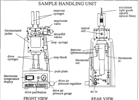
m.2.1 General aspects of stopped-flow spectrophotometry
18
26
26
29
32
33
34
34
36
36
37
38
41
47
49
50
56
62
65
68
72
84
III.2.2Solving experimental problems for running experiments under
strict anaerobic conditions 86
m.2.2.1 Did you buy an anaerobic stopped-flow?
How to make a test. 86
m.2.2.2 Determination of the oxygen ingress rate 87
m.2.2.3Trying to localise the oxygen entrance point 88
m.2.2A Measuring volumes 90
m.2.2.5 Conclusions 91
m.2.3 Stopped-flow experiments on the reduction of cytochrome C3
from Desulfovibrio gigas by sodium dithionite 92
m.2A Stopped-flow experiments on the interaction between cytochromeC3,
FMN and flavodoxin from Desulfovibrio gigas 94
m.3 Kinetic model 97
mAKinetic studies of the reduction of cytochrome C3 by sodium dithionite 108
mA.1 Application of the kinetic model 108
mA.2 Results and Discussion 110
mA.3Conclusions 119
m.5 Kinetic studies of the interaction between cytochrome C3 and FMN
or cytochrome C3 and flavodoxin from Desulfovibrio gigas 122
m.5.1 Introduction 122
m.5.2Results 126
m.5.2.1 Reduction of FMN and flavodoxin by sodium dithionite 127
m.5.2.2 Electron transfer between cytochrome C3 and FMN 129
m.5.2.2.1 Oxidised C3 vs. (FMN+dithionite) 129
III.5.2.2.2Oxidised (c3+FMN) vs. dithionite 130
m.5.2.2.3 Reduced C3 vs. oxidised FMN 132
m.5.2.3Electron transfer between cytochrome C3 and flavodoxin
in the presence of excess sodium dithionite 133
m.5.2.3.1 Oxidised C3 vs. (flavodoxin+dithionite) 133
Ill.5.2.4 Electron transfer between cytochromeC3 and flavodoxin
in the absence of excess sodium dithionite 136
Ill.5.2.4.1 OxidisedC3vs. reduced flavodoxin 136
Ill.5.2.4.2 ReducedC3 vs. oxidised flavodoxin 138
Ill.5.3 Application of the kinetic model 140
Ill.5.3.1 Reaction with fully reduced flavodoxin 141
Ill.5.3.2 Reaction with flavodoxin semiquinone 144
Ill.5.3.3 Reaction with FMN 148
Ill.5.4 Future developments 151
Ill.5.4.1 Experimental progress 152
Ill.5.4.1.1 Improving the quality of the data 152
Ill.5.4.I.2 Running experiments at different pH values 153
Ill.5.4.I.3 Running experiments at different ionic strengths 153
Ill.5.4.I.4 Running experiments in the presence of calcium
or phosphate ions 154
Ill.5.4.2 Improvements in data analysis 155
III.5.5 Conclusions 156
Chapter IV: CONCLUDING REMARKS
IV. 1 Thermodynamic properties of cytochromes C3
IV.2 Kinetic properties of cytochromes C3
IV.3 Proposed model for energy transduction mediated by cytochromeC3 in
sulfate-reducing bacteria
REFERENCES
APPENDICES
Appendix A: Sequential model
Appendix B: Individual model
Appendix C: Integrated rate equations
160
163
170
174
188
190
I INTRODUCTION
I.1General characteristics and activities of sulfate-reducing bacteria
1.2 Considerations about the bioenergetic metabolism of sulfate-reducing bacteria 1.3 Tetrahaem cytochrome123
I INTRODUCTION
1.1 General characteristics and activities ofsulfate-reduclngbacteria
SuIfate-reducing bacteria are a very heterogeneous group of anaerobic prokaryotes which
share the common ability of using sulfate or sulfur as terminal acceptor for the electrons that
result from the oxidation of both organic and inorganic compounds in their bioenergetic
processes. Work in recent years has demonstrated that there is an enormous diversity of
morphological and metabolic characteristics among these bacteria, which could be the result
of an early evolutionary divergence. This diversity enables sulfate-reducing bacteria to live in
many different environments and increases their economic interest and their importance as
objects of scientific investigation. Since suIfate-reducers play an important role in
contamination of petroleum products and in anaerobic corrosion of iron and steel, these
organisms are of great economic interest in many industrial sectors. Their environmental
impact is also very important in highly polluted ecosystems.
These bacteria were difficult to grow and manipulate in the laboratory but newly developed
experimental techniques are leading to the discovery of a great variety of metabolic processes
and are revealing a diversity of microbial forms not previously apparent in this group of
organisms. Moreover, significant advances in the understanding of their bioenergetics,
genetics and molecular biology are leading to an increase in their environmental and industrial
importance.
The early taxonomic organisation of suIfate-reducing bacteria was very simple. According to
Postgate and Campbell (CampbelI and Postgate, 1965 and Postgate and CampbelI, 1966) only
two genera were considered on the basis of morphological characteristics, the ability to form
spores, the C+G content and the presence of certain pigments. These genera were
Desulfovibrio and Desulfotomaculum. Although most of the organisms responsible for
dissimilatory sulfate reduction still fit into these two categories, it was demonstrated later that
sulfate-reducers are much more diverse than previously recognised and at least 14 new genera
with different morphological and metabolic capabilities are presently recognised (Widdel and
Hansen 1991 ; Singleton, 1993). Antigenic studies and comparison of 16S ribosomal RNA
1.2 Considerations about the bioenergetic metabolism ofsulfate-reducingbacteria
Sulfate-reducing bacteria extract the energy necessary for their growth from the anaerobic
respiration of suIfate. In this process, sulfate is used as the terminal acceptor for the electrons
and protons that result from the oxidation of foodstuffs. Note that, since sulfate reduction is
being used for bioenergetic purposes and not for the incorporation of sulfur into biological
compounds, sulfate respiration is a dissimilatory rather than an assimilatory process.
Itwill be mentioned below that it would be impossible for the bacteria to grow on lactate and
sulfate in the absence of oxidative phosphorylation coupled to the overall process of sulfate
reduction. This is because the ATP obtained by substrate level phosphorylation in the process
of lactate oxidation is completely consumed in the activation of sulfate. Also, the finding that
some sulfate-reducing bacteria can grow chemolithotrophically on H2 plus sulfate as the sole
energy source clearly established that the reduction of sulfate to sulfide is coupled to ATP
phosphorylation (Badziong et al., 1978 ; Badziong and Thauer, 1978).
1.2.1 Electron donors
Most of the substrates used by sulfate-reducing bacteria as carbon and energy sources are
low-molecular-weight compounds which are typical fermentation products that result from
the anaerobic bacterial degradation of carbohydrates, proteins and other components of dead
biomass (Widdel, 1988). The number of compounds which are known to be metabolised by
these bacteria increased enormously during the past two decades and now amounts to over
125. Also, great progress has been made in the knowledge of the pathways and the enzymes
that are used in the degradation of the substrates by sulfate-reducing bacteria (Hansen, 1994).
Examples of electron donors used by sulfate-reducing bacteria are hydrogen, ethanol, lactate,
pyruvate and malate (Widdel, 1988 ; Singleton, 1993 ; Woordouw, 1995). However, many
other organic compounds, including crude oil components (Rueter et aI., 1994) and metallic
iron, (Odom, 1993) can serve as electron donors for sulfate reduction.
It has been proposed that sulfate-reducers can be organised in two major metabolic groups
(Widdel 1988): one carries out incomplete oxidation of lactate or other organic substrates to
acetate and CO2 , whereas the other is capable of completely oxidising a variety of
compounds, including acetate, fatty acids and aromatic compounds, to CO2, Apparently the
acetate unit (acetyl coenzyme A). Although some of them can utilise alcohols and aldehydes
of low molecular weight (Turner et aI., 1987;Widdel, 1988 ;Hensgens et aI., 1993 ;Barata et
al., 1993 ; Hansen, 1994) they are nutritionally less versatile than those of the second group.
The complete oxidisers are able to use most of the electron donors utilised by the incomplete
oxidisers and also other substrates which have not been observed so far to be used by the
members of the first group. Such compounds, besides acetate, include branched fatty acids,
acetone, phenyl-substituted organic acids, phenolic compounds and indole. Most complete
oxidisers grow significantly more slowly than incomplete oxidisers (Widdel 1988).
Chemolithotrophic growth of Desulfovibrio spp. in the presence of
sol-
with H2 as the soleenergy source was reported and explained by Badziong and Thauer, 1978. Under these
conditions, acetate and CO2 are required as the carbon source (Badziong et aI., 1979). Some
sulfate-reducers have the ability to grow autotrophically using C02 as the sole carbon source
(Brysch et aI., 1987). Hydrogen is oxidised by periplasmic hydrogenases and a proton
gradient results from this process because hydrogen oxidation and sulfate reduction occur in
different cellular compartments (Badziong and Thauer, 1980). Since in energetic terms 2
moles of ATP are necessary for the activation of sulfate (see below) the net synthesis of 1
mole of ATP per mole of
sol-
reduced requires the phosphorylation of 3 moles of ATP asthe result of the proton gradient(cf. Figure 1.1).
Periplasm
8H+
\
Cytoplasm
NET: 4 H
2+
80i-
+
ADP
+
P
j - - '8
2-+
ATP
Figure 1.1. Bioenergetic processes occurring in sulfate-reducing bacteria growing on hydrogen and
sulfate, Hydrogen oxidation and suIfate reduction take place in different cellular compartments, generating a
When lactate and pyruvate are used as electron donors their oxidation is incomplete because
CO2 and acetate result from this process. Lactate is first oxidised to pyruvate by an NAD(P)
independent membrane bound lactate dehydrogenase that is not well characterised (Hansen,
1993). Subsequently, pyruvate is oxidised to acetate and CO2 via acetyl coenzyme A and
acetylphosphate intermediates. In this process, 1 mole of ATP is produced by substrate level
phosphorylation (Figure 1.2). Since 2 electrons and 2 protons are released in each oxidation
step and 8 electrons are required for the reduction of sulfate to sulfide, two moles of lactate
are oxidised per mole of suIfate. The overall balance shows that there is no net production of
ATP in this process, unless proton-driven ATP synthesis occurs in addition to substrate level
phosphorylation. However, the mechanism for the generation of the proton gradient is not
obvious because production and consumption of electrons and protons take place in the same
cellular compartment. Odom and Peck, 1981 proposed the hydrogen cycling hypothesis to
explain the formation of the proton gradient under these conditions (see below).
Periplasm
Cytoplasm
2 lactate
f-.4e'+4H+
2 pyruvate
2 P;-1--.4e-+ 4H+
~2C02
2
acr:~ADP
b
2 ATP2 acetate
SO
2-4 2ATP
2 ADP+2P;
4Hp
NET: 2 lactate
+
SOi---.
2 acetate+
2 CO2+
S2-Figure 1.2. Oxidation of lactate to acetate and COz is coupled to suifate reduction. Since the 2 ATP
formed by substrate level phosphorylation in the conversion of 2 acetylphosphate (acetyl-P) into 2 acetate are consumed in the activation of sulfate, there is no net production of ATP, unless proton-driven ATP synthesis occurs in addition to the processes represented in the picture.
It should be noted that, if pyruvate only is used as electron donor, the ATP formed by
substrate level phosphorylation is sufficient to sustain growth. However, oxidation of
pyruvate is similarly thought to be coupled to proton translocation (Peck 1993). In some
organisms, pyruvate can support growth in the absence of sulfate. The mechanism is not
clearly established, but it probably involves the pyruvate phosphoroclastic system because
organisms (Peck, 1993).
Some strains can grow on malate and sulfate, possibly oxidising malate to pyruvate and CO2
(Postgate, 1984 ; Hansen, 1993). In the absence of sulfate, Desulfovibrio gigas converts 3 mol
of malate into 2 mol of succinate plus 1 mol of acetate (Chen et aI., 1995). Acetate is formed
from decarboxylation of pyruvate and succinate results from the reduction of fumarate, which
works as the electron acceptor in the absence of sulfate. These authors observed
NADH-linked fumarate reductase activity, supporting the involvement of pyridine nucleotides in the
electron pathway toward the reduction of sulfate or fumarate.
1.2.2 Hydrogen cycling hypothesis
To explain the bioenergetics of growth when lactate is used as electron donor, adorn and
Peck, 1981 proposed a mechanism known as the hydrogen cycling. According to this
hypothesis, the electrons produced by oxidation of lactate and pyruvate cannot be used
directly for sulfate reduction. Instead, they combine to form hydrogen in a reaction catalysed
by cytoplasmic hydrogenase. Then, hydrogen diffuses to the periplasmic space where it is
decomposed again into electrons and protons by a periplasmic hydrogenase (Figure 1.3).
Periplasm
ETC
8e-Cytoplasm
4e-
~+
2Tt.te
2 pyruvate
2COz
2 acetyl-P
V
2 AD P}--~2ATP
2 acetate
Figure 1.3.Hydrogen cycling hypothesis. The electrons and protons produced by the oxidation of lactate in the cytoplasm combine to form hydrogen in a reaction catalysed by the cytoplasmic hydrogenase (dark Hase). Hydrogen diffuses to the periplasmic space where it is re-oxidised back to electrons and protons in a reaction catalysed by the periplasmic hydrogenase (light Hase). This process generates a proton gradient that is utilised
The protons produced in the periplasm from the oxidation of molecular hydrogen contribute
to the proton gradient used by the ATPsynthase to generate ATP. The electrons that result
from the same reaction are transported across the membrane, through a transmembrane
electron transfer complex, to be used in the cytoplasm for sulfate reduction. The hydrogen
cycling hypothesis requires the presence of both cytoplasmic and periplasmic hydrogenases in
Desulfovibriospp. Since only the periplasmic hydrogenases are present in all sulfate-reducers
belonging to this genus, there might be alternative mechanisms for the generation of the
coupled proton gradient, for instance involving the membrane bound lactate dehydrogenase or
the membrane-bound NADH or NADPH dehydrogenases (Hansen, 1994; Woordouw, 1995).
Note that the mechanism proposed by Badziong and Thauer, 1980 to explain the bioenergetics
of growth in the presence of sulfate with hydrogen as sole energy source (cf. Figure I.1),
corresponds to the second half of the mechanism of hydrogen cycling. However, since
external hydrogen is being utilised, only the periplasmic hydrogenases are necessary for this
bioenergetic mechanism.
Independently of the general validity of the hydrogen cycling model, an important question
concerns the involvement of pyridine nucleotides in linking substrate oxidation to sulfate
reduction. Recent work has shown a role for NADH and NADPH as redox carriers in the
degradation of certain substrates (Hansen, 1994 ; Chen et aI., 1995 ; Fareleira et aI., 1997). If
NADH is reoxidised by the final electron acceptor via H2, an efficient removal of the
hydrogen gas could change the unfavourable transfer of reducing equivalents from NADH to
H+ into a thermodynamically favourable process (Fareleira et aI., 1997). Moreover, an
alternative mechanism could be envisaged for the production of hydrogen in the cytoplasm in
those organisms that lack the cytoplasmic hydrogenase.
1.2.3 Electron acceptors
The electrons and protons resulting from the oxidative processes mentioned above are used to
reduce sulfate to sulfide via an electron transport system. Interestingly, many electron transfer
proteins of sulfate-reducing bacteria are soluble and cytoplasmic, which raises some questions
about the mechanism of oxidative phosphorylation coupled to electron transfer (see below).
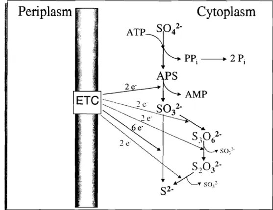
Periplasm
wCytoplasm
Figure 1.4. Steps involved in dissimilatory sulfate reduction. The activation of sulfate (Sot) by ATP results in adenylylsulfate (APS) formation. APS is reduced to sulfite (S03 2") and AMP is released. The reduction of sulfite to sulfide (S2') is either a 6-electron step (dark reaction) or involves trithionate (S30/') and thiosulfate
(S2ol") as intermediates. The reactions of the trithionate pathway are shown in grey. ETC: electron transfer complex.
The first step is thermodynamically unfavourable, requiring activation by ATP. This reaction
is catalysed by ATP sulfurylase and generates adenylylsulfate (APS) and inorganic
pyrophosphate (PPj). Further hydrolysis of PPj drives the reaction forward. Since both
phosphoanhydride bonds of ATP are hydrolysed in this process, this is energetically
equivalent to the consumption of two moles of ATP. Ithas also been suggested that, in some
species of the genus Desulfotomaculum, PPj could be used for ATP production via
phosphorylation of acetate to form acetylphosphate (Liu and Peck, 1981 ; Liu et al., 1982).
The second step in the process of sulfate reduction is the reduction of APS to bisulfite, a
reaction catalysed by APS reductase, which is a soluble, cytoplasmic enzyme, containing
non-haem iron and flavin centres.
Finally, there is the reduction of bisulfite to sulfide. This reaction is catalysed by dissimilatory
bisulfite reductase, which is a high-molecular-weight heterooligomer containing iron-sulfur
clusters and sirohaem. Depending on the spectral characteristics, four different enzymes have
been described so far: desulfoviridin, desulforubidin, desulfofuscidin and P582 (LeGall and
reduction of bisulfite to sulfide. Two major mechanisms have been proposed: the trithionate
pathway (see reactions in grey in Figure 104) and direct reduction of sulfite to sulfide
involving six electrons in a single step. The main arguments for and against the trithionate
pathway are discussed by LeGall and Fauque, 1988. This question is not resolved but
physiologically it seems likely that a single enzyme catalyses the overall reaction with
formation of relatively stable intermediates. However, it is also possible that the absence of
free intermediates is limited toDesulfovibriospecies, whereas an active trithionate pathway is
operative inDesulfotomaculum (LeGall and Fauque, 1988).
It is worth mentioning that suIfate-reducing bacteria can use electron acceptors other than
sulfate. One example is the process of fumarate respiration mentioned above, in which the
electrons from the oxidation of malate and pyruvate are transferred to fumarate, generating
succinate (Chen et aI., 1995).
Some organisms can reduce nitrate or nitrite to ammonia (Mitchell, et aI., 1986 ; Seitz and
Cypionka, 1986 ; Dalsgaard and Bak, 1994).
Under microaerophilic conditions, some Desulfovibrio spp. can even use oxygen as an
electron acceptor (Dilling and Cypionka, 1990 ; Chen et aI., 1993a,b). Apparently, the ATP
produced in this process can be used for cell maintenance but not for growth.
SuIfate-reducers can use Fe(III) (Coleman et aI., 1993) and heavy metals such as Cr(VI)
(Fude et aI., 1994 ; Lovley and Phillips, 1994) and U(VI) (Lovley et aI., 1993) as electron
acceptors contributing to their reduction and immobilisation. It is thought that cytochromeC3
works as the metal reductase in some of these cases (Lovley et aI., 1993 ; Lovley and Phillips,
1994).
As mentioned above, D. vulgaris can grow on pyruvate in the absence of sulfate, producing
acetate, CO2 and stoichiometric amounts of hydrogen (Postgate, 1952). Protons are used as
electron acceptors in this system. Interestingly, growth in lactate in the absence of sulfate is
negligible or absent, unless H2-utilising methanogenic bacteria are also present (Bryant et aI.,
1977). These bacteria contribute to the lowering of the hydrogen pressure in the media, thus
facilitating growth of the sulfate-reducers, Under these circumstances, all reducing
equivalents generated from oxidation of lactate to acetate and CO2 appear as methane. In this
case, the energy for growth in lactate is provided by substrate level phosphorylation, because
1.3 Tetrahaem cytochromeC3
Tetrahaem cytochrome C3 is one of the proteins involved in the bioenergetic metabolism of
sulfate-reducing bacteria. It is produced in large quantities by these organisms and it is
probably the best characterised multi-haem cytochrome.
The name 'cytochrome C3' has been given to many multihaem c-type cytochromes isolated
from sulfate-reducers which have approximately 25 aminoacid residues per haem and display
bis-histidinyl axial coordination of the haems. In this work, cytochrome C3 refers to the
tetrahaem protein only. This cytochrome was first isolated in 1954 (Postgate,1954 and
Ishimoto et aI., 1954) from two Desulfovibrio vulgaris strains and was the first cytochrome
discovered in an anaerobic, non-photosynthetic organism. It is a soluble protein with a
molecular weight of ea. 15 000 and is present in all species ofDesulfovibrio. All sequenced
cytochromes C3 have a typical N-terminal signal peptide that indicates a periplasmic
localisation. The four haems have bis-histidinyl axial coordination and exhibit different and
low redox potentials. They are also low spin in both oxidation states. Cytochromes C3 isolated
from different organisms can have very different aminoacid compositions and, consequently,
the isoelectric points and the distribution of charges on the protein can vary appreciably from
one molecule to another. Interestingly, the architecture of the haem core as well as the general
folding of the protein are highly conserved, despite the low sequence homology observed
between most of these molecules (Coutinho and Xavier, 1994).
1.3.1 Physiological role
Since the presence of cytochrome C3 is necessary for the reduction in vitro of low molecular
weight electron transfer proteins such as ferredoxin, flavodoxin and rubredoxin by hydrogen
in the presence of hydrogenase, this cytochrome has been proposed as a coupling factor for
the enzyme hydrogenase (Bell et aI., 1978). Computer modelling studies indeed suggested
that cytochrome C3 may specifically dock with flavodoxin (Stewart et aI., 1988), ferredoxin
(Cambillau et aI., 1988) and rubredoxin (Stewart et aI., 1989). However, localisation of the
cytochrome in the periplasmic space would preclude their direct interaction in vivo because
the small redox proteins are localised in the cytoplasm. This compartmentalisation of the
redox proteins raises some questions about the physiological relevance of these complexes. A
transmembrane localisation of cytochrome C3 would resolve this enigma but it is highly
1988). However, studies with artificial liposomes modified with cytochrome C3 showed that
the electron transport rate across the membrane was very much accelerated and coupled to
rapid proton influx in the presence of the cytochrome (Tabushi et aI., 1984). These authors
suggested that a cytochrome C3dimer could form an electron-channel across the membrane of
the liposome. Peck, 1993 also proposed that a cytochrome C3 dimer could mediate the
transmembrane electron transfer between hydrogen oxidation in the periplasm and APS
reduction in the cytoplasm. Furthermore, the homology between tetrahaem cytochromeC3and
the hexadecahaem cytochromeC3, which was proposed to be part of a transmembrane electron
transfer complex (Rossi et aI., 1993), may provide some physiological relevance for the
studies of complex formation between tetrahaem cytochrome C3 and the small cytoplasmic
redox proteins.
It has been shown recently that, in the presence of cytochrome C3, the periplasmic
hydrogenase maintains the maximum hydrogen uptake activity at physiological pH, whereas
in the absence of the cytochrome this activity decreases to ca. 40% (Louro et aI., 1997a).
According to the hydrogen cycling hypothesis, the reaction of hydrogen oxidation that takes
place in the periplasm is crucial for the bioenergetic metabolism of sulfate-reducers, because
it is the only energy production step for cells growing with hydrogen as sole energy source.
Louro et aI., 1997a proposed that cytochrome C3 coupled to hydrogenase performs energy
transduction, acidifying the protons produced in the reaction of hydrogen oxidation at the
expense of electronic energy, in the absence of a membrane confinement. The role played by
cytochrome C3in this mechanism is fundamental: its peculiar thermodynamic properties make
it capable of accepting both electrons and protons from hydrogenase, thus favouring the
reaction of hydrogen oxidation and, by giving electrons and protons to different acceptors,
this cytochrome conserves and increases the charge separation achieved by hydrogenase.
1.3.2 Structure
The structures of several cytochromes C3 have been solved by X-ray crystallography:
cytochrome C3from Desulfovibrio vulgaris (Dvc-) strain Hildenborough (Matias et aI., 1993),
Dvc, strain Miyazaki (Higuchi et aI., 1984), cytochrome C3 from Desulfovibrio gigas (Dgc-)
(Matias et aI., 1996), cytochrome C3 from Desulfovibrio desulfuricans (Ddc-) strain ATCC
27774 (Morais et aI., 1995) and cytochrome C3 from Desulfomicrobium baculatum (Dmbc-)
now, only the oxidised form of the cytochrome has been accessible to crystallographic studies
because this protein is readily oxidised in the presence of small amounts of oxygen. Recently,
the structure in solution of the reduced form ofDvc, Hildenborough has been solved by NMR
techniques (Messias et al., 1998) opening interesting new perspectives for studies of
structure-function correlation.
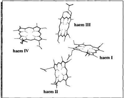
~
..<')"'~- "...,.,
..,
haem I haem IV
haem 11
haemIII
Figure 1.5.Haem core of cytochrome C3 fromDesulfovibrio gigas, The haems are numbered according to
the order of attachment to the polypeptide chain. Picture taken from the X-ray structure determined by Matias et al., 1996.
In all structures, the four haems are ligated to the apoprotein through sequence segments of
the type Cys-X-X-(X-X)-Cys-His in which the cysteine residues are covalently bound through
thioether linkages to the vinyl sidechains of the porphyrin ring and the histidine is the fifth
axial ligand of the iron. The sixth axial histidine ligand to the iron is provided by a different
region of the polypeptide chain. As stated above, the haem core is highly conserved: all
cytochromes C3 display similar iron to iron distances and the same relative orientations of the
haem planes. The haem core of cytochrome C3 from D. gigas is shown in Figure 1.5. All the
haems are well exposed to the solvent, which is easily understood given the small number of
aminoacid residues of this protein. Structural invariants other than the haem ligations include
the conservation of a threonine close to haem IV, a glycine close to haem Il and a
phenylalanine close to haems I and Ill. Depending on the sequence alignment chosen, a few
other residues may be regarded as conserved. The conserved phenylalanine was thought to
play an important role in the electron transfer process but site-directed mutagenesis of this
residue in Dvc, has shown that fast intramolecular electron transfer is still observed and that
the thermodynamic properties, namely the positive cooperativity observed between haems I
present a segment with a high content of lysine residues close to haem IV in the C-terminal
region. This highly positively charged region has been postulated as the docking site for the
physiological redox partners (Stew art et aI., 1988).
Sequence alignments were proposed assuming that all cytochromes have a similar haem core
and postulating sequence insertions and deletions in order to maximise the homologies
between the various segments (Kissinger, 1989 ; Moore and Pettigrew, 1990 ; Palma et aI.,
1994; Magro et aI., 1997). The lowest homology (ea. 25%) is found between the cytochromes
from Dsm. baculatumandD.vulgaris.
1.3.3 Thermodynamic properties
The thermodynamic properties of cytochrome C3 are very complex and, since their modelling
is one of the main objectives of this thesis (cf. chapter II), only the general properties are
mentioned here. As stated above, the four haems in cytochrome C3 have different
environments, thus displaying different redox potentials. Moreover, since the distances
between them are small, the redox potential of each of the four haems is affected by the redox
state of the neighbouring haems. This effect is accounted for by the definition of haem-haem
interacting potentials (Santos et aI., 1984a). Only negative cooperativities would be expected
if the interactions were simply of an electrostatic nature. However, at least two of the haems
show positive cooperativity between them, in all cytochromes C3 studied so far (Louro et aI.,
1997b). This positive cooperativity probably arises from a localised conformational change
linked to the redox process. In addition to that, the redox potentials of the haems depend upon
the protonation state of an ionisable residue, thus displaying a redox-Bohr effect. It was
demonstrated (Turner et aI., 1994, 1996) that the pH-dependence of the redox potentials of the
haems can be accounted for by the definition of four haem-proton interacting potentials. Since
all these interacting potentials correspond to positive cooperativities, they can be explained by
simple electrostatics.
It should be noted that, although the structure of the haem core is strictly conserved, the order
of reduction of the four haems is not the same for all cytochromes C3 (Coutinho and Xavier,
1994 ; Pereira et aI., 1998), nor is the positive cooperativity always observed between the
same pair of haems. However, for all cases studied upto now, the interplay between all
haem-haem and haem-haem-proton cooperativities results in a concerted two-electron step which is
(Louro et aI., 1997b). These thermodynamic properties give cytochrome C3the ability to work
as an energy transducing system (Louro et aI., 1996a), but kinetic control of the various steps
is essential to avoid unproductive pathways.
1.3.4 Kinetic properties
Cytochrome C3 has four redox centres and, therefore, both intramolecular and intermolecular
electron transfer processes must be considered for the analysis of the kinetic properties of this
molecule. NMR studies have shown that the first order rate constant for the intramolecular
electron exchange is higher than 105S·I (Santos et aI., 1984a). Since this process is faster than
the bimolecular electron exchange with the redox partners or artificial reducing agents, the
system is under thermodynamic control. Therefore, independently of the electron entrance
gate, the electrons are distributed inside the molecule according to the relative redox
potentials of the haems. Thus, the correct modelling of the kinetic properties of cytochrome C3
has to rely on an accurate thermodynamic description of the system.
The reduction of cytochrome C3 by redox proteins or exogenous electron donors usually
exhibits a biphasic kinetic profile. Most of the second order rate constants determined for the
reduction process from stopped-flow studies with sodium dithionite or with redox proteins are
of the order of 106_107M·ls·1(Favaudon et aI., 1978; Tabushi et aI., 1983; Capeillere-Blandin
et al.. 1986; Catarino et aI., 1991). The second order rate constants for the oxidation of
reduced cytochrome C3 by molecular oxygen or by oxidised flavodoxin are smaller, having
values of the order of 104_105 M·1s·1 (Capeillere-Blandin et aI., 1986; De Francesco et aI.,
1994). It is apparent from the values of the rate constants reported in the literature that factors
such as the pH and the ionic strength affect the electron transfer rates and also the relative
amplitudes of the fast and slow phases. These rate constants are also strongly dependent on
the formal charge of the reducing agent.
1.4 Thesis plan
Apart from the general introduction and the concluding remarks, this thesis is divided into two
main chapters, one dealing with the thermodynamic and the other with the kinetic properties
of cytochrome C3. Both aspects are fundamental to the global understanding of the function of