Effect of a single session of transcranial direct-current
stimulation on balance and spatiotemporal gait
variables in children with cerebral palsy:
A randomized sham-controlled study
Luanda A. C. Grecco1,2,3, Natália A. C. Duarte1,3, Nelci Zanon3,4, Manuela Galli5, Felipe Fregni2, Claudia S. Oliveira1
ABSTRACT | Background: Transcranial direct-current stimulation (tDCS) has been widely studied with the aim of
enhancing local synaptic eficacy and modulating the electrical activity of the cortex in patients with neurological
disorders. Objective: The purpose of the present study was to determine the effect of a single session of tDCS regarding immediate changes in spatiotemporal gait and oscillations of the center of pressure (30 seconds) in children with cerebral palsy (CP). Method: A randomized controlled trial with a blinded evaluator was conducted involving 20 children with CP between six and ten years of age. Gait and balance were evaluated three times: Evaluation 1 (before the stimulation), Evaluation 2 (immediately after stimulation), and Evaluation 3 (20 minutes after the stimulation). The protocol consisted of a 20-minute session of tDCS applied to the primary motor cortex at an intensity of 1 mA. The participants were randomly allocated to two groups: experimental group – anodal stimulation of the primary motor cortex; and control group – placebo
transcranial stimulation. Results: Signiicant reductions were found in the experimental group regarding oscillations
during standing in the anteroposterior and mediolateral directions with eyes open and eyes closed in comparison with
the control group (p<0.05). In the intra-group analysis, the experimental group exhibited signiicant improvements in gait velocity, cadence, and oscillation in the center of pressure during standing (p<0.05). No signiicant differences were found in the control group among the different evaluations. Conclusion: A single session of tDCS applied to the primary
motor cortex promotes positive changes in static balance and gait velocity in children with cerebral palsy.
Keywords: cerebral palsy; physical therapy; movement; balance; electric stimulation; motor cortex.
This study was registered with the Brazilian Registry of Clinical Trials (RBR-9B5DH7).
HOW TO CITE THIS ARTICLE
Grecco LAC, Duarte NAC, Zanon N, Galli M, Fregni F, Oliveira CS. Effect of a single session of transcranial direct-current stimulation on balance and spatiotemporal gait variables in children with cerebral palsy: A randomized sham-controlled study. Braz J Phys Ther. 2014 Sept-Oct; 18(5):419-427. http://dx.doi.org/10.1590/bjpt-rbf.2014.0053
Introduction
Transcranial direct-current stimulation (tDCS) is
a widely studied innovative technique consisting of
the application of low-intensity monophasic electrical
current to the scalp. The electrical current lows from the electrodes and penetrates the skull, reaching the cerebral cortex. Although most of the current is dissipated among the overlying tissues, a suficient amount of current reaches the structures of the cortex,
modifying the membrane potential of the cells and
modulating cortex activity1,2. It has been suggested
that the effects of tDCS stem from persistent changes
that resemble long-term potentiation and can lead to
enhanced synaptic eficacy3.
There has been an increase in the number of studies
stating that tDCS applied to the motor cortex can be
used for the treatment of neurological disorders in
children, such as cerebral palsy (CP)4. CP results in
diminished activation of the central nervous system during the execution of movements5. A reduction in motor cortex excitability in children is associated with poor motor development6. Neurophysiological analyses have revealed global alterations in cortex
1Programa de Pós-Graduação em Ciências da Reabilitação, Universidade Nove de Julho (UNINOVE), São Paulo, SP, Brazil
2Laboratory of Neuromodulation, Center of Clinical Research Learning, Spaulding Rehabilitation Hospital, Harvard Medical School, Boston, MA, United States
3Centro de Neurocirurgia Pediátrica (CENEPE), São Paulo, SP, Brazil
4Departamento de Neurocirurgia, Universidade Federal de São Paulo (UNIFESP), São Paulo, SP, Brazil 5Dipartimento di Bioingegneria, Politecnico di Milano, Milan, Italy
excitability in children with CP, with a reduction in the activation of corticospinal and somatosensory
circuits7. The reduction in somatosensory activation may be the neurological basis for poor tactile, proprioceptive and kinesthetic awareness in children
with CP8. While there is no cure for the brain
lesion associated with this condition, sequelae
can be minimized through neurorehabilitation methods9. Studies involving functional magnetic resonance in children with CP have demonstrated
that rehabilitation resources are capable of promoting
the activation of the primary motor cortex9, which is
an important area of the brain capable of facilitating cerebral reorganization10.
Ninety percent of children with CP exhibit impaired gait due to diminished cortex excitability, excessive muscle weakness, abnormal joint kinematics,
and diminished postural reactions11. Moreover,
inadequate postural control limits motor development
in these children12,13.
Kaski et al.14 found that anodal tDCS induces
changes in the excitability of the motor cortex referring to the lower limbs, with improvements in
both balance and gait. The hypothesis of the present study is that a single session of anodal tDCS applied
to the primary motor cortex in children with CP can
momentarily potentiate motor patterns through the
enhancement of cortex excitability and activation of corticospinal circuits. The authors believe that the facilitation of cortical excitability of the primary motor cortex may enhance motor control and velocity of motor responses in children with CP. In CP, deicits
in spatiotemporal gait parameters and postural stability are notorious and generate a functional
impairment of the child. Additionally the evaluation
of the static balance and gait analysis are consecrated
and scientiically valid techniques. For these reasons,
the stabilometry and analysis of spatio-temporal parameters of gait were selected as outcomes of
this study. The expected outcomes are an increase in gait velocity and reductions in the oscillation of
the center of pressure (CoP) during standing in the
anteroposteior and mediolateral directions. However,
the changes would likely be lost after a few minutes due to the limitation of tDCS to a single session.
The aim of the present study was to determine the effect of a single session of tDCS applied to the
primary motor cortex regarding immediate changes
in spatiotemporal gait and oscillations of the CoP
during standing in children with CP classiied at levels I to III of the Gross Motor Function Classiication System (GMFCS).
Method
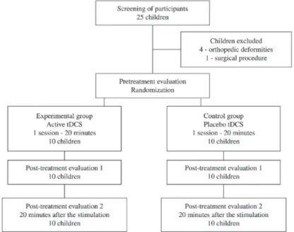
The present randomized, sham-controlled,
cross-sectional study (Figure 1) was carried out in compliance with the ethical standards of the
Declaration of Helsinki and received approval from the Human Research Ethics Committee of Universidade Nove de Julho (UNINOVE), São Paulo, SP, Brazil, under process number 69803/2012.
This study is registered with the Brazilian Registry of Clinical Trials (process RBR-9B5DH7). All
guardians signed a statement of informed consent agreeing to the participation of their children.
Children with a diagnosis of spastic CP were recruited from specialized clinics. The inclusion
criteria were classiication at levels I, II and III of the GMFCS15,16, independent gait for at least 12 months,
age six to ten years, and degree of understanding
compatible with the procedures proposed. The
following were the exclusion criteria: having
undergone any surgical procedure or neurolytic block
in the previous 12 months, orthopedic deformity, epilepsy, metal implants in the skull or hearing aids. Following the application of the eligibility criteria, 20 children were selected for the study.
The participants were randomized into the
experimental and control groups based on the order
of inclusion into the study. A randomization list
was generated using blocks of six (for every six participants, three were randomly allocated to each group) and four (for every four participants, two were
randomly allocated to each group) to minimize the risk of imbalance in the size of the groups.
The procedures were carried out in a single day.
Following Evaluation 1 (pretreatment evaluation/ before stimulation), the children received 20 minutes of either active (experimental group) or sham (control group) tDCS. The children received tDCS at rest
and seated comfortably. A responsible therapist
accompanied the stimulation session. Evaluation 2 (post-treatment evaluation/after stimulation)
was performed immediately following tDCS and
Evaluation 3 (twenty minutes after stimulation) was performed after 20 minutes of rest. Three researchers carried out the procedures – two performed the evaluations and one performed the tDCS. The evaluations and tDCS were carried out in separate rooms to ensure the blinding of the examiners. Only
the researcher in charge of the application of the tDCS was aware of the allocation of the children to
the experimental and control groups.
Transcranial direct-current stimulation tDCS is the application of a low-intensity direct current on the scalp using two electrodes (anode and
cathode). A suficient amount of current penetrates the overlying tissues and reaches the structures of the motor cortex, modifying the neuronal
membrane potential. Anodal stimulation enhances
cortex excitability. The tDCS device (Soterix Medical Inc., USA) included two non-metallic
sponge surface electrodes measuring 5 × 5 cm2 and moistened with saline solution. The children in the
experimental group received anodal stimulation of the primary motor cortex and those in the control group received placebo transcranial stimulation. The anode was positioned over the primary motor cortex of the dominant hemisphere following the 10-20 international system of electrode placement
for electroencephalography17 and the cathode was positioned in the supra-orbital region contralateral to the anode. The current was applied to the primary
motor cortex for 20 minutes, during which the children remained seated. The tDCS device has a
button that allows the operator to control the intensity of the current. Stimulation was gradually increased
until reaching 1 mA and gradually reduced in the inal 10 seconds. For sham stimulation, the electrodes were
positioned in the same manner and the stimulator was
switched on for 30 seconds. This procedure gave the children in the control group the initial sensation, but they did not receive electrical stimulation for the remainder of the session. This is considered a valid control procedure in studies involving tDCS18.
Evaluation procedures
The evaluation of spatiotemporal gait variables (gait velocity, cadence, step length, stride length and step width) was performed using the SMART-D 140® system (BTS Engineering, Italy) with eight infrared cameras, the SMART-D INTEGRATED WORKSTATION® with 32 analog channels and a synchronized video system. After the determination of the anthropometric measures (height, mass, lower limb length, distance between the femoral condyles or diameter of the knee, distance between the malleolus or diameter of the ankle, distance between the anterior iliac spines, and thickness of the pelvis), passive markers were placed at speciic reference points directly on the skin for the evaluation of each segment of the body. The markers were placed over C7 and the sacrum as well as bilaterally over the acromion, anterosuperior iliac spine, greater trochanter, femoral epicondyle, femoral wand, tibial head, tibial wand, lateral malleolus, lateral aspect of the foot at the ifth metatarsal head and at the heel (only for static offset measurements), as described by Davis et al.19.
The Davis marker-set was chosen as the protocol of choice to acquire the movement of lower limbs and trunk based on Ferrari et al.20. After the child was
familiarized with the process, at least six trials were
performed along a 5-meter catwalk at a pace self-selected by each child. Three consistent trials of each lower limb were considered for analysis. All readings
to ensure the reliability of the data collection. In the
present study, only spatiotemporal and kinematic gait variables were identiied and computed. The
following spatiotemporal parameters were analyzed:
• velocity (m/s): mean velocity of progression; • cadence: number of steps in a time unit (steps/
min);
• stride length (m): longitudinal distance between
successive points of heel contact of the same foot; • step length (m): longitudinal distance between the
point of initial contact of one foot and the point of
initial contact of the contralateral foot;
• step width (m): distance between the rear end of the right and left heel centerlines along the
mediolateral axis;
• stance phase: % of gait cycle that begins with initial contact and ends at toe-off of the same limb.
Mean and standard deviation values of gait velocity, cadence, step length, stride length, and step
width were used for the statistical analysis.
Static balance was evaluated with the use of a force plate (Kistler model 9286BA), which allows
stabilometric analysis through readings of oscillations
of the CoP. The acquisition frequency was 50 Hz,
captured by four piezoelectric sensors positioned at
the extremities of the platform, which measured 40 ×
60 cm. The data were recorded and interpreted using
the SWAY software program (BTS Engineering) integrated to and synchronized with the SMART-D 140®. The child was instructed to remain in a quiet standing position on the platform, barefoot, arms alongside the body, gaze ixed on a point marked at a distance of one meter at the height of the glabellum,
with heels aligned and an unrestricted foot base. The
children classiied at level III of the GMFCS15,16
used their usual gait assistance device, which was
positioned off the force plate. The platform used has
dimensions (600X400X35mm) that do not require the child to make great postural adjustments to position the gait assistance device off the platform. The children were instructed to keep the assistance device off the platform. The positioning of the device should allow a comfortable posture. The exact location of the device was marked on the loor with a white ribbon. The positioning was used in the three Evaluations to allow same condition assessment and comparative
analysis21.
Readings of displacement from the CoP on the
X (anteroposterior) and Y (mediolateral) axes were
performed under two conditions: eyes open and
eyes closed. Three acquisitions of 30 seconds were obtained for each condition and the average of the acquisitions was used in the statistical analysis. The
outputs of the force platform allowed us to compute the CoP time series in the anteroposterior direction and the mediolateral direction. The output of the
platform was processed to compute quantitative
parameters in the time domain. The anteroposterior
and mediolateral coordinates of the CoP trajectory underwent post-acquisition iltering using a low-pass ilter with a cut-off frequency of 10 Hz. In the analysis, we identiied and computed the range of
CoP displacement in the anteroposterior direction
(RANGEAP index) and the mediolateral direction (RANGEML index), expressed in mm21.
Statistical analysis
The Kolmogorov-Smirnov test was used to determine the adherence of the data to the Gaussian curve. Parametric distribution was demonstrated, the data were expressed as mean and standard deviation values. To verify the effect of transcranial stimulation (active and placebo) over the three Evaluations in each group, intragroup analysis was
performed. Intergroup analysis was performed to
verify a possible effect obtained by the experimental group (active stimulation). With these goals, two-way analysis of variance (ANOVA) was used with the Bonferroni post hoc test, considering the variables: anteroposterior oscillations (open and closed eyes), mediolateral oscillations (open and closed eyes), and spatiotemporal gait parameters (gait velocity, cadence, step length, stride length, and step width). The level of signiicance was set to 0.05. The data
was tabulated and processed using Statistical Package
for the Social Sciences (SPSS, v.19.0).
Results
Twenty children with CP were randomly allocated
to the experimental group (active tDCS applied to the primary motor cortex) or control group (sham tDCS). No statistically significant differences between groups were found regarding the baseline data (age, anthropometric data, gait velocity, cadence, and
static balance). Table 1 displays the anthropometric
characteristics and functional classiication of the
children studied. All children tolerated the stimulation
without complaints. Adverse effects were uncommon
(three children) and restricted to redness and tingling
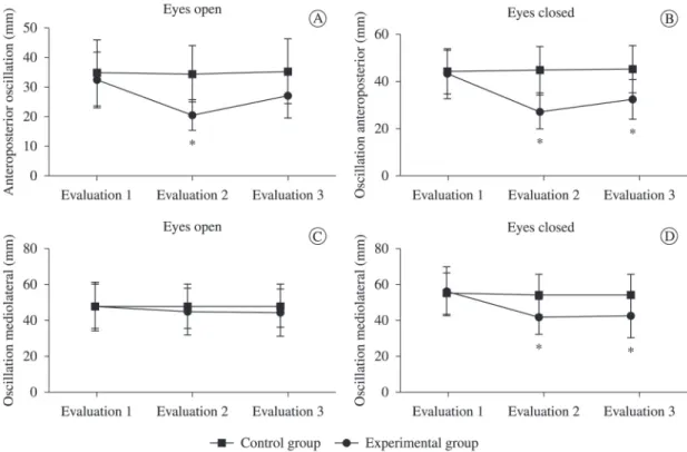
Figure 2 is a description of the results obtained in
the oscillations of the CoP. The experimental group
showed a reduction in anteroposterior sway with
eyes open in Evaluation 2 [F (2,36)=15.1, p=0.001], anteroposterior sway with eyes closed in Evaluations 2 [F (2,18)=29.3, p=0.001] and 3 [F (2,18)=17.8, p=0.001], and mediolateral sway with eyes closed in Evaluations 2 [F (2,18)=49.9, p=0.001] and 3 [F (2,18)=42.6, p=0.001]. The effects obtained also exhibited signiicant reductions in anteroposterior oscillation with eyes open (Pretreatment vs.
Post-treatment 1 – effect: –11.8 mm, p<0.001; Pretreatment vs. Post-treatment 2 – effect: –5.2 mm, p=0.003), anteroposterior oscillation with eyes closed (Pretreatment vs. Post-treatment 1 – effect: –15.7 mm, p<0.001; Pretreatment vs. Post-treatment 2 – effect: –10.6 mm, p<0.001), mediolateral oscillation with eyes open (Pretreatment vs. Post-treatment 1 – effect: –2.7 mm, p<0.001; Pretreatment vs. Post-treatment 2 – effect: –3.1 mm, p<0.05), and mediolateral oscillation with eyes closed (Pretreatment vs. Post-treatment 1 – effect: –14.6 mm, p<0.001;
Table 1. Anthropometric characteristics and functional classiication of the participants. Experimental group
(n=10)
Control group (n=10)
Age (years)* 7.2 (1.8) 7.8 (1.5)
Body mass (Kg)* 26.3 (3.2) 27.1 (2.6)
Stature (cm)* 125.8 (7.2) 126.1 (8.2)
Body mass index (Kg2/m)* 16.8 (1.2) 17.1 (1.1)
GMFCS (I\ II\ III)** (3\4\3) (3\4\3)
Topography (hemiparesis\diparesis)** (4\6) (3\7)
GMFCS – Gross Motor Functional Classiication System. *Data expressed as mean (standard deviation); **numbers indicate frequency (n) of children in each group.
Pretreatment vs. Post-treatment 2 – effect: –14.2 mm, p<0.001). In contrast, no significant differences among evaluations were found in the control group regarding gait variables or oscillations of the CoP.
The control group showed no statistical difference in the intragroup analysis (p>0.05).
Table 2 describes the results obtained in the
spatiotemporal gait variables. In experimental group,
the statistical analysis showed an increase in walking
speed in Evaluation 2 [F (2,18)=36.1, p=0.001], step length in Evaluation 2 [F (1,9)=19.3, p=0.017], and stride length in Evaluation 2 [F (2,36)=17.0, p=0.001] compared with the control group. No signiicant differences were identiied in the control
group (p>0.05). Figure 3 illustrates the results of gait speed and cadence.
Discussion
tDCS currently occupies an important place in studies addressing neuromotor rehabilitation due to its potential in optimizing the results of physical therapy14,21-24. The authors of the present study were curious about the possible effects of tDCS performed in an isolated fashion regarding changes in postural stability and whether children would be able to
tolerate the current. No previous studies were found
addressing the effects of tDCS on postural control
and gait in children with CP. Therefore, the aim of the present investigation was to determine the
immediate effect of a single session of tDCS applied
to the primary motor cortex in children classiied at levels I to III of the GMFCS. To enhance the validity of the study, the experimental group (active tDCS)
was compared to a control group (sham tDCS) and
double-blind procedures (participants and examiners)
were employed.
Three-dimensional gait analysis25,26 and stabilometry26,27 are considered fundamental
assessment tools for the adequate quantiication of the effects of interventions aimed at improvements in gait and static balance. These sensitive methods allow the identiication of small changes within a
short span of time and were therefore selected for
the present study. The experimental group exhibited signiicant differences in the evaluations after the application of active tDCS regarding gait velocity
and oscillations of the CoP in comparison with the
evaluation held prior to stimulation.
The present study offers important findings.
The experimental group exhibited an increase in gait velocity immediately following tDCS, but this
Table 2. Performance at evaluation 1 (before stimulation), evaluation 2 (after stimulation), and evaluation 3 (twenty minutes after stimulation) of outcome of variables spatiotemporal gait.
Experimental group Control group
Evaluation 1 Evaluation 2 Evaluation 3 Evaluation 1 Evaluation 2 Evaluation 3 Gait velocity (m/s) 0.75 (0.19) 1.04 (0.21) 0.85 (0.27) 0.78 (0.23) 0.80 (0.20) 0.78 (0.21)
Cadence 104.6 (28.5) 98.3 (43.4) 90.6 (52.4) 103.5 (25.1) 105.3 (25.9) 104.2 (25.8)
Step length 0.33 (0.10) 0.40 (0.09) 0.34 (0.08) 0.35 (0.09) 0.34 (0.10) 0.34 (0.08)
Stride length 0.83 (0.01) 0.91 (0.07) 0.81 (0.06) 0.78 (0.10) 0.79 (0.07) 0.79 (0.10)
Step Width 0.15 (0.09) 0.16 (0.02) 0.15 (0.08) 0.16 (0.11) 0.16 (0.11) 0.16 (0.06)
increase was not maintained for more than 20 minutes
after the end of the stimulation. Although this was a
cross-sectional study involving a single session of tDCS, the results suggest that the momentary increase in cortex activation may have exerted an inluence on motor control and gait. As the primary motor cortex was only stimulated for 20 minutes during rest, the authors did not expect the changes to be maintained in medium or long term. However, the indings could
encourage future studies to combine tDCS with motor rehabilitation therapies to determine whether this
technique can assist in improving gait and postural
control in children with CP.
Gait velocity has an important relationship with the cadence. However, in this study there was an increase in walking velocity without increasing cadence. The authors believe that this fact can be explained by an increased step length.
Analyzing a population of elderly individuals (n=9) with leukoaraiosis, an ischemic lesion of the cerebral white matter that results in gait and balance disorders,
Kaski et al.24 found that a single session of anodal stimulation in combination with gait and balance
training had repercussions in the form of improvements in gait velocity, stride length, step length variability,
and balance. In a study by Kaski et al.14, 30 healthy
individuals received either active or sham tDCS to either the primary motor cortex or prefrontal cortex prior to walking on a moving platform (a mobile sled moved with a maximum velocity of 1.4 m/s). The group that received active tDCS exhibited an increase in gait velocity. Thus, anodal stimulation was capable of inducing changes in the excitability of the motor cortex of the lower limbs, thereby potentiating locomotion control. All of these previous indings underscore the potential of anodal stimulation of the motor cortex regarding the facilitation of motor recovery.
Balance deicit resulting in frequent falls is one
of the most limiting aspects of CP13,26,27. Regarding
oscillations of the CoP, two important indings were identiied in the present study: 1) the similarity in the results with and without visual restriction; and 2) although a small number of participants were classiied at level III of the GMFCS (three per group), the effects apparently involved these children, who require gait-assistance devices.
Visual compensation is an important aspect of postural stability. In children with CP, oscillations are greater with eyes closed due to the lack of visual
compensation. The results suggest that there was
a momentary improvement in postural stability.
Although there are no studies that address the effects
of tDCS on static balance, the authors believe that greater effectiveness of the proprioceptive system
may have resulted from the stimulation of the cortical area. Thus, the motor responses were effective in minimizing the oscillations with visual restriction.
Similar results on the effect size of the oscillations
of the CoP, with and without visual restriction, are observed only with more dynamic interventions, such
as the use of ankle-foot orthoses26.
All clinical effects observed following the application of tDCS are directly related to cortex modulations
resulting from stimulation dependent on the polarity
of the current. Anodal stimulation increases cortex excitability, favoring the depolarization of the neuronal membrane, whereas the cathode has an inhibitory
effect through the hyperpolarization of the neuronal membrane28,29. A number of studies have demonstrated
that tDCS is successful in achieving these effects, but
some papers suggest that anodal stimulation applied to
the primary motor cortex seems to have an effect that
is dependent on the learning task and the formation of memory. These neurophysiological aspects and the
clinical indings described in the results and discussion
sections of this paper suggest that tDCS may be an important tool for potentiating the effects of neuromotor
rehabilitation. Although the present investigation has limitations, such as not being a prospective study and not involving a broader stimulation protocol, important
preliminary findings are described herein30. Such indings can offer a direction for the development
of further studies that address the use of tDCS in combination with physical therapy to treat locomotion and postural disorders in children with CP.
Conclusion
Based on the present indings, a single session of tDCS applied to the primary motor cortex in children with CP was capable of causing signiicant reduction
in anteroposterior oscillation with eyes open and eyes closed and in mediolateral oscillation with eyes closed in comparison with the control group (tDCS
sham). Moreover, increases in gait velocity, step length, and stride length were also observed after stimulation. However, the results were not maintained for more than 20 minutes after the end of stimulation.
Acknowledgements
References
1. Miranda PC, LomarevM, Hallett M. Modeling the current distribution during transcranial direct current stimulation.
Clin Neurophysiol. 2006;117(7):1623-9. http://dx.doi.
org/10.1016/j.clinph.2006.04.009. PMid:16762592
2. Wagner T, FregniF, Fecteau S, Grodzinsky A, ZahnM,
Pascual-Leone A. Transcranial direct current stimulation:
a computer-based human model study. Neuroimage.
2007;35(3):1113-24. http://dx.doi.org/10.1016/j.
neuroimage.2007.01.027. PMid:17337213
3. Liebetanz D, Nitsche MA, Tergau F, Paulus W. Pharmacological approach to the mechanisms of transcranial DC-stimulation-induced after-effects of
human motor cortex excitability. Brain. 2002;125(10):2238
-47. http://dx.doi.org/10.1093/brain/awf238. PMid:12244081
4. Yook SW, Park SH, Seo JH, Kim SJ, Ko MH. Suppression of seizure by cathodal transcranial direct current stimulation in an epileptic patient - a case report -. Ann
Rehabil Med. 2011;35(4):579-82. http://dx.doi.org/10.5535/
arm.2011.35.4.579. PMid:22506177
5. Shin YK, Lee DR, Hwang HJ, You SJ, Im CH. A novel
EEG-based brain mapping to determine cortical activation
patterns in normal children and children with cerebral palsy during motor imagery tasks. NeuroRehabilitation.
2012;31(4):349-55. PMid:23232157.
6. Pitcher JB, Schneider LA, Burns NR, Drysdale JL, Higgins RD, Ridding MC, et al. Reduced corticomotor excitability
and motor skills development in children born preterm. J Physiol. 2012;590(22):5827-44. http://dx.doi.org/10.1113/
jphysiol.2012.239269. PMid:22966161
7. Rose S, Guzzetta A, Pannek K, Boyd R. MRI structural
connectivity, disruption of primary sensorimotor pathways, and hand function in cerebral palsy. Brain Connect. 2011;1(4):309-16. http://dx.doi.org/10.1089/
brain.2011.0034. PMid:22432420
8. Kurz MJ, Wilson TW. Neuromagnetic activity in the somatosensory cortices of children with cerebral palsy.
Neurosci Lett. 2011;490(1):1-5. http://dx.doi.org/10.1016/j.
neulet.2010.11.053. PMid:21184811
9. Dinomais M, Lignon G, Chinier E, Richard I, Ter
Minassian A, Tich SN. Effect of observation of simple
hand movement on brain activations in patients with unilateral cerebral palsy: an fMRI study. Res Dev
Disabil. 2013;34(6):1928-37. http://dx.doi.org/10.1016/j.
ridd.2013.03.020. PMid:23584173
10. Kesar TM, Sawaki L, Burdette JH, Cabrera MN, Kolaski K, Smith BP, et al. Motor cortical functional geometry in cerebral palsy and its relationship to disability.
Clin Neurophysiol. 2012;123(7):1383-90. http://dx.doi.
org/10.1016/j.clinph.2011.11.005. PMid:22153667
11. Chagas PSC, ManciniMC, Barbosa A, SilvaPTG. Analysis
of the interventions used for gait promotion in children with cerebral palsy: a systematic review of the literature. Rev Bras Fisioter.2004;8(2):155-63.
12. De Kegel A, Dhooge I, Peersman W, Rijckaert J, Baetens T, Cambier D, et al. Construct validity of the
assessment of balance in children who are developing
typically and in children with hearing impairments. Phys
Ther. 2010;90(12):1783-94. http://dx.doi.org/10.2522/
ptj.20100080. PMid:21030662
13. GreccoLA, Tomita SM, Christovão TC, Pasini H, Sampaio
LM, Oliveira CS. Effect of treadmill gait training on static and functional balance in children with cerebral palsy: a randomized controlled trial. Braz J Phys Ther.
2013;17(1):17-23. PMid:23538455.
14. Kaski D, Quadir S, Patel M, Yousif N, Bronstein AM.
Enhanced locomotor adaptation after effect in the
“broken escalator” phenomenon using anodal tDCS.
J Neurophysiol. 2012;107(9):2493-505. http://dx.doi.
org/10.1152/jn.00223.2011. PMid:22323638
15. Palisano R, Rosenbaum P, Walter S, Russell D, Wood E,
Galuppi B. Development and reliability of a system to classify gross motor function in children with cerebral palsy. Dev Med Child Neurol. 1997;39(4):214-23.
http://dx.doi.org/10.1111/j.1469-8749.1997.tb07414.x.
PMid:9183258
16. Hiratuka E, Matsukura TS, Pfeifer LI. Cross-cultural adaptation of the gross motor function classification
system into Brazilian-Portuguese (GMFCS).Rev Bras Fisioter. 2010;14(6):537-44. http://dx.doi.org/10.1590/
S1413-35552010000600013. PMid:21340249
17. Homan RW, Herman J, Purdy P. Cerebral location
of international 10-20 system electrode placement. Electroencephalogr Clin Neurophysiol. 1987;66(4
):376-82. http://dx.doi.org/10.1016/0013-4694(87)90206-9.
PMid:2435517
18. Adeyemo BO, Simis M, Macea DD, FregniF. Systematic
review of parameters of stimulation, clinical trial design characteristics, and motor outcomes in non-invasive brain
stimulation in stroke. Front Psychiatry. 2012;3:88. http://
dx.doi.org/10.3389/fpsyt.2012.00088. PMid:23162477
19. Davis RB 3rd, Ounpuu S, Tyburski D, Gage JR. A gait
analysis data collection and reduction technique. Hum Mov Sci. 1991;10(5):575-87. http://dx.doi.
org/10.1016/0167-9457(91)90046-Z.
20. Ferrari A, Benedetti MG, PavanE, Frigo C, Bettinelli D, Rabuffetti M, et al. Quantitative comparison of five current protocols in gait analysis. Gait Posture. 2008;28(2):207
-16. http://dx.doi.org/10.1016/j.gaitpost.2007.11.009.
PMid:18206374
21. Cimolin V, Galli M, Rigoldi C, Grugni G, VismaraL,
MainardiL, et al. Fractal dimension approach in postural
control of subjects with Prader-Willi Syndrome.J Neuroeng
Rehabil. 2011;8(1):45.
http://dx.doi.org/10.1186/1743-0003-8-45. PMid:21854639
22. Kadaba MP, Ramakrishnan HK, Wootten ME. Measurement
of lower extremity kinematics during level walking. J
Orthop Res. 1990;8(3):383-92. http://dx.doi.org/10.1002/
jor.1100080310. PMid:2324857
23. Khedr EM, Shawky OA, El-Hammady DH, Rothwell JC, Darwish ES, MostafaOM, et al. Effect of anodal versus cathodal transcranial direct current stimulation on stroke rehabilitation: a pilot randomized controlled trial.
Neurorehabil Neural Repair. 2013;27(7):592-601. http://
dx.doi.org/10.1177/1545968313484808. PMid:23609526
24. Kaski D, Dominguez RO, Allum JH, Bronstein AM.
Improving gait and balance in patients with leukoaraiosis
physical training: an exploratory study.Neurorehabil Neural Repair. 2013;27(9):864-71. http://dx.doi.
org/10.1177/1545968313496328. PMid:23897903
25. Domagalska M, Szopa A, Syczewska M, Pietraszek S, Kidoń
Z, OnikG. The relationship between clinical measurements and gait analysis data in children with cerebral palsy. Gait Posture. 2013;38(4):1038-43. http://dx.doi.org/10.1016/j.
gaitpost.2013.05.031. PMid:23810569
26. Wren TA, Lening C, Rethlefsen SA, Kay RM. Impact of gait
analysis on correction of excessive hip internal rotation
in ambulatory children with cerebral palsy: a randomized controlled trial. Dev Med Child Neurol. 2013;55(10):919
-25. http://dx.doi.org/10.1111/dmcn.12184. PMid:23738949
27. Roque AH, Kanashiro MG, Kason S, GreccoLAC, Salgado ASI, Oliveira CS. Analysis of static balance in children with cerebral palsy spastic diparetic type with and without the use of orthoses. Fisioter Mov. 2012;25(2):311-6. http://
dx.doi.org/10.1590/S0103-51502012000200008.
28. Nobre A, MonteiroFF, GolinMO, Biasotto-Gonzalez D, Corrêa JC, Oliveira CS. Analysis of postural oscillation
in children with cerebral palsy. Electromyogr Clin
Neurophysiol. 2010;50(5):239-44. PMid:20718335.
29. Auvichayapat P, AuvichayapatN. Basic knowledge of transcranial direct current stimulation. J Med Assoc Thai.
2011;94(4):518-27. PMid:21591542.
30. Thibaut A, Chatelle C, GosseriesO, Laureys S, Bruno MA. Transcranial direct current stimulation: a new tool for neurostimulation. Rev Neurol (Paris). 2013;169(2):108
-20. http://dx.doi.org/10.1016/j.neurol.2012.05.008.
PMid:22959705
Correspondence
Luanda André Collange Grecco
Rua Diogo de Faria, 775, cj 91, Vila Clementino CEP 04037-002, São Paulo, SP, Brasil
![Table 2 describes the results obtained in the spatiotemporal gait variables. In experimental group, the statistical analysis showed an increase in walking speed in Evaluation 2 [F (2,18)=36.1, p=0.001], step length in Evaluation 2 [F (1,9)=19.3,](https://thumb-eu.123doks.com/thumbv2/123dok_br/19016258.469445/6.765.55.681.611.990/describes-obtained-spatiotemporal-variables-experimental-statistical-evaluation-evaluation.webp)
