1 Curso de Fisioterapia, Faculdade de Medicina de Ribeirão Preto (FMRP), Universidade de São Paulo (USP), Ribeirão Preto, SP, Brasil 2 Departamento de Biomecânica, Medicina e Reabilitação do Aparelho Locomotor, FMRP, USP, Ribeirão Preto, SP, Brasil
3 Departamento de Neurociências e Ciências do Comportamento, FMRP, USP, Ribeirão Preto, SP, Brasil Received: 08/11/2013 Revised: 12/18/2013 Accepted: 02/17/2014
a r t i c l e
Balance and muscle power of children
with Charcot-Marie-Tooth
Tais R. Silva¹, Amanda Testa¹, Cyntia R. J. A. Baptista², Wilson Marques Jr3, Ana C. Mattiello-Sverzut²
ABSTRACT | Background: In certain diseases, functional constraints establish a greater relationship with muscle power than muscle strength. However, in hereditary peripheral polyneuropathies, no such relationship was found in the literature. Objective: In children with Charcot-Marie-Tooth (CMT), to identify the impact of muscle strength and range of movement on the static/dynamic balance and standing long jump based on quantitative and functional variables. Method: The study analyzed 19participants aged between 6 and 16 years, of both genders and with clinical diagnoses of CMT of different subtypes.Anthropometric data, muscle strength of the lower limbs (hand-held dynamometer), ankle and knee range of movement, balance (Pediatric Balance Scale) and standing long jump distance were obtained by standardized procedures. For the statistical analysis, Pearson and Spearman correlation coeficients were used. Results: There was a strong positive correlation between balance and the muscle strength of the right plantar lexors (r=0.61) and dorsilexors (r=0.59) and a moderate correlation between balance and the muscle strength of inversion (r=0.41) and eversion of the right foot (r=0.44). For the long jump and range of movement, there was a weak positive correlation with right and left plantar lexion (r=0.20 and r=0.12, respectively) and left popliteal angle (r=0.25), and a poor negative correlation with left dorsilexion (r=–0.15). Conclusions: The data on the patients analyzed suggests that the maintenance of distal muscle strength favors performance during balance tasks, while limitations in the range of movement of the legs seem not to be enough to inluence the performance of the horizontal long jump.
Keywords: Charcot-Marie-Tooth disease; strength; balance; range of movement; assessment; physical therapy.
HOW TO CITE THIS ARTICLE
Silva TR, Testa A, Baptista CRJA, Marques Jr W, Mattiello-Sverzut AC. Balance and muscle power of children with
Charcot-Marie-Tooth. Braz J Phys Ther. 2014 July-Aug; 18(4):334-342. http://dx.doi.org/10.1590/bjpt-rbf.2014.0055
Introduction
Charcot-Marie-Tooth disease (CMT) is a hereditary polyneuropathy with various subtypes. The common clinical phenotype is the impairment of motor and sensory peripheral nerves due to a demyelinating
and axonal degenerative process¹. The predominant distal muscle weakness may cause signiicant motor
dysfunction in ambulation, participation in daily life, socio-cultural activities in both children and adults. It is important to note that weakness of the ankle
dorsilexors occurs in association with shortening
of the plantar lexormuscles and the development of foot deformities2.
The main clinical hypothesis for the development of foot deformities focuses on the intimate relationship between the imbalance in the strength of the invertor and evertor muscles of the feet and overload of the
plantar lexormuscles, in contrast to the weakness
of the dorsilexor group3. The latter is considered the
main manifestation of the disease and contributes to foot deformity (e.g. pes cavus), ankle contracture,
poor motor function and dificulty in walking in
affected children and adults2.
It is believed that losses in the range of motion (ROM) of distal muscles in patients with CMT compromise muscle power as they impair the stretching-shortening cycle. In the case of the
horizontal jump (i.e.standing long jump), 50% of the
muscle performance is attributed to the ankle4. Thus,
the ROM in the lower limbs can be correlated with the performance in a standing long jump test used to infer muscle strength.
M u s c l e s t r e n g t h , R O M , a n d d i f f e r e n t
neuromuscular demands in the lower extremity are
factors that modify the limits of postural stability
and may inluence the performance of a speciic
CMT disease: balance and muscle power
therapy procedures in CMT disease can be targeted and assertive if based on the understanding of the actual contributions of the variables involved in static and dynamic balance.
It is important to study CMT hereditary polyneuropathy because its incidence is relatively
high, affecting 1 in every 2,500 individuals2.
Although the initial symptoms of the disease usually
appear in the irst or second decade of life with slow
progression over the subsequent decades, adults are the target population in the majority of studies6-8.
Interventional studies involving drugs are still in progress because there is no effective therapy for CMT disease1, and the use of orthoses presents
controversial results8. In addition, studies that
focused on clarifying the contribution of the major
deicits (i.e. musculoskeletal, neuromuscular, and
biomechanical) to balance in children with CMT are limited. Thus, it becomes relevant to investigate the behavior of the biomechanical variables during the initial phase of the disease. This is a preliminary step to the proposal of physical therapy interventions that can potentially help in the rehabilitation of these children and adolescents.
In children and adults, the triad of muscle weakness, joint hyper/hypomobility, and compensatory
biomechanical disorders can cause signiicant motor dysfunctions of distal-proximal predominance
with loss of balance, ambulation, and hampered participation in activities of daily living2. Similarly,
the relationship between passive ROM with horizontal jump, measured using the standing long jump test, and balance, assessed with the Pediatric Balance
Scale (PBS), were tested. Briely, the aim of this study was to evaluate the inluence of passive ROM
and strength of the major muscle groups of the lower limbs on the static/dynamic balance and horizontal jump capacity of children with CMT disease.
Method
This study included a total of 19 child and adolescent participants who were admitted to the Neurogenetic Disorders Outpatient Clinic of the
Hospital das Clínicas da Faculdade de Medicina de Ribeirão Preto da Universidade de São Paulo
(HCFMRP/USP), Ribeirão Preto, São Paulo state,
Brazil, between 2011 and 2012 with a conirmed
CMT diagnoses. The participants were of both
genders, aged between 5 and 16 years, were able to
walk independently, and had no diseases associated with CMT disease that affect the cardiorespiratory system.
Consent was obtained from the parents or guardians
who illed out the informed consent form previously
approved by the Research Ethics Committees of the
HCFMRP/USP (Number 4334/2011).
In a standardized manner, anthropometric data, goniometry, muscle strength (hand-held dynamometer - Lafayette Instrument Co., Lafayette, UK), lower limb power (long jump test), and static/ dynamic balance (Pediatric Balance Scale) were obtained from all participants.
The passive ROM was measured for the knees
(popliteal angle) and ankles (plantar lexion and dorsilexion), according to the method described by
Marques9. The measurements were performed using a
universal goniometer (CARCI - Indústria e Comércio de Aparelhos Cirúrgico e Ortopédicos Ltda.).
The muscle strength (in kilogram-force) of the
hip extensors, knee extensors, and dorsiflexors, plantar flexors, supinators, and pronators of the
foot were measured three times using a hand-held dynamometer, alternating between the right and left lower limbs to prevent fatigue. The highest value was used for analysis. To ensure that the dynamometer was kept perpendicular to the segment being tested and as distal as possible, an assistant stabilized the participant during the measurements, and the following bodily positions were adopted: supine, lower limbs in the anatomical position and feet out of the stretcher to measure the muscle strengths of the
dorsilexors, plantar lexors, supinators and pronators; the prone position with knee lexed to 90° to measure the muscle strength of the hip extensors; and the sitting position with knee lexed to 90° to measure the muscle strength of the knee extensors. The voice
command “force” was used during the tests while the evaluator prevented any range of motion to ensure an
isometric contraction for ive seconds.
The standing long jump test, also called the horizontal jump or broad jump, is easy to apply and requires only chalk or pencil to mark the ground and a plastic tape measure or self-retracting tape measure to measure the distance jumped. The participants were positioned behind a line marked on the ground with the feet slightly apart and were asked to jump the greatest horizontal distance possible by bendingthe legs and using the impulse generated by swinging the arms10. This strategy allowed balance
to be restored or maintained through the transfer of angular momentum from the arms to the rest of the body. Three attempts were made, and the highest value was used for analysis. The result was given in centimeters, measuring the distance between the
starting line and the mark achieved by the calcaneus on the ground.
The PBS was used to measure functional balance because it was suitable for school-aged children with mild to moderate motor disability, according to Franjoine et al.11. The test lasted approximately 15 minutes and did not require the
use of specialized equipment, and it provided clinical data for the measurement of functional balance tasks. The brazilian version of the PBS described by Ries et al.12 was used to apply the test. The following
materials were used: a chair with back support, adjustable height, and arm rests; markers for the feet; stopwatch; tape measure; and step stool. The participants were instructed, through demonstrations, how they were to perform the tests. A preliminary trial of each proposed task was allowed for each tested item.
The PBS consisted of 14 items that required
the child to perform static and dynamic balance
tasks. Each item was be scored from 0 to 4, with 4 corresponding to a better ability to perform the required task. The scores on each of the 14 tasks were summed, and the inal score was determined from this number, with a maximum value of 56.
Higher scores were associated with greater ability to perform the required task and therefore with better balance. In healthy children from the age of seven, the
maximum score of 56 should normally be achieved,
and there is no mention in the literature regarding the
classiication of lower scores11.
To meet the study objective, which was to correlate the dynamometry data of lower limbs with balance and range of motion data of the lower limbs with the horizontal impulse measured by the standing long
jump test, the Pearson correlation coeficient (r) and
the Spearman correlation coeficient, which quantify
the association between two quantitative variables,
were used. These coeficients ranged from –1 and 1. A value 0 of (zero) indicated that there was no linear
correlation; 1 indicated perfect linear correlation; and
–1 also indicated a perfect negative linear correlation
(i.e., when one of the variables increased, the other
decreased). Values closer to 1 or –1 indicated stronger
linear correlation between the two variables. The
classiication of the Spearman correlation coeficients
was performed based on the study by Hulley et al.13,
and the classification of the Pearson correlation
coeficients was performed based on the study by
Pagano and Gauvreau14. The following correlations
were tested: muscle strength × balance and, standing long jump × ROM.
Results
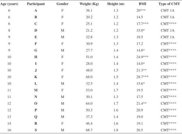
The anthropometric data and the classiication
of the participants are shown in Table 1. Among the 19 patients in the study, nine were males and
ten were females; the mean age was 10.11 years (standard deviation: 2.64), mean weight was 40.59 kg (standard deviation: 15.37), and mean height was 1.43 m (standard deviation: 0.18). Considering the
normative values provided by the World Health Organization (WHO)15, nine participants had a
body mass index (BMI) appropriate for their age,
while four participants wereunderweight, two were overweight, and four were obese.
The lower limb muscle strength, passive ROM, standing long jump test, and PBS scores obtained
are shown in Table 2.
The isometric muscle strength values were not proportional to the age of the participants. The
dorsiflexor, invertor, and evertor muscle groups
presented the lowest isometric muscle strength
values, with the dorsilexion strength being zero in
participants C and K.
For balance, which was determined using the PBS, high scores were observed for the participants with
CMT (scores ranged between 51 and 56), indicating
a good overall performance. However, considering the PBS items separately, the most challenging
tasks were identiied as follows: standing with eyes
closed, standing with one foot in front, standing on one foot, retrieving an object from the ground, and reaching forward.
The ROM data indicated that bilateral ankle joint
mobility was preserved except in three cases in which
there was limitation (participants H, N, and R), with
dorsilexion being less than 10 degrees, and in three
cases with lack of mobility (participants K, M, and
O), with dorsilexion being equal to or less than
zero. The bilateral popliteal angle was preserved
in most participants (except for values lower than 140°) (Table 2).
For the standing long jump test, there was no increase in performance with age, and the values
from 7 (A, H, I, K, L, O, Q) of the 19 participants
were lower than the values described as normative16 (Table 2).
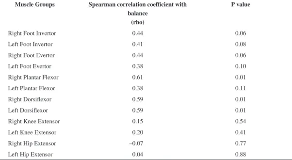
Correlations between PBS and lower limb muscle strength
The results of the Spearman test indicated a strong positive correlation between balance and the strength
CMT disease: balance and muscle power
and left dorsilexors (r=0.59; p=0.01) and a moderate
correlation between balance and the strength of the
following muscle groups: right invertors (r=0.44; p=0.06), left invertors (r=0.41; p=0.08), and right evertors (r=0.44; p=0.06) – Table 3.
Correlations between the standing long jump test and passive ROM of the lower limbs
The values obtained in the correlation of the standing long jumptest with the ROM of the lower limbs showed a weak positive correlation between
the ROM of right plantar lexion (r=0.20; p=0.41), left plantar lexion (r=0.12; p=0.61), and left popliteal angle (r=0.25; p=0.31). There was a weak negative correlation for left dorsilexion (r=–0.15; p=0.54), and no correlation was found for right dorsilexion (r=0.09; p=0.69) or right popliteal angle (r=0.00; p=1.00), as shown in Table 4. Therefore, the data
indicated no correlation between ankle and knee joint ROM with muscle strength as demonstrated in the standing long jump test.
Discussion
This study demonstrated that participants with CMT presented weakness in the following muscle
groups: foot evertors, invertors, dorsilexors and plantar lexors. With the exception of dorsilexion,
the ROMs were preserved. Overall, balance was
preserved; however, there was a deicit in speciic
items of the PBS. The standing long jump test indicated that muscle strength was preserved in the majority of the participants, with certain
exceptions.
Although, by definition, the sensorimotor impairment in CMT disease is symmetrical, variations
in muscle strength, lexibility, and motor coordination
have been observed. Thus, some correlations were found only for the strength and ROM of either the right or left side. These correlations suggest that
the preserved strength of dorsilexors and plantar lexors positively inluenced performance in tasks
that required balance. The ROMs obtained did not seem to have affected the muscle strength.
Table 1. Anthropometric data and classiication of participants according to the type of CMT.
Age (years) Participant Gender Weight (Kg) Height (m) BMI Type of CMT
6 A F 36.1 1.3 20*** CMT 1A
6 B F 20.2 1.2 14.5 CMT 1A
6 C F 25.1 1.2 17.7*** CMT****
8 D M 21.2 1.2 15.0* CMT 1A
9 E M 32.8 1.3 18.5 CMT 1A
9 F F 30.9 1.3 17.2 CMT****
9 G M 27.7 1.4 14.8* CMT****
10 H F 51.0 1.4 24.9*** CMT****
10 I F 28.0 1.4 14.0* CMT****
10 J F 48.0 1.5 21.3** CMT****
10 K F 68.0 1.5 28.7*** CMT****
10 L M 32.5 1.4 15.6* CMT****
11 M F 53.0 1.7 19.5 CMT****
11 N M 30.1 1.3 17.5 CMT****
12 O M 64.0 1.7 21.4** CMT****
12 P M 50.3 1.6 20.9 CMT****
13 Q M 37.3 1.4 19.0 CMT****
14 R F 46.4 1.6 19.1 CMT****
16 S M 68.7 1.8 20.5 CMT****
* BMI – underweight; ** BMI – overweight; *** BMI – obesity; **** CMT subtype unspeciied.
v
a TR, T
esta A, Baptista C
R
JA, Mar
ques Jr W
,
Mattiello-S
v
erzut A
C
Table 2. Lower limb muscle strength, goniometry, long jump, and Pediatric Balance Scale (PBS) scores.
Participant Age (years)
Muscle Strength (Kgf) Goniometry (degrees) Long
Jump
(cm) PBS
IR IL ER EL FPR FPL DFR DFL EKR EKL EHR EHL PFR PFL DR DL PAR PAL
A 6 6 5 7 6 17 20 6 5 12 8 13 13 50 42 10 12 190 145 38 55
B 6 4 4 7 6 14 14 7 6 9 10 13 10 65 60 20 20 145 155 60 54
C 6 2 3 2 2 6 8 0 0 14 10 10 12 45 45 10 10 150 140 49 51
D 8 4 6 5 4 18 20 8 5 12 10 14 11 40 45 22 20 140 150 102 55
E 9 6 7 6 7 9 10 9 7 16 17 13 11 50 40 10 0 154 150 115 55
F 9 7 3 4 5 7 10 2 1 17 18 19 17 50 50 10 10 150 150 99 54
G 9 4 5 7 5 21 18 11 9 14 14 16 17 50 45 20 20 155 140 113 56
H 10 8 9 7 10 18 18 7 6 10 10 9 11 32 36 8 10 134 132 59 56
I 10 11 9 7 8 13 15 10 8 24 22 14 13 35 40 22 22 130 140 18 53
J 10 12 12 13 11 19 22 12 12 20 20 16 18 34 34 20 18 145 140 94 56
K 10 4 5 2 2 12 14 0 0 18 19 16 15 50 52 -10 0 140 130 62 51
L 10 6 4 8 10 9 15 6 5 9 9 17 14 40 35 10 17 130 130 63 53
M 11 9 9 10 12 11 9 10 10 20 23 20 24 42 52 0 0 138 138 94 56
N 11 7 5 5 5 15 15 6 4 18 16 13 13 50 40 5 10 140 145 108 56
O 12 7 8 6 6 13 16 7 7 18 17 11 11 36 30 0 0 136 140 83 56
P 12 8 6 7 7 20 20 13 11 15 16 11 11 40 50 20 10 120 120 107 56
Q 13 5 4 5 6 18 10 3 2 11 10 10 10 40 50 10 10 150 136 60 56
R 14 9 12 3 5 24 20 2 2 25 19 14 12 50 40 5 10 128 142 88 55
S 16 12 12 8 9 22 22 26 20 29 28 29 29 50 50 15 12 155 150 180 56
IR= Inverter Right Foot, IL= Inverter Left Foot, ER= Evertor Right Foot, EL= Evertor Left Foot; FPR= Plantar lexor Right; FPE= Plantar lexor Left, DFR= Dorsilexor Right; DFL= Dorsilexor Left; EKR= Extensor Knee Right; EKL= Extensor Knee Left; EHR= Extensor Hip Right; EHL= Extensor Hip Left; PFR= Plantar Flexion Right; PFL= Plantar Flexion Left, DR= Dorsilexion Right, DL= Dorsilexion Left, PAR= Popliteal Angle Right, PAL= Popliteal Angle Left; 1st= First Trial; 2nd= Second Trial, 3rd= Third Trial; PBS= Pediatric Balance Scale.
Br
az J Ph
y
s Ther
. 2014 Jul
y
-A
CMT disease: balance and muscle power
Muscle strength and balance
Balance is an essential factor in the coordination of motor responses, movements, and postural adjustments. For balance to be effective, several factors, such as the vestibular system, proprioceptive information, visual perception, muscle strength,
and joint lexibility need to operate eficiently and
harmoniously in the body17. The muscles surrounding
the ankle are essential to maintaining balance because they provide proprioceptive information and correct small postural oscillations, in addition to correcting possible destabilization through muscular torque, thereby regulating the center of gravity and keeping the center of mass located between the feet18.
Typically, the natural history of various subtypes of CMT involves, among other manifestations, the progressive reduction of distal muscle strength, which
can impair the maintenance of the center of mass over the base of support, both dynamically and statically2.
The ankle strategy is the most frequently used strategy to maintain balance, and it requires the
preservation of the plantar lexor, dorsilexor, evertor,
and invertor muscle strength19. This strategy is more
effective when perturbations to balance are slow and
small and the supporting surface is irm (i.e., during
static balance)19. The ankle dorsilexion produced
during the ankle strategy is crucial to maintaining balance after a destabilization because when the forefoot is lifted, a counter-movement force is created, which helps to re-balance the body20. Thus,
the reduction in dorsilexor muscle strength observed in the participants may explain the deicit found in
the maintenance of static balance.
In the study, the participants presented data consistent with the data reported in the literature2,3,5,
Table 4. Pearson coeficient of correlation (r) for passive range of motion of the lower limbs and the long jump test.
Measured range of motion of lower limbs
Pearson correlation coefficient (r) with the long jump test
P value
Right Plantar Flexion 0.20 0.41
Left Plantar Flexion 0.12 0.61
Right Dorsilexion 0.09 0.69
Left Dorsilexion –0.15 0.54
Right Popliteal Angle 0.00 1.00
Left Popliteal Angle 0.25 0.31
Table 3. Spearman coeficient of correlation (rho) and p value for muscle strength in the lower limbs and the Pediatric Balance Scale (PBS).
Muscle Groups Spearman correlation coefficient with balance
(rho)
P value
Right Foot Invertor 0.44 0.06
Left Foot Invertor 0.41 0.08
Right Foot Evertor 0.44 0.06
Left Foot Evertor 0.38 0.10
Right Plantar Flexor 0.61 0.01
Left Plantar Flexor 0.38 0.11
Right Dorsilexor 0.59 0.01
Left Dorsilexor 0.59 0.01
Right Knee Extensor 0.15 0.54
Left Knee Extensor 0.20 0.41
Right Hip Extensor –0.07 0.77
Left Hip Extensor 0.04 0.88
such as reduced muscle strength, especially in the
evertor and dorsilexor muscles and shortening of the plantar lexor muscles. A study conducted by
Nyström et al.21 established reference values for
lower limb isometric muscle strength according to the age and body weight of healthy participants. Thus, the data obtained in the current study were compared with the reference values obtained by Nyström et al.21 using the weight and height of
the participants because the reference values by age could lead to misinterpretation. It was found that most participants with CMT presented with isometric muscular strength compatible with their
body weight and height. Exceptions were found for the dorsilexor muscles of participants C, E, and N.
Normative data for comparison were not found for the foot invertor and evertor muscles nor for the plantar
lexor muscles. However, it is worth noting that in 9 of
the 19 participants, the muscle strength of invertors
and evertors was lower than 5 KgF, which suggests a strength deicit in these muscle groups.
For the participants in this study, who presented with reduced distal muscle strength, the tasks involving static balance were more affected than the tasks involving dynamic balance because static postures required greater ROM and higher torque of the ankle musculature22.
The balance deicits found in the participants of
this study were not disabling, considering that the
PBS score was close to the maximum (between 51 and 56). Because several factors positively
or negatively affect balance17, it is possible that
mechanisms compensating for the distal muscle strength deficits were used (e.g., use of the hip strategy and upper limb assistance). Furthermore, proprioception and stabilization mechanisms, such as muscle stiffness, are key factors in the establishment of balance23. Anticipatory control is another factor
that could have been triggered by the patients to obtain static and dynamic balance control22,23.
The positive correlation observed between
the isometric muscle strength of the dorsilexors, plantar lexors, evertors, and invertors with balance
suggests that the maintenance of muscle strength of these muscle groups may positively affect balance. Ribeiro et al.24 associated ankle muscle
strength with balance in the elderly and, similar to Sundermier et al.25, who evaluated children,
corroborated the current study, concluding that
plantar lexor and dorsilexor strength was positively
associated with balance.
ROMandstanding long jump
The ROM available for a joint can also be deined as flexibility, which is an important element of physical itness26. Flexibility can be achieved by
active muscle contraction, referred to as dynamic
lexibility, or by passive motion caused by a force external to the joint. Gender, anthropometric
measurements, body composition, genetic, and pathological characteristics, in addition to the growth and development processes26, all inluence
lexibility. The participants with CMT in this study presented with a joint ROM with relative lexibility
and a preserved motion arc, which established a weak correlation with performance in the standing long jump test.
The standing long jump test results of the participants were compared to the normative data described by Condon and Cremin16, who studied this variable in 534 children aged 4 to 15 years. The
age-matched comparison with participants of the current
study demonstrated that 7 (A, H, I, K, L, O, Q) of
the 19 participants presented lower values than were described as normative.
During the performance of the standing long jump test, the additional impulse imparted to the jump by the swinging of the arms might have increase the distance jumped and the takeoff speed27.
In this study, all participants were instructed to perform the test movement using the technique of propelling themselves with the arms. Ashbya and Heegaard27 indicated that the arm swing enhanced
the force-producing ability of the lower extremity extensor muscles, slowing the contraction speed
at key moments in the jump. To maintain balance throughout the jump, measures such as anticipatory control or even the employment of counterproductive mechanisms that reduce the jumping distance with free arm motion may have been adopted27.
Considering that children with CMT are aware of
their balance deicits, it is possible that they adopted
anticipatory control measures using the free arms. Thus, the restricted use of the arms by certain
participants may explain in part the lower jumping performance of participants A, H, I, K, L, O and Q, which was signiicantly lower than the overall mean
of the jumps considered.
The standing long jump test, while considered a
motor task or skill, is a complex motor pattern that
requires the coordinated performance of all body segments, where the impulse and the landing must be performed with both feet. The horizontal jump
CMT disease: balance and muscle power
isokinetic measures of lower limb strength, and is indicated as a good predictor of performance in the standing long jump10.
The lack of correlation or even the weak correlation found between ROM and the standing long jump test may be attributed to the fact that most participants in this study had relatively preserved ROM in distal muscles. A group of affected participants without preserved ROM should be evaluated to assess the
inluence of passive ROM on the standing long jump,
which was a limitation of the present study.
The sample size, heterogeneity of the CMT subtypes, different levels of motor maturation, and different anthropometric characteristics are common limitations in studies of this nature. Based on anthropometric data, the participants were
classiied in all categories of BMI, and 21% of them were obese, which may have inluenced the results. BMI does not seem to negatively affect lexibility,
in contrast to propulsion tests28. Obese individuals
suffer disadvantages in more challenging balance activities, such as standing on one foot29. For muscle
strength, a recent review30 indicated that although
obese individuals presented with higher absolute values compared to their normal-weight peers, obesity had no impact on the intrinsic properties of the muscle to generate force. Thus, the interference of BMI on the data obtained in the present study was considered minimal.
The results of this study may assist the physical therapist in making decisions during clinical practice
because they suggest that preserved dorsilexor and plantar lexor muscle strength is associated with better
static and dynamic balance performance. Similarly, the maintenance and/or gain of joint mobility,
especially of dorsilexion through stretching, may
promote good functional performance and muscle strength as demonstrated in the standing long jump test. Thus, in the treatment of children and adolescents with CMT disease, the maintenance and/or gain of
strength and lexibility of the dorsilexor and plantar lexor muscles should be prioritized.
Conclusion
The maintenance of distal muscle strength in children with CMT contributes to their performance of balance tasks. The losses found in passive ROM
of the lower limbs seem not to have been suficient
to affect muscle strength in the horizontal long jump.
Acknowledgments
To Elisangela Aparecida da Silva Lizzi, who was responsible for the statistical analysis; to the patients and their guardians and to the Fundação de Amparo à Pesquisa do Estado de São Paulo (FAPESP), process
number 2012/15521-3 and 2012/15522-0, Brazil, for
their support in the development of this study.
References
1. Pareyson D, Marchesi C. Diagnosis, natural history, and
management of Charcot–Marie–Tooth disease. Lancet Neurol. 2009;8:654-67. http://dx.doi.org/10.1016/ S1474-4422(09)70110-3
2. Burns J, Crosbie J, Hunt A, Ouvrier R. The effects of pes cavus on foot pain and plantar pressure. Clin
Biomech. 2005;20:877-82. PMid:15882916. http://dx.doi. org/10.1016/j.clinbiomech.2005.03.006
3. Tachdjian MO. The neuromuscular system-deformities of the foot and ankle. In: Tachdjian MO. Pediatric
orthopedics. 2nd ed. Philadelphia: WB Saunders; 1990. p. 1937-57.
4. Robertson DG, Fleming D. Kinetics of standing broad
and vertical jumping. Can J Sport Sci. 1987;12(1):19-23. PMid:3594313.
5. Cote KP, Brunet ME, Gansneder BM, Shultz SJ. Effects of pronated and supinated foot postures on static and
dynamic postural stability. J Athl Training. 2005;40(1):41-6. PMid:15902323 PMCid:PMC1088344.
6. Maggi G, Bragadin MM, Padua L, Fiorina E, Bellone E, Grandis M, et al. Outcome measures and a rehabilitation treatment in patients affected by Charcot-Marie-Tooth
Neuropathy: a pilot study. Am J Phys Med Rehabil. 2011 Aug 8;90:628-637. PMid:21681064. http://dx.doi. org/10.1097/PHM.0b013e31821f6e32
7. Rose KJ, Burns J, Wheeler DM, North KN. Interventions for increasing ankle range of motion in patients with neuromuscular disease. Cochrane Database Syst
Rev. 2010;(2):CD006973. PMid:20166090.
8. Sackley C, Disler PB, Turner-Stokes L, Wade DT, Brittle N, Hoppitt T. Rehabilitation interventions for foot drop in neuromuscular disease. Cochrane Database of Syst
Rev. 2009;(2):CD003908. PMid:19588347.
9. Marques AP. Ângulos articulares de membros inferiores.
In: Marques AP. Manual de goniometria. 2ª. ed. São Paulo: Manole; 2003. p. 41-7. PMid:12591094.
10. Wakai M, Linthorne NP. Optimum take-off angle in
the standing Long Jump. Hum Mov Sci. 2005;24:81-96. PMid:15949583. http://dx.doi.org/10.1016/j. humov.2004.12.001
11. Franjoine MR, Gunther JS, Taylor MJ. Pediatric Balance
Scale: A Modiied Version of the Berg Balance Scale
for the School-Age Child with Mild to Moderate
Motor Impairment. Pediatr Phys Ther. 2003;15(2):114-28. PMid:17057441. http://dx.doi.org/10.1097/01. PEP.0000068117.48023.18
12. Ries LGK, Michaelsen SM, Soares PSA, Monteiro VC, Allegretti KMG. Cross-cultural adaptation and reliability analysis of the Brazilian version of Pediatric
Balance Scale (PBS). Rev Bras Fisioter. 2012;16(3):205-15. PMid:22699691. http://dx.doi.org/10.1590/ S1413-35552012005000026
13. Hulley SB, Cummings SR, Browner WS, Grady D, Hearst N, Newman TB. Delineando a pesquisa clínica: uma
abordagem epidemiológica. 2ª. ed. Porto Alegre: Editora Artmed; 2003.
14. Pagano M, Gauvreau K. Princípios de bioestatística. 2ª. ed. São Paulo: Editora Thomson; 2004.
15. World Health Organization - WHO [homepage
Internet]. Geneva: WHO; 2006-2013 [cited 2013 July 18]. Available from: http://apps.who.int/bmi/index. jsp?introPage=intro_3.html.
16. Condon C, Cremin K. Static Balance Norms in Children.
Physiother Res Int. 2014 Mar;19(1):1-7. http://dx.doi. org/10.1002/pri.1549
17. De Weerdt W, Spaepen A. Equilíbrio. In: Durward BR, Baer GD, Rowe J. Movimento Funcional Humano. São
Paulo: Manole; 2001. p. 204.
18. Kuo AD, Zajac FE. A biomechanical analysis of muscle strength as limiting factor in standing
posture. J Biomech. 1993;(26):137-50. http://dx.doi. org/10.1016/0021-9290(93)90085-S
19. Horak FB, Shupert CL, Mirka A. Components of postural dyscontrol in the elderly: a review.
Neurobiol Aging. 1989;10:727-38. http://dx.doi. org/10.1016/0197-4580(89)90010-9
20. Wolfson LI, Whipple R, Amerman P, Kleinberg A. Stressing the postural response: a quantitative method
for testing balance. J Am Geriatr Soc. 1986;34:845-50. PMid:3782696.
21. Nyström EM, Kroksmark A-K, Beckung E. Isometric
muscle torque in children 5 to 15 years of age: normative data. Arch Phys Med Rehabil. 2006;87:1091-9. PMid:16876555. http://dx.doi.org/10.1016/j. apmr.2006.05.012
22. Robinovitch SN, Heller B, Lui A, Cortez J. Effect of strength and speed of torque development on balance recovery
with the ankle strategy. J Neurophysiol. 2002;88:613-20. PMid:12163514.
23. Van der Linden MH, Van der Linden SC, Hendricks HT, Van Engelen BGM, Geurts ACH. Postural instability in Charcot-Marie-Tooth type 1A patients is strongly
associated with reduced somatosensation. Gait
Posture. 2010;31:483-8. PMid:20226674. http://dx.doi. org/10.1016/j.gaitpost.2010.02.005
24. Ribeiro F, Teixeira F, Brochado G, Oliveira J. Impact of low cost strength training of dorsi- and plantar lexors on balance
and functional mobility in institutionalized elderly people.
Geriatr Gerontol Int. 2009;9:75-80. PMid:19260983. http://dx.doi.org/10.1111/j.1447-0594.2008.00500.x 25. Sundermier L, Woollacott M, Roncesvalles N, Jensen J. The
development of balance control in children: comparisons of EMG and kinetic variables and chronological and
developmental groupings. Exp Brain Res. 2001;136:340-50. http://dx.doi.org/10.1007/s002210000579
26. Melo SIL, Guth VJ, Sousa ACS, Sacomori C, Martins ACV, Lucca L. Estudo comparativo de amplitudes de movimentos
articulares em crianças diferentes gêneros entre os 7 e os 12 anos de idade. Motricidade. 2011;7(1):13-20. http:// dx.doi.org/10.6063/motricidade.7(1).116
27. Ashbya BM, Heegaard JH. Role of arm motion in the
standing long jump. J Biomech. 2002;35:1631-7. http:// dx.doi.org/10.1016/S0021-9290(02)00239-7
28. Dumith SC, Ramires VV, Souza MA, Moraes DS, Petry FG, Oliveira ES, et al. Overweight/obesity and physical fitness among children and adolescents. J Phys Act
Health. 2010;7(5):641-8. PMid:20864760.
29. Goulding A, Jones IE, Taylor RW, Piggot JM, Taylor D. Dynamic and static tests of balance and postural sway in boys: effects of previous wrist bone fractures and high
adiposity. Gait Posture. 2003;17:136-41. http://dx.doi. org/10.1016/S0966-6362(02)00161-3
30. Mafiuletti NA, Ratel S, Sartorio A, Martin V. The impact
of obesity on in vivo human skeletal muscle function.
Curr Obes Rep. 2013;2:251-60. http://dx.doi.org/10.1007/ s13679-013-0066-7
Correspondence
Cyntia Rogean de Jesus Alves de Baptista Universidade de São Paulo
Faculdade de Medicina de Ribeirão Preto
Departamento de Biomecânica, Medicina e Reabilitação do Aparelho Locomotor
Avenida Bandeirantes, 3900
CEP 14049-900, Ribeirão Preto, SP, Brasil