Can the adapted arcometer be used to assess the
vertebral column in children?
Juliana A. Sedrez, Cláudia T. Candotti, Fernanda S. Medeiros, Mariana T. Marques, Maria I. Z. Rosa, Jefferson F. Loss
ABSTRACT | Background: The adapted arcometer has been validated for use in adults. However, its suitability for use in children can be questioned given the structural differences present in these populations. Objective: To verify the concurrent validity, repeatability, and intra- and inter-reproducibility of the adapted arcometer for the measurement of the angles of thoracic kyphosis and lumbar lordosis in children. Method: Forty children were evaluated using both sagittal radiography of the spine and the adapted arcometer. The evaluations using the arcometer were carried out by two trained evaluators on two different days. In the statistical treatment, the intraclass correlation coeficient (ICC), Pearson’s product moment correlation, Spearman’s rho, the paired t test, and Wilcoxon’s test were used (α=.05). Results: A moderate and signiicant correlation was found between the x-ray and the adapted arcometer regarding thoracic kyphosis, but no correlation was found regarding lumbar lordosis. Repeatability and intra-evaluator reproducibility of the thoracic kyphosis and lumbar lordosis were conirmed, which was not the case of inter-evaluator reproducibility. Conclusion: The adapted arcometer can be used to accompany postural alterations in children made by the same evaluator, while its use for diagnostic purposes and continued evaluation by different evaluators cannot be recommended. Further studies with the aim of adapting this instrument for use in children are recommended.
Keywords: physical therapy; evaluation; spine; children; validity of tests.
HOW TO CITE THIS ARTICLE
Sedrez JA, Candotti CT, Medeiros FS, Marques MT, Rosa MIZ, Loss JF. Can the adapted arcometer be used to assess the vertebral column in children? Braz J Phys Ther. 2014 Nov-Dec; 18(6):538-543. http://dx.doi.org/10.1590/bjpt-rbf.2014.0060
Introduction
The early identiication of spinal alterations is
fundamental, particularly in childhood, because during this phase such alterations are unconsolidated and may therefore be delayed or even reverted1. To classify postural alterations and follow up any treatment, an accurate assessment of the spinal curvature is essential, given that treatments are generally based on the degree of curvature and its progression2.
Generally, physiotherapeutic postural evaluation employs methods based on observation that do not
permit objective quantification of the degree of
alteration, which constitutes a limitation in clinical
practice. The need for early quantitative identiication of postural alterations, without overexposing the
patient to radiation, has encouraged the development
of non-invasive instruments designed to objectively
measure the curvature of the spine and postural alterations3-5.
The choice of assessment instrument should be
based on scientiic parameters, such as precision,
accuracy, concurrent validity, repeatability, reproducibility, and the diagnostic capacity of the measurements provided. In addition, the choice should also consider practical parameters, such as ease of transport and ease of use of the instrument, in order to ensure that the patient can be assessed quickly and comfortably6. The arcometer proposed
by D’Osualdo et al.7 in 1997 for the assessment of
the thoracic spine incorporates most of these features.
Recently, Chaise et al.5 proposed modiications to
the structure of the original instrument and to the method used to calculate the spinal curvature and were, thus, also able to validate its use in the lumbar spine5. Although the original instrument was assessed
in a younger sample7, the concurrent validity and
intra- and inter-evaluator reproducibility of the
adapted arcometer have only been conirmed in an
adult population5.
However, given the structural differences between adults and children, such as the size of the trunk and the magnitude of the spinal curvature, the applicability
Escola de Educação Física, Universidade Federal do Rio Grande do Sul (UFRGS), Porto Alegre, RS, Brazil Received: 12/20/2013 Revised: 05/17/2014 Accepted: 06/26/2014
of this instrument in this speciic population may be questioned. Hence, the objective of this study was to
verify the concurrent validity, repeatability, and inter- and intra-evaluator reproducibility of the adapted arcometer when assessing the angles of sagittal curvature in the spines of children.
Method
The sample consisted of 40 individuals, 15 female and 25 male, average age 10.7±2.7 years, average body mass 38.7±13.1 kg, and average height 1.39±0.17 m. The sample size was calculated using GPower Software with effect size of 0.5, a probability error of 5%, and power test of 95%, resulting in a recommendation of 34 individuals. Six children were added to ensure suficient sample size during the data collection period. With the child’s agreement, the
parents signed an informed consent form authorizing participation in the study, which was approved by the
Ethics Committee of Universidade Federal do Rio Grande do Sul (UFRGS), Porto Alegre, RS, Brazil, under the number 19685.
The assessment consisted of two procedures: a panoramic X-ray examination of the vertebral column
and an evaluation using the adapted arcometer5. The
X-ray was carried out in the sagittal plane, while the child stood still with the shoulders and elbows
lexed at 90 degrees. Based on the X-ray, the angles
of the thoracic and lumbar curvatures were calculated
using the two-line Cobb method8,9. To obtain the
Cobb angle (CA) of the thoracic curvature, the upper vertebral plateau of T1 and the lower vertebral plateau of T12 were marked, and for the CA of the lumbar curvature, the upper vertebral plateau of L1 and the lower vertebral plateau of L5 were marked.
Two independent evaluators carried out all of the
procedures to obtain the CA for each participant on
two different occasions. Based on the assumption
in the literature that ive degrees is considered the
mean error when measuring the CA10, in those cases
in which the measurements obtained for a particular
participant varied by more than ive degrees, either
between the evaluators or between the measurements obtained by the same evaluator, a new evaluation was performed. The mean values of the angles obtained were used in the statistical analyses.
To evaluate the thoracic kyphosis and lumbar lordosis with the adapted arcometer, as with the
X-ray examination, the child stood still with the shoulders and elbows lexed at 90 degrees. The spinal process of T1 and T12, and L1 and L5 respectively, were identiied by means of palpation. The upper
rod (FA) and the lower rod (FB) of the adapted
arcometer were positioned on the palpated spinal
process and the central rod (f) was positioned on
the apex of the curvature. Figure 1 illustrates the
position of the adapted arcometer when evaluating thoracic kyphosis. Based on the measurements obtained with the adapted arcometer, the angles of the sagittal curvature of the spine were calculated using trigonometry, according to the method described by
Chaise et al.5.
Two trained evaluators (evaluator A and evaluator B) performed the evaluations with the adapted
arcometer on two different days, with a minimum
interval of one day and maximum interval of ten
days. Evaluator A assessed the children twice on the
same day (to verify the repeatability) while evaluator
B assessed the children twice on two different days
(to verify intra-evaluator reproducibility). For the concurrent validity, the Cobb angle results of the
thoracic and lumbar spine were used together with the
results obtained by evaluator A in the irst evaluation,
and to verify the inter-evaluator reproducibility, the results from the second evaluation of evaluator A were compared with those obtained by evaluator B in the
irst evaluation (Figure 2). The statistical treatment was conducted using SPSS version 17 software.
The normality of the data was assessed using the
Shapiro-Wilk test. The paired t-test or Wilcoxon
test was used to verify the differences between
measurements. Intraclass Correlation Coeficient (ICC), Pearson’s product-moment correlation or Spearman’s rho was used to calculate the correlation
between measurements. The correlation rates were
classiied as trivial (.00 to .10), small (.10 to .30), moderate (.30 to .50), large (.50 to .70), very large
Figure 1. The adapted arcometer being used to measure thoracic
kyphosis. H1: distance between T1 spinal process and the apex of the curvature. H2: distance between the apex of the curvature and T12 spinal process. FA, f and FB: upper rod, central rod and
(.70 to .90), and practically perfect (.90 to 1.00)11. The
level of signiicance adopted in all the tests was .05.
Results
The results of the evaluations for thoracic kyphosis and lumbar lordosis carried out using the adapted
arcometer showed no signiicant difference when
compared with the evaluations based on X-rays
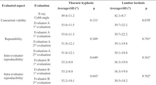
(Table 1). Regarding the tests of repeatability and
intra- and inter-evaluator reproducibility, there were
no signiicant differences in terms of either thoracic
kyphosis or lumbar lordosis (Table 1).
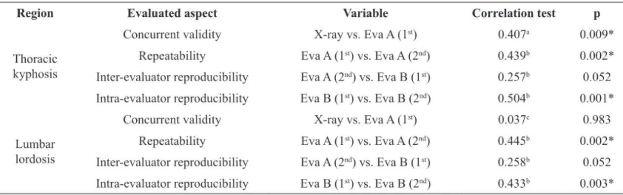
When the correlation between the measurements
obtained with the adapted arcometer and those obtained with X-rays were evaluated, there was
only a moderate correlation for thoracic kyphosis, while for lumbar lordosis the correlation was not
statistically signiicant. Similarly, the inter-evaluator reproducibility was not statistically signiicant for
either thoracic kyphosis or lumbar lordosis. The correlations between the remaining evaluations can
be classiied as moderate (Table 2).
Discussion
The aim of the present study was to verify the validity, repeatability, and intra- and inter-evaluator reproducibility of the adapted arcometer when used to measure the angles of sagittal curvature in the spine of children. To achieve this, the study conducted by
Chaise et al.5 with adults was used as reference. In that
Table 1. Average values and standard deviations (SD) of the different evaluations made with X-ray and adapted arcometer.
Evaluated aspect Evaluation Thoracic kyphosis Lumbar lordosis
Average±SD (°) p Average±SD (°) p
Concurrent validity
X-ray
Cobb angle 49.4±11.2
0.131a
42.1±8.7
0.070b Evaluator A
1st evaluation 53.6±11.5 39.7±22.2
Repeatability
Evaluator A
1st evaluation 53.6±11.5
0.349a
39.7±22.2
0.791b Evaluation A
2nd evaluation 51.8±12.1 39.1±19.8
Inter-evaluator reproducibility
Evaluation A
2nd evaluation 51.8±12.1
0.640a
39.1±19.8
0.361b Evaluator B
1st evaluation 53.2±8.8 36.3±19.0
Intra-evaluator reproducibility
Evaluator B
1st evaluation 53.2±8.8
0.643a
36.3±19.0
0.762b Evaluator B
2nd evaluation 53.2±10.1 30.5±18.2
aPaired t test; bWilcoxon test.
study, the adapted arcometer was found to provide valid and reproducible results in both the intra- and inter-evaluations5. By contrast, in the present study,
when used to evaluate children, the adapted arcometer did not present good levels of concurrent validity or inter-evaluator reproducibility, which indicates it is inappropriate for use in the diagnosis of postural alterations in the spine of children and for clinical follow-up when performed by different evaluators.
Despite this, the instrument presented adequate
repeatability and intra-evaluator reproducibility, which indicates that it is appropriate for use in the clinical follow-up conducted by the same evaluator.
Despite the existence of non-invasive methods,
when attempting to determine the position of the spine, the X-ray will probably remain the most accurate method and, therefore, the gold standard diagnosis and treatment follow-up method12. However, the X-ray depends on advanced technological resources and is often inappropriate for routine use, as the
individual is exposed to physical risk13. Consequently,
a variety of methods has been used to evaluate spinal curvature. This evaluation is equally important for diagnostic purposes, to accompany postural
alterations to the spine, and assess the eficacy of
treatments. Among the non-invasive instruments and
methods used are DIPA (Digital Image-based Postural Assessment), which is a postural evaluation software
based on photogrammetry14, kypholordometry15,16,
Moiré’s topography17, the flexible ruler6,18,19, the
plumbline distance20,21, the Inclimed21, and the arcometer7.
Two studies in the literature consider the validation
aspects of the arcometer. D’Osualdo et al.7, the
irst to describe the method in their evaluation of
children with different degrees of kyphosis, obtained
excellent correlations for validity (r=.98), intra-evaluator reproducibility (r=.99), and inter-intra-evaluator
reproducibility (r=.99) and consequently suggest that
the arcometer can be used to accompany postural alterations to the thoracic spine. The second study,
by Chaise et al.5, proposed structural modiications to
the original instrument that provided a greater degree of freedom in upper and lower rods, thus allowing them to present different lengths. The alteration to
the length of the rods led to the modiication of the
method of calculating the angle of the curvature, which could then be carried out considering two
distinct arcs. With these modiications, Chaise et al.5
improved the original proposal and thus also managed to validate the instrument for use in measuring lumbar
curvature. However, the very strong and signiicant
correlation found for the validity of thoracic curvature
(r=.94, p<0.01) and the strong and significant
correlation found for the validity of lumbar curvature
(r=.71, p<0.01) were only veriied in an adult sample.
Given that in the present study there is a considerable difference in the age, body mass, and height of the sample in relation to that of
Chaise et al.5, these characteristics may explain the
divergent results obtained between the studies, since the evaluators were previously duly trained in both the palpation technique and the collection protocol
with the adapted arcometer. Moreover, the greater
variability in terms of body posture and the greater
flexibility of the spine in the young, could also partially explain the contrasting results in this and the cited papers with older subjects, since the position used in both exams was the same.
Furthermore, if the estimated error, due to
variation in the execution of the protocol (palpation, positioning the rods, etc), is considered the same in
adults and children, the repercussion of the error in the calculated angle will be proportionally much
greater in children. For example, when measuring an adult, a 1 cm error represents less than 10% of the
Table 2. Statistical results referring to the correlations between the different evaluations.
Region Evaluated aspect Variable Correlation test p
Thoracic kyphosis
Concurrent validity X-ray vs. Eva A (1st) 0.407a 0.009* Repeatability Eva A (1st) vs. Eva A (2nd) 0.439b 0.002* Inter-evaluator reproducibility Eva A (2nd) vs. Eva B (1st) 0.257b 0.052 Intra-evaluator reproducibility Eva B (1st) vs. Eva B (2nd) 0.504b 0.001*
Lumbar
lordosis
Concurrent validity X-ray vs. Eva A (1st) 0.037c 0.983 Repeatability Eva A (1st) vs. Eva A (2nd) 0.445b 0.002* Inter-evaluator reproducibility Eva A (2nd) vs. Eva B (1st) 0.258b 0.052 Intra-evaluator reproducibility Eva B (1st) vs. Eva B (2nd) 0.433b 0.003*
Eva A – evaluator A; Eva B – evaluator B; 1st – irst evaluation; 2nd – second evaluation; aPearson’s r; bICC; cSpearman’s rho; *signiicant
distance between the rods, while in children the same
error could represent more than 40%, due to the size of the trunk. Moreover, when using the arc tangent
to calculate angles, the smaller the value using this trigonometric function the greater the impact any error will have on the estimated angle. In adults, the numbers used as input in the arc tangent function
will be approximately 1 unit, while in children it will be approximately 0.5. If we have 0.1 of variance in 1 unit (from 1.0 to 1.1), the angle calculated using the arc tangent will change from 45.0° to 47.7°. By contrast, the same variation of 0.1 in 0.5 (from 0.5 to 0.6), the angle calculated using the arc tangent will change from 26.5° to 30.9°. These differences arise
from variations in the positions of the rods when placed on the spine. Therefore, due to the variations that occur over short lengths of the trunk, there is a
clear need to ind a more appropriate procedure that can be used in children. For example, when using
the adapted arcometer in clinical practice, the risk of error could be reduced by registering the length of the rods and maintaining the same length during a second evaluation. This issue is particularly important when one considers the intrinsic postural variability of children and adolescents. It should be noted that
the results assessed herein refer to a speciic range of
thoracic and lumbar curvatures. Thus, the fact that this study did not evaluate straighter or more accentuated curvatures may be considered a limitation.
Conclusion
While the adapted arcometer can be used to
quantify the thoracic and lumbar curvatures of adults in the sagittal plane, to date it has not been possible to validate and establish inter-evaluator reproducibility for its use in children, making it unsuitable for diagnostic purposes and in the follow up of postural alterations performed by different evaluators in this population. However, as the adapted arcometer has been shown to have intra-evaluator reproducibility it can be used by the same evaluator in the clinical situation to monitor spinal curvature in children.
Nevertheless, further studies designed to adapt this
instrument for use in children are necessary.
References
1. Schivinski CIS, Richiardi J, Reis JTS, Antonelli M, Ribeiro
MAGO. Intervenção precoce da fisioterapia no péctus
excavatum: dois casos clínicos em pediatria. Saúde Soc.
2011;20(1):257-62.
2. Vrtovec T, Pernus F, Likar B. A review of methods for quantitative evaluation of spinal curvature. Eur Spine J.
2009;18(5):593-607.
http://dx.doi.org/10.1007/s00586-009-0913-0. PMid:19247697
3. Bone CM, Hsieh GH. The risk of carcinogenesis from radiographs to pediatric orthopaedic patients.
J Pediatr Orthop. 2000;20(2):251-4. http://dx.doi.
org/10.1097/01241398-200003000-00023. PMid:10739292
4. LerouxMA, Zabjek K, Simard G, Badeaux J, Coillard
C, Rivard CH. A noninvasive anthropometric technique
for measuring kyphosis and lordosis: an application
for idiopathic scoliosis. Spine (Phila Pa 1976).
2000;25(13):1689-94.
http://dx.doi.org/10.1097/00007632-200007010-00012. PMid:10870144
5. ChaiseFO, CandottiCT, Torre ML, Furlanetto TS, Pelinson
PP, Loss JF. Validation, repeatability and reproducibility of a noninvasive instrument for measuring thoracic and lumbar curvature of the spine in the sagittal plane. Rev Bras Fisioter. 2011;15(6):511-7. http://dx.doi.org/10.1590/
S1413-35552011005000031. PMid:22045292
6. De Oliveira TS, Candotti CT, La Torre M, Pelinson
PPT, Furlanetto TS, Kutchak FM, et al. Validity and reproducibility of the measurements obtained using the
flexicurve instrument to evaluate the angles of thoracic
and lumbar curvatures of the spine in the sagittal plane.
Rehabilitation Research and Practice. 2012;2012:1-9. http://
dx.doi.org/10.1155/2012/186156
7. D’Osualdo F, Schierano S, Iannis M. Validation of clinical measurement of kyphosis with a simple instrument, the arcometer. Spine (Phila Pa 1976). 1997;22(4):408-13.
http://dx.doi.org/10.1097/00007632-199702150-00011.
PMid:9055369
8. Harrison DE, Cailliet R, Harrison DD, Janik TJ, Holland B. Reliability of centroid, Cobb, and Harrison posterior
tangent methods: which to choose for analysis of thoracic
kyphosis. Spine (Phila Pa 1976). 2001;26(11):e227-34.
http://dx.doi.org/10.1097/00007632-200106010-00002.
PMid:11389406
9. Harrison DE, Harrison DD, Cailliet R, Janik TJ, Holland B. Radiographic analysis of lumbar lordosis: centroid,
Cobb, TRALL, and Harrison posterior tangent methods. Spine (Phila Pa 1976). 2001;26(11):eE235-42. http://dx.doi.
org/10.1097/00007632-200106010-00003. PMid:11389407
10. Mac-ThiongJM, Pinel-GirouxFM, de Guise JA, Labelle H. Comparison between constrained and non-constrained
Cobb techniques for the assessment of thoracic kyphosis
and lumbar lordosis. Eur Spine J. 2007;16(9):1325-31. http://
dx.doi.org/10.1007/s00586-007-0314-1. PMid:17426991
11. Kotrlik JW, Williams HA. The incorporation of effect size in information technology, learning and performance research. Inf Technol Learn Perform J. 2003;21(1):1-7.
12. ChenYL, Lee YH. A non-invasive protocol for the determination of lumbosacral vertebral angle. Clin
Biomech (Bristol, Avon). 1997;12(3):185-9. http://dx.doi.
org/10.1016/S0268-0033(97)00076-4. PMid:11415692
13. DoodyMM, Lonstein JE, Stovall M, Hacker DG, Luckyanov
N, LandCE. Breast cancer mortality after diagnostic
radiography: findings from the U.S. Scoliosis Cohort
http://dx.doi.org/10.1097/00007632-200008150-00009.
PMid:10954636
14. Furlanetto TS, CandottiCT, Comerlato T, Loss JF. Validating
a postural evaluation method developed using a Digital Image-based Postural Assessment (DIPA) software. Comput Methods Programs Biomed. 2012;108(1):203
-12. http://dx.doi.org/10.1016/j.cmpb.2012.03.012.
PMid:22522063
15. Baraúna MA, Canto RST, Sanchez HM, Bustamante JCF, Ventura-Silva RA, Malusá S. Validade e confiabilidade intra-indivíduo do cifolordômetro na avaliação da
convexidade torácica. Rev Bras Fisioter. 2005;9(3):318-25.
16. Souza FR, Ferreira F, Narciso FV, MakhoulCMB, Canto RST, Barauna MA. Evaluation of lumbar concavity using a radiographic method and kypholordometry. Rev Bras Fisioter. 2009;13(2):103-9. http://dx.doi.org/10.1590/
S1413-35552009005000016
17. Takasaki H. Moiré topography.Appl Opt. 1970;9(6):1467
-72. http://dx.doi.org/10.1364/AO.9.001467. PMid:20076401
18. Hart DL, Rose SJ. Reliability of a noninvasive method for measuring the lumbar curve. J Orthop Sports
Phys Ther. 1986;8(4):180-4. http://dx.doi.org/10.2519/
jospt.1986.8.4.180. PMid:18802227
19. Teixeira FA, Carvalho GA. Reliability and validity of
thoracic kyphosis measurements using the flexicurve
method. Rev Bras Fisioter. 2007;11(3):173-7. http://dx.doi.
org/10.1590/S1413-35552007000300005
20. Zaina F, Atanasio S, Ferraro C, Fusco C, Negrini A, Romano M, et al. Review of rehabilitation and orthopedic conservative approach to sagittal plane diseases
during growth: hyperkyphosis, junctional kyphosis,
and Scheuermann disease. Eur J Phys Rehabil Med.
2009;45(4):595-603. PMid:20032919.
21. Zaina F, Donzelli S, LusiniM, Negrini S. How to measure
kyphosis in everyday clinical practice: a reliability
study on different methods. Stud Health Technol Inform.
2012;176:264-7. PMid:22744505.
Correspondence Jefferson Fagundes Loss
Universidade Federal do Rio Grande do Sul Escola de Educação Física
Rua Felizardo, 750,