Occurrence of Neuroblastoma among
p.R337H Carriers
Ana Luiza Seidinger
1☯, Fernanda Paschoal Fortes
1☯, Maria José Mastellaro
2, Izilda
Aparecida Cardinalli
3, Lilian Girotto Zambaldi
4, Simone Santos Aguiar
2,5, José
Andrés Yunes
1,6*
1Molecular Biology Laboratory, Boldrini Children’s Center, Campinas, Sao Paulo, Brazil,2Pediatric Oncology Department, Boldrini Children’s Center, Campinas, Sao Paulo, Brazil,3Pathology Department, Boldrini Children’s Center, Campinas, Sao Paulo, Brazil,4Cytogenetics Laboratory, Boldrini Children’s Center, Campinas, Sao Paulo, Brazil,5Center for Research in Pediatrics, Faculty of Medical Sciences, State University of Campinas, Campinas, Sao Paulo, Brazil,6Medical Genetics Department, Faculty of Medical Sciences, State University of Campinas, Campinas, Sao Paulo, Brazil
☯These authors contributed equally to this work. *[email protected]
Abstract
The high incidence of adrenocortical tumors and choroid plexus carcinoma in children from
South and Southeastern regions of Brazil is associated with the germline p.R337H mutation
of
TP53
gene. The concomitant occurrence of neuroblastoma and adrenocortical tumors in
pediatric patients harboring the p.R337H mutation at our institution prompted us to investigate
the putative association between p.R337H and pediatric neuroblastoma. Genomic DNA
sam-ples from 83 neuroblastoma patients referred to a single institution during the period of 2000
–
2014 were screened for the p.R337H mutation. Available samples from carriers were
investi-gated for both nuclear p53 accumulation and loss of heterozigosity in tumor. Clinical data
were obtained from medical records in order to assess the impact of 337H allele on
manifes-tation of the disease. Seven out 83 neuroblastoma patients (8.4%) were carriers of the
TP53
p.R337H mutation in our cohort. Immunohistochemical analysis of p.R337H-positive tumors
revealed nuclear p53 accumulation. Loss of heterozigosity was not found among available
samples. The presence of 337H allele was associated with increased proportion of stage I
tumors. Our data indicate that in addition to adrenocortical tumors, choroid plexus carcinoma,
breast cancer and osteosarcoma, genetic counseling and clinical surveillance should
con-sider neuroblastoma as a potential neoplasia affecting p.R337H carriers.
Introduction
TP53
is a tumor suppressor gene involved in the etiology of a variety of tumors [
1
]. Germline
mutations in this gene are usually found in families presenting Li-Fraumeni syndrome (LFS) or
Li-Fraumeni-like syndrome (LFLS). These clinical conditions predispose individuals to a wide
spectrum of early-onset cancers, including soft tissue and bone sarcomas, central nervous
sys-tem (CNS) tumors, adrenocortical tumors (ACT), breast cancer and leukemia [
2
–
13
].
OPEN ACCESSCitation:Seidinger AL, Fortes FP, Mastellaro MJ, Cardinalli IA, Zambaldi LG, Aguiar SS, et al. (2015) Occurrence of Neuroblastoma amongTP53p.R337H Carriers. PLoS ONE 10(10): e0140356. doi:10.1371/ journal.pone.0140356
Editor:Rossella Rota, Ospedale Pediatrico Bambino Gesù, ITALY
Received:December 30, 2014
Accepted:September 24, 2015
Published:October 9, 2015
Copyright:© 2015 Seidinger et al. This is an open access article distributed under the terms of the
Creative Commons Attribution License, which permits unrestricted use, distribution, and reproduction in any medium, provided the original author and source are credited.
Data Availability Statement:All relevant data are within the paper and its Supporting Information files.
Funding:Brazilian National Counsel of Technological and Scientific Development (www.cnpq.br), grant number 401991/2010—JAY; Coordination for the
Improvement of Higher Education Personnel (www. capes.gov.br), PROCAD grant number 247/2007—
The germline mutation p.R337H of
TP53
gene has an unusually high prevalence in Brazil,
reaching 0.3% of the healthy population from Southern region [
14
,
15
]. Although its
tumori-genic effect initially appeared to be tissue-specific, being associated with only ACT [
16
], we
and others found evidences indicating its association with a broader spectrum of human
malig-nancies, e.g. breast cancer, choroid plexus carcinoma, osteosarcoma, phyllodes tumors of the
breast and LFLS families [
17
–
24
].
In a preliminary study at our institution we identified two carriers of p.R337H mutation
presenting concomitant ACT and neuroblastoma (NB) (data presented hereafter), indicating a
putative role for p.R337H on NB tumorigenesis. The surveillance program developed in
South-ern Brazil for the early diagnosis of cancer among children carriers of the p.R337H, reported,
as expected, occurrence of ACT and choroid plexus carcinoma, and less frequently
glioblas-toma multiforme, Burkitt lymphoma and neuroblasglioblas-toma [
15
].
Neuroblastoma is an embryonal tumor of the sympathetic nervous system, derived from
primordial neural crest cells. Together with ganglioneuroblastoma and ganglioneuroma,
neu-roblastoma constitutes the group of neuroblastic tumors. NB is the most immature, and
malig-nant form of neuroblastic tumor and it arises almost exclusively in infants and young children.
The most frequent identified primary sites are adrenal medulla and paravertebral sympathetic
ganglia. NB is a remarkably heterogeneous neoplasia, presenting spontaneous regression and
differentiation in some infants, while children with high-risk disease often present resistance to
therapy [
25
].
NB is not commonly associated with
TP53
mutations [
26
]. Recently, a single nucleotide
polymorphism (SNP) that maps to 3
’
UTR of
TP53
(rs78378222) was found to be associated
with neuroblastoma susceptibility [
27
]. This germline variant impairs proper termination and
polyadenylation of
TP53
transcripts and besides NB, it was also found to confer susceptibility
to cutaneous basal cell carcinoma, prostate cancer, glioma and colorectal adenomas [
28
].
Although the role of
TP53
on neuroblastoma tumorigenesis is still under debate, our
prelim-inary findings prompted us to investigate the association between the highly prevalent p.
R337H mutation and pediatric neuroblastoma. In addition, we investigated the presence of
SNP rs78378222 in our cohort and the impact of 337H allele on clinical manifestation and
prognosis of this disease.
Material and Methods
Patients
The subjects included in the current study comprised pediatric patients diagnosed and treated
for neuroblastoma at a single institution located in Campinas, São Paulo, Brazil, during the
year 2000 through July 2014. During this period, 178 patients were diagnosed with
neuroblas-toma and classified according to International Neuroblasneuroblas-toma Staging System (INSS) and
Shi-mada criteria [
29
,
30
]. The 83 patients enrolled in this study were selected only on the basis of
availability of samples for the p.R337H mutation investigation. The samples studied included
both peripheral blood mononuclear cells (MNCs) and/or tumor samples. The cancer family
history,
MYCN
status, demographic and clinical data were obtained from patients
’
medical
records.
Ethics Statement
This study was approved by the Ethical Research Committee of the Faculty of Medical Sciences
at the State University of Campinas (approval number 1121/2008), which waived the signature
of informed consent because the work was conducted using retrospective samples from tumor
bank.
Screening for p.R337H mutation
Genomic DNA was isolated from peripheral blood or tumor samples by using a standard
phe-nol:chlorophorm extraction method [
31
] followed by a PCR to amplify the exon 10 of
TP53
gene. The PCR product was digested with the restriction enzyme
Hha
I (Fermentas Inc.), which
yields 2 fragments (293 bp and 154 bp) in the wild-type amplicon but only 1 fragment when
the p.R337H mutation is present [
16
]. The presence of p.R337H mutation was confirmed by
Sanger sequencing by using the BigDye Terminator Cycle Sequencing Ready Reaction Kit
(Applied Biosystems) in an ABI PRISM 310 automated sequencer (Applied Biosystems).
Loss of heterozygosity analysis
Paired DNA samples from germline and tumor tissue were investigated for loss of
heterozigos-ity (LOH). p.R337H allelic discrimination was achieved through custom made TaqMan SNP
genotyping. Assay standardization and validation in more than 30,000 samples will be
pre-sented elsewhere (Caminha IP et al. in preparation). Briefly, genomic DNA was subjected to
qPCR using primers that flank the region of the p.R337H mutation: 5
’
-CCTCCTCTGTTGC
TGCAGATC-3
’
and 5
’
-CCTCATTCAGATCTCTCGGAAC-3
’
in conjunction with two MGB
probes: 5
’
-VIC-CGTGAGC
G
CTTCGAG-3
’
and 5
’
-FAM-CGTGAGC
A
CTTCGAG-3
’
, that
bind to the wild-type and mutant allele, respectively. The reaction consisted of 6
μ
L of 2x
Taq-Man
1Universal PCR Master Mix (Life Technologies), 0,2
μ
M of each probe (Life
Technolo-gies), 0,9
μ
M of each primer in a final volume of 12
μ
L. The cycling conditions consisted of
95°C for 10 minutes, followed by 40 repetitions of a two-step cycle (15 seconds at 95°C and 1
minute at 60°C) in a Step One Real-Time PCR System (Life Technologies). Since a probe for
each allele was included in the reaction, heterozygous samples show signal amplification for
both probes. Homozygous samples or samples that have lost the heterozygosity show signal
amplification from only one probe.
Screening for mutations within the
TP53
DNA-binding domain
Tumor samples from patients with p.R337H were screened for other possible mutations in
exons 5 to 9 of
TP53
gene according to IARC protocol, available in
http://p53.iarc.fr/
ProtocolsAndTools.aspx
. PCR products were sequenced based on Sanger method by using the
BigDye Terminator Cycle Sequencing Ready Reaction Kit (Applied Biosystems) in an ABI
PRISM 310 automated sequencer (Applied Biosystems). Sequences obtained were compared to
NCBI reference sequence NG_017013.2. Suspected mutations were confirmed by amplifying a
new PCR product from the same DNA sample, followed by a new sequencing reaction.
Genotyping for SNP rs78378222
This hypomorphic allele is characterized by an A to C transversion at the 3
’
UTR of
TP53
(NM_000546.5:c.
1175A
>
C). In order to genotype the patients included in our cohort, we
amplified the correspondent region with primers rs78378222F: 5
’
- GTAAAACGACGGC
CAGTGGGTCAACATCTTTTACATTC-3
’
and rs78378222R: 5
’
- TAATACGACTCACTA
Immunohistochemistry
After dewaxing and rehydration, 5-
μ
m-thick tumor sections were treated with H
2O
2to reduce
endogenous peroxidase activity and then underwent wet heat-mediated antigen retrieval with
TRIS-ethylene diamine tetra acetic acid, pH 8.9. Sections were incubated with mouse
monoclo-nal anti-p53 antibody (clone DO-7; Dako A/S). This antibody is specific for an N-termimonoclo-nal
epi-tope and reacts with both wild-type and mutant human p53 proteins. Ten NB tumors negative
for p.R337H were stained in parallel as negative controls. Colon adenocarcinoma sections,
known to stain positively for p53, were stained in parallel as positive controls. Results were
observed by using the standard avidin-biotin complex method with the Dako LSAB System
horseradish peroxidase kit in a Nikon Eclipse E200 microscope (Nikon Instruments).
Statistical Analysis
For statistical purposes, the primary site of disease was categorized as adrenal medulla or
non-adrenal. The association between p.R337H presence and gender, age at diagnosis, tumor stage
at diagnosis, primary site of disease,
MYCN
amplification and outcome was assessed by the
Mann Whitney test, Chi-square test for trend or Fisher Exact test. Statistical analyses were
per-formed by using Graphpad Prism 5.0 (GraphPad Software Inc., San Diego, CA).
Results
TP53
findings among NB patients
Patients diagnosed with adrenocortical tumor at our institution are invited to do the p.R337H
testing due the strong association of this mutation with ACT at our geographic region [
15
–
17
,
19
,
22
]. Two p.R337H heterozygous ACT patients were found to present synchronous
neu-roblastoma (Figs
1
and
2
). A complete description of clinical characteristics of these two
patients was summarized in
S1 Table
. This finding led us to hypothesize that p.R337H could be
actively associated to NB tumorigenesis.
We investigated therefore the presence of p.R337H in an additional group of 81 pediatric
patients diagnosed exclusively with neuroblastoma. In this group, we identified other 5 carriers
of this mutation. The presence of p.R337H mutation in patients with NB was confirmed by
using at least two methodologies. The 7 patients harboring the p.R337H mutation (2 diagnosed
with ACT plus NB and 5 with solely NB) accounted for 8.4% of tested patients. None of 77
tested patients from our cohort (p.R337H positive n = 7 / p.R337H negative n = 70) was found
to carry the hypomorphic allele (C) for SNP rs78378222.
TP53
mutations in NB may arise as a consequence of tumor progression or be induced by
chemotherapy [
32
,
33
]. In our cohort, only two out seven p.R337H positive tumors were
sub-jected to chemotherapy before mutation genotyping. Nevertheless, the mutation was also
detected in blood samples from these two patients, confirming that the mutation was germline
and was not acquired in consequence of a cytotoxic exposure (
Table 1
). Direct sequencing of
exons 5
–
9 of
TP53
gene revealed no additional mutation on p.R337H positive tumors
(
Table 1
).
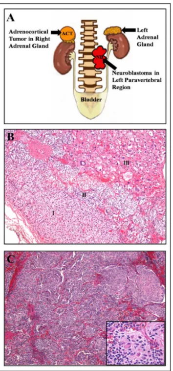
Fig 1. Morphological and histological characterization of tumors from patient #1, presenting concomitant NB and ACT.A) Schematic representation based on patient #1 image files demonstrating adrenocortical tumor in right adrenal gland and the neuroblastoma in left paravertebral region. B) Right adrenocortical tumor from patient #1 measuring 2,0 x 1,8 x 0,8 cm. The microscopic examination revealed in I) cortical region of non-tumoral adrenal; II) medullary region of non-tumoral adrenal; III) adrenocortical tumor characterized by atypical large pleomorphic cells with acidophilic cytoplasm, presenting Fuhrman nuclear atypia grade 3 (H&E 100x magnification). C) Histological examination of left paravertebral tumor from patient #1. The neuroblastoma presented dimensions of 4,3 x 3,2 x 1,5 cm and was characterized by small round primitive cell clusters (H&E staining, 40x magnification). In detail, neuroblasts at different maturation stages embedded in neurofibrillary stroma (H&E, 400x magnification).
Demographical and clinical data of NB patients
Table 2
summarizes the clinical and demographical data for NB patients included in the
pres-ent study, according to p.R337H status. No significant association was observed between p.
R337H presence and gender, age at diagnosis, primary site of disease,
MYCN
amplification and
outcome. p.R337H was statistically associated with increased proportion of stage I tumors.
These findings should be considered with caution, due to the small number of p.R337H
patients analyzed.
The cancer family history was investigated at the time of diagnosis. The interview was
con-ducted by an oncologist and data obtained were based only on patient
’
s report, with no further
confirmation by pathology report. Among p.R337H negative patients with available
informa-tion (n = 61), 39 patients meninforma-tioned sporadic cases in family members, while no family met
the criteria for LFS or LFLS (
Table 3
). Among the p.R337H positive patients (n = 7), 4 reported
sporadic cases in family members and one met the criteria for LFLS (Birch) associated with the
parental side segregating the p.R337H mutation (
Table 3
). Among the negative patients, the
most frequent sites reported were uterus (n = 6), leukemia (n = 6), skin (n = 5), breast (n = 4)
and prostate (n = 4). Among the positive individuals, the most frequent sites reported were
breast (n = 2), esophagus (n = 2) and lung (n = 2).
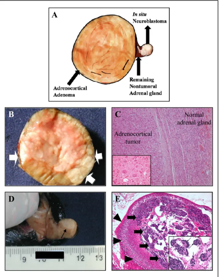
neuroblastoma. B) The macroscopic examination of tumor demonstrates a solid and well-circumscribed adrenocortical tumor, completely surrounded by a thin fibrous capsule (arrow). The cut surface shows a yellowish, shiny, and homogeneous neoplasm with slightly lobular architecture. C) The left half of the picture shows the adrenocortical tumor surrounded by a thin and fibrous capsule (HE, 100x). Insert a detail demonstrating tumor cells of large size, with huge and eosinophilic cytoplasm, large nuclei, with central and conspicuous nucleoli. (HE, 400x). D)In situneuroblastoma can be identified as a slightly pale region indicated by an arrow. E) Microscopic analysis from the pale region of remainder adrenal gland shows well-preserved cortical parenchyma cells (arrowheads). The inner medullary zone is heavily infiltrated byin situneuroblastoma (arrows) (HE, 40x). Insert, a detail showing tumor composed of small
round blue cells with scant cytoplasm, and dark, hyperchromatic nuclei with frequent formation of Homer-Wright rosettes (thin arrows) (HE, 100x).
doi:10.1371/journal.pone.0140356.g002
Table 1. Characteristics of p.R337H positive patients.
Patient Diagnosis Tissue screened for p.R337H
Codon 337 status
DBD mutation
IHC p53 tumor
Cytotoxic therapy before DNA analysis of tumor?
1 Neuroblastoma IV + ACT Blood Arg/His No NB–+ Yes
Tumor (NB) Arg/His ACT–++++
2 Neuroblastomain situ
adrenal + ACT
Blood Arg/His NA NB–NA NA
ACT–++
3 Neuroblastoma III Tumor Arg/His No +++ No
4 Neuroblastoma III Tumor His/(His?)a No ++ No
5 Neuroblastoma IV Blood Arg/His No +++ Yes
Tumor Arg/His
6 Neuroblastoma I Tumor Arg/His No ++++ No
7 Neuroblastoma III Blood Arg/His No ++++ No
Tumor Arg/His
NB–Neuroblastoma; ACT–adrenocortical tumor; NA–material not available for analysis; IHC–immunohistochemistry; DBD–DNA-binding domain. Immunohistochemistry was scored as 0 (no cells positive), + (up to 25% of cells positive), ++ (26% to 50% of cells positive), +++ (51% to 75% of cells positive), or ++++ (>75% of cells positive).
aOnly the mutant allele was found in this tumor. It was not possible to distinguish between homozygosity or loss of heterozygosity with retention of the mutated allele in the tumor.
Discussion
Since its description in 2001 by Ribeiro and colleagues [
16
], continuous efforts have been
devoted to better understand how the p.R337H mutation contributes to carcinogenesis.
Although initially controversial, its association with a broad spectrum of tumors is now
well accepted. Strongly associated with ACT and CPC, the 337H allele is thought to be
responsible for the high incidence of these tumors in the Southern and Southeastern regions of
Brazil [
16
,
19
,
20
,
22
]. Besides ACT and CPC, osteosarcoma and breast cancer, including
phyl-lodes tumors of the breast, are also associated with p.R337H, in a lesser extent though
[
18
,
19
,
21
,
23
,
24
]. In the present work, we describe the identification of p.R337H carriers among
neuroblastoma pediatric patients. Seven out of 83 patients tested (8.4%) were carriers of 337H
allele. This frequency is about 28 to 42 times higher than the estimated 0.2 to 0.3% frequency
for p.R337H in people not selected by cancer diagnosis from our region [
14
,
15
], suggesting
that carriers of the p.R337H are at increased risk of developing NB than the general population.
Although cancer in general does not arise from a single gene defect, populations in which p.
R337H was identified should consider neuroblastoma as a potential neoplasia affecting carriers.
Fig 3. Immunohistochemical p53 staining on p.R337H positive NB tumors.A, B) Representative images of nuclear p53 accumulation on neuroblastoma cells from patients 3 and 7, respectively (100x magnification).
In accordance with our findings, Custódio and colleagues have also identified a neuroblastoma
patient in a surveillance program for p.R337H carriers in Southern Brazil (
Table 4
) [
15
].
Nuclear p53 accumulation on NB cells suggests p53 inactivation in p.R337H-positive
tumors (
Fig 3
). Paired analysis of germline and tumor tissues revealed no LOH in available
cases (
Table 1
). LOH with retention of the mutated allele was identified in virtually all cases of
ACT and CPC associated with p.R337H [
19
]. On the other hand, the mechanism of breast
car-cinogenesis associated with p.R337H mutation appears to be not related to the classical two-hit
model involving tumor suppressor genes, since LOH at the mutation locus is not common in
these cases [
24
]. It is important to note that p.R337H is a dominant negative mutation that
Table 2. Clinico-biological data of neuroblastoma patients according to p.R337H status.
Positive p.R337H n (%) Negative p.R337H n (%) pvalue
Overall number 7 76
-Gender Male 3 (43%) 40 (53%) 0.7F
Female 4 (57%) 36 (47%)
Age at diagnosis Median 27 months 22 months 0.8M
Stage at diagnosis I 2 (28.5%) 4 (5.2%) 0.04X
IIA/IIB 0 5 (6.6%)
III 3 (43%) 23 (30.2%)
IV 2 (28.5%) 42 (55.3%)
IVS 0 2 (2.7%)
Site of primary disease Adrenal 3 (43%) 36 (47%) 1a,F
Non-Adrenal 4 (57%) 40 (53%)
MYCNstatus Normal 3 (43%) 37 (49%) 1b,F
Amplified 3 (43%) 33 (43%)
Unknown 1 (14%) 6 (8%)
Outcome Alive 5 (71%) 32 (42%) 0.28c,F
Death 2d(29%) 42 (55%)
Lost follow up 0 2 (3%)
Median time follow up 55 mo 28 mo
FFisher Exact test MMann Whitney test XChi-square test for trend
aFor statistical purposes, the primary site of disease was categorized as adrenal or non-adrenal. bThe statistical analysis included only patients with knownMYCNstatus.
cThe statistical analysis did not include patients who were lost of follow up. dPatient 4 and 7 inTable 1.
doi:10.1371/journal.pone.0140356.t002
Table 3. Cancer family history of the 83 neuroblastoma patients according to p.R337H status.
Positive p.R337H (n = 7) Negative p.R337H (n = 76)
No information available 0 15
Absence of cancer history 2 (29%) 22 (36%)
Sporadic casesa 4 (57%) 39 (64%)
LFS/LFLS 1 (14%) 0 (0%)
a
Presence of at least one member of the family with cancer, without criteria for a cancer predisposition syndrome though.
affects the oligomerization domain of p53, so it can interfere with normal function of wild-type
allele through the impaired tetramer conformation of the protein [
34
]. Moreover, different
mechanisms of allele inactivation, as promoter methylation and
cis
-acting elements, may also
play an important role on allelic imbalance, thus rendering LOH not the exclusive mechanism
responsible for reducing expression of the wild-type allele [
35
–
37
].
Neuroblastoma is a very heterogeneous malignancy that affects almost exclusively infants
and young children. The clinical behavior of NB ranges from spontaneous regression to
aggres-sive tumors that do not respond to current therapies [
29
]. The presence of p.R337H mutation
was statistically associated with increased proportion of stage I tumors. However, due the small
number of patients analyzed, this should be taken as a preliminary finding.
Although we had no access to the majority of pathology reports, data obtained on cancer
family history of p.R337H positive NB patients showed that only one out of 7 families
pre-sented features consistent with LFS/LFLS (
Table 3
). This finding is in accordance with the
broad phenotypic variation observed among families with p.R337H, i.e., a large proportion of
families without any history of cancer while some families presenting with clear LFS/LFLS [
15
–
18
,
22
]. This phenotypic variation highlights the importance of penetrance modifying factors,
such as low-penetrant mutations and polymorphisms.
Recently, a polymorphism that maps to 3
’
UTR of
TP53
(rs78378222) was found to be
asso-ciated with neuroblastoma susceptibility (
Table 4
) [
27
]. None of the NB patients included in
our cohort was found to carry the hypomorphic allele at this locus. From our extensive
litera-ture revision on
TP53
polymorphisms studied in the context of NB, we found rs1042522 as the
most commonly SNP studied among NB patients (
Table 4
). This polymorphism results in
either an arginine (R) or proline (P) at codon 72 (R72P) and although it has been extensively
studied, its clinical significance is still unclear according to NCBI SNP database (available in
http://www.ncbi.nlm.nih.gov/snp/?term=rs1042522
). Interestingly, the allele that codifies for
an arginine was consistently overrepresented among neuroblastoma patients (
Table 4
).
Whether R72 is a risk modifying factor for NB remains to be determined.
To our knowledge, 34 patients with neuroblastic tumors harboring
TP53
mutations have
been described until now (summarized in
Table 5
). Considering the cases with somatic
alter-ations, a significant proportion of mutations may have arisen as a consequence of tumor
Table 4. Literature review onTP53polymorphisms in neuroblastoma.
TP53polymorphisms in neuroblastoma
Study design Polymorphism Location Nucleotide change
Amino acid change
Minor allele frequency
(MAF)a MAF amongNB Reference
Cohort of 41 NB patients
rs1042522 exon 4 (codon 72)
CCC—CGC Pro to Arg 0.457 0.1375 [45]
Cohort of 286 NB patients
rs1042522 exon 4 (codon 72)
CCC—CGC Pro to Arg 0.457 0.20 [46]
Cohort of 2804 NB patients
rs35850753 5’UTR
Δ133p53
G—A - 0.0056 0.036 [27]
rs78378222 3’UTR A—C - 0.0026 0.027
Cohort of 2101 NB patients
rs8079544 intron 1 G—A - 0.0783 0.068
Cohort of 1809 NB patients
rs1042522 exon 4 (codon 72)
CCC—CGC Pro to Arg 0.457 0.25
a
The minor allele frequency for a given SNP is based on its frequency in a default global population. The current default global population is 1000 Genome phase 1 genotype data from 1094 worldwide individuals, released in the May 2011 dataset.
Table 5. Literature review onTP53mutations in association with neuroblastoma.
NB studies
Study design TP53region screened
TP53
mutant / total
Mutation Germline / Somatic Diagnosis Reference
This study codon 337 7/83 R337H Germline Neuroblastomaa
-Case report Whole exome 1 R248Q Germline mosaicism
and homozygous at tumors
Benign myofibroblastic proliferation–9 mo; Sarcoma NOS–11 mo;
Neuroblastoma–1y 4 mo
[38]
Cohort of NB patients exons 2–11 0/40 - - - [47]
Cohort of NB patients exons 2–11 2/86 C135Y Acquired in relapsed tumor
Neuroblastoma [32]
G204V Acquired in metastatic tumor
Neuroblastoma
Cohort of NB patients exons 5–9 0/48 - - - [48]
Cohort of NB patients exons 2–11 0/38 - - - [49]
Cohort of NB patients exons 5–9 2/20 V172V No germline tissue available for analysis
Neuroblastoma [50]
D259Y Somatic Neuroblastoma
Cohort of NB patients exons 5–8 0/29 - - - [51]
Case report exons 5–8 1 C277F Acquired after
chemotherapy
Neuroblastoma–3y [33]
Case report exons 2–11 1 R248W Germline ACT–10 mo and neuroblastoma–10
mo
[39]
Not specified 1 Codon
248
Not specified Neuroblastoma
Case report Not specified 1 R248W Germline Ganglioneuroblastoma–1y 6 mo;
ACT–1 y 6 mo; Turner syndrome (45,X)
[41]
Cohort of NB patients Whole exome 2/240 D281N Somatic Neuroblastoma–5y [52]
P219S Germline Neuroblastoma–3y 6 mo
Cohort of NB patients exons 5–8 0/30 - - - [53]
Cohort of NB patients exons 5–8 0/44 - - - [54]
Cohort of NB patients exons 4–9 6/41 F270L Acquired after chemotherapy and present at relapse
Neuroblastoma–1y 7 mo [45]
V157G Acquired after chemotherapy
Neuroblastoma–7 mo
D259Y At relapse Neuroblastoma–2y 3 mo D259Y Acquired after
chemotherapy
Neuroblastoma–5y 9 mo
V203M At diagnosis Neuroblastoma–15y C238Y Acquired after
chemotherapy
Neuroblastoma–16y
Cohort of NB patients exons 4–8 3/40 R273L Not specified Neuroblastoma–12y [55]
R283C Not specified Neuroblastoma–1y 10 mo R283C Not specified Neuroblastoma–3 mo
Case report Not specified 1 I162F Germline Neuroblastoma–8 mo; ACT–8 mo [56]
TP53/ LFS studies Study design TP53region
screened
NB / total Mutation Germline / Somatic Diagnosis Reference
Cohort of R337H(+) newborn carriers
codon 337 1/461b R337H Germline Neuroblastoma [15]
progression or induced by chemotherapy. With respect to 18 patients with germline mutations,
we found that p.R337H is the most common inherited
TP53
mutation associated with NB
(n = 7). It is intriguing that among the other 11 germline cases described, four (36%) had
muta-tions at codon 248 [
38
–
41
]. Noteworthy, one of them presented concomitant NB and ACT and
another patient presented ganglioneuroblastoma and ACT [
39
,
41
]. Whether neuroblastic
tis-sue present a marked susceptibility to alterations at codon 248 of p53 remains to be
investi-gated. Tissue-specificity of
TP53
mutations has been considerably discussed. Missense
TP53
mutations located in the DNA-binding loop that contact the minor groove of DNA were
asso-ciated with brain tumors, whereas mutations in the non DNA-binding loops,
β
-sheets and
olig-omerization domain were associated with adrenocortical tumors [
42
]. Mutations affecting
TP53
splicing sites were strongly associated with Wilm
’
s tumor, while null mutations were not
associated with a specific type of tumor, but were associated with early onset tumors, in
partic-ular brain tumors [
26
,
42
]. Apparently, mutations affecting different domains of the protein
may exert different impact on protein function or protein-protein interactions, culminating in
different tissue-susceptibility to cancer.
From this perspective, it is well accepted that p.R337H predisposes carriers to a broad
spec-trum of cancer. The specspec-trum of tumors found in p.R337H families may, eventually, overlap
that found in LFS or LFLS [
17
,
18
,
20
]. However, in the majority of cases, this spectrum is not
identical [
15
,
16
,
43
,
44
]. Therefore, the clinical criteria for LFS and LFLS are not suitable for
sus-pecting p.R337H. The challenge in identify individuals carrying the p.R337H mutation and,
therefore, family members at high risk of developing cancer, has motivated several efforts to
define the exact spectrum of tumors associated to this mutation. The present work shows that
in addition to ACT, CPC, breast cancer and osteosarcoma, genetic counseling and clinical
sur-veillance should consider neuroblastoma as a potential disease affecting p.R337H carriers.
Supporting Information
S1 Table. Clinical data of patients diagnosed with synchronous ACT and NB.
(DOC)
Table 5. (Continued)
NB studies
Study design TP53region screened
TP53
mutant / total
Mutation Germline / Somatic Diagnosis Reference
Cohort of LFS individuals All exons, splice junctions and promoter region
3/148c Not specified
Germline Neuroblastoma [26]
Families with osteosarcoma
Not specified 1/17 familiesd
R273H Germline Neuroblastoma–1y [57]
Cohort of patients with two primary malignant neoplasms and no LFS
exons 5–9 1/59 R248W Germline Neuroblastoma–1 y; Breast
carcinoma–32 y
[40]
NB–neuroblastoma; LFS–Li-Fraumeni Syndrome ain two cases NB were concomitant with ACT
bof these 461 carriers, 11 developed adrenocortical tumors, 2 choroid plexus tumors, 1 glioblastoma multiforme, 1 Burkitt lymphoma and 1 neuroblastoma cAll 148 cancer cases were diagnosed in carriers ofTP53germline mutations
dThere is no information on the total number of individuals (carriers) analyzed.
Author Contributions
Conceived and designed the experiments: ALS MJM JAY. Performed the experiments: ALS
FPF IAC LGZ. Analyzed the data: ALS FPF MJM IAC LGZ SSA JAY. Contributed reagents/
materials/analysis tools: SSA JAY. Wrote the paper: ALS JAY.
References
1. Vogelstein B, Lane D, Levine AJ. Surfing the p53 network. Nature. 2000; 408: 307–10. PMID: 11099028
2. Li FP, Fraumeni JF Jr. Soft tissue sarcomas, breast cancer, and other neoplasms.A familial syndrome? Ann InternMed. 1969; 71: 747–752.
3. Li FP, Fraumeni JF Jr, Mulvihill JJ, Blattner WA, Dreyfus MG, Tucker MA, et al. A cancer family syn-drome in twenty-four kindreds. Cancer Res. 1988; 48: 5358–5362. PMID:3409256
4. Malkin D, Li FP, Strong LC, Fraumeni JF Jr, Nelson CE, Kim DH, et al. Germ line p53 mutations in a familial syndrome of breast cancer, sarcomas, and other neoplasms. Science. 1990; 250: 1233–1238. PMID:1978757
5. Srivastava S, Zou ZQ, Pirollo K, Blattner W, Chang EH. Germ-line transmission of a mutated p53 gene in a cancer-prone family with Li-Fraumeni syndrome. Nature. 1990; 348: 747–749. PMID:2259385
6. Eeles RA, Bartkova J, Lane DP, Bartek J. The role of TP53 in breast cancer development. CancerSurv. 1993; 18: 57–75.
7. Birch JM, Hartley AL, Tricker KJ, Prosser J, Condie A, Kelsey AM, et al. Prevalence and diversity of constitutional mutations in the p53 gene among 21 Li-Fraumeni families. Cancer Res. 1994; 54: 1298– 304. PMID:8118819
8. Eeles RA. Germline mutations in the TP53 gene in breast and other cancers, University of London 2000.
9. Chompret A, Abel A, Stoppa-Lyonnet D, Brugiéres L, Pagés S, Feunteun J, et al. Sensitivity and predic-tive value of criteria for p53 germline mutation screening. J Med Genet. 2001; 38: 43–47. PMID: 11332399
10. Bougeard G, Sesboüé R, Baert-Desurmont S, Vasseur S, Martin C, Tinat J, et al. Molecular basis of the Li-Fraumeni syndrome: An update from the French LFS families. J Med Genet. 2008; 45: 535–538. doi: 10.1136/jmg.2008.057570PMID:18511570
11. Tinat J, Bougeard G, Baert-Desurmont S, Vasseur S, Martin C, Bouvignies E, et al. 2009 Version of the Chompret criteria for Li Fraumeni syndrome. J Clin Oncol. 2009; 27: e108–e109, author reply e110.
12. Malkin D. Li-fraumeni syndrome. Genes Cancer 2011; 2: 475–484. doi:10.1177/1947601911413466 PMID:21779515
13. Bougeard G, Renaux-Petel M, Flaman JM, Charbonnier C, Fermey P, Belotti M, et al. Revisiting Li-Fraumeni syndrome from TP53 mutation carriers. J Clin Oncol. 2015; 33: 2345–52. doi:10.1200/JCO. 2014.59.5728PMID:26014290
14. Palmero EI, Schüler-Faccini L, Caleffi M, Achatz MI, Olivier M, Martel-Planche G, et al. Detection of R337H, a germline TP53 mutation predisposing to multiple cancers, in asymptomatic women participat-ing in a breast cancer screenparticipat-ing program in Southern Brazil. Cancer Lett. 2008; 261: 21–25. doi:10. 1016/j.canlet.2007.10.044PMID:18248785
15. Custódio G, Parise GA, Kiesel Filho N, Komechen H, Sabbaga CC, Rosati R, et al. Impact of neonatal screening and surveillance for the TP53 R337H mutation on early detection of childhood adrenocortical tumors. J Clin Oncol. 2013; 31: 2619–2626. doi:10.1200/JCO.2012.46.3711PMID:23733769
16. Ribeiro RC, Sandrini F, Figueiredo B, Zambetti GP, Michalkiewicz E, Lafferty AR, et al. An inherited p53 mutation that contributes in a tissue-specific manner to pediatric adrenal cortical carcinoma. Proc Natl Acad Sci U S A. 2001; 98: 9330–9335. PMID:11481490
17. Achatz MI, Olivier M, Le Calvez F, Martel-Planche G, Lopes A, Rossi BM, et al. The TP53 mutation, R337H, is associated with Li-Fraumeni and Li-Fraumeni-like syndromes in Brazilian families. Cancer Lett. 2007; 245: 96–102. PMID:16494995
18. Assumpção JG, Seidinger AL, Mastellaro MJ, Ribeiro RC, Zambetti GP, Ganti R, et al. Association of
the germline TP53 R337H mutation with breast cancer in southern Brazil. BMC Cancer. 2008; 8: 357. doi:10.1186/1471-2407-8-357PMID:19046423
19. Seidinger AL, Mastellaro MJ, Fortes FP, Assumpção JG, Cardinalli IA, Ganazza MA, et al. Association
20. Custodio G, Taques GR, Figueiredo BC, Gugelmin ES, Oliveira Figueiredo MM, Watanabe F, et al. Increased incidence of choroid plexus carcinoma due to the germline TP53 R337H mutation in south-ern Brazil. PLoS One. 2011; 6: e18015. doi:10.1371/journal.pone.0018015PMID:21445348
21. Giacomazzi J, Koehler-Santos P, Palmero EI, Graudenz MS, Rivero LF, Lima E, et al. A TP53 founder mutation, p.R337H, is associated with phyllodes breast tumors in Brazil. Virchows Arch. 2013; 463: 17– 22. doi:10.1007/s00428-013-1439-8PMID:23794094
22. Giacomazzi J, Selistre SG, Rossi C, Alemar B, Santos-Silva P, Pereira FS, et al. Fraumeni and Li-Fraumeni-like syndrome among children diagnosed with pediatric cancer in Southern Brazil. Cancer. 2013; 119: 4341–4349. doi:10.1002/cncr.28346PMID:24122735
23. Cury NM, Ferraz VE, Silva WA Jr. TP53 p.R337H prevalence in a series of Brazilian hereditary breast cancer families. Hered Cancer Clin Pract. 2014; 12: 8. doi:10.1186/1897-4287-12-8PMID:24625245
24. Giacomazzi J, Graudenz MS, Osorio CA, Koehler-Santos P, Palmero EI, Zagonel-Oliveira M, et al. Prevalence of the TP53 p.R337H mutation in breast cancer patients in Brazil. PLoS One. 2014; 9: e99893. doi:10.1371/journal.pone.0099893PMID:24936644
25. Brodeur GM, Hogarty MD, Mosse YP, Maris JM. Neuroblastoma. In: Pizzo PA, Poplack DG (editors). Principles and practice of pediatric oncology. 6th ed. Philadelphia: Lippincott Williams & Wilkins; 2011. p. 886–922.
26. Birch JM, Alston RD, McNally RJ, Evans DG, Kelsey AM, Harris M, et al. Relative frequency and mor-phology of cancers in carriers of germline TP53 mutations. Oncogene. 2001; 20: 4621–4628. PMID: 11498785
27. Diskin SJ, Capasso M, Diamond M, Oldridge DA, Conkrite K, Bosse KR, et al. Rare variants in TP53 and susceptibility to neuroblastoma. J Natl Cancer Inst. 2014; 106: dju047. doi:10.1093/jnci/dju047 PMID:24634504
28. Stacey SN, Sulem P, Jonasdottir A, Masson G, Gudmundsson J, Gudbjartsson DF, et al. A germline variant in the TP53 polyadenylation signal confers cancer susceptibility. Nat Genet. 2011; 43: 1098– 1103. doi:10.1038/ng.926PMID:21946351
29. Brodeur GM, Pritchard J, Berthold F, Carlsen NL, Castel V, Castelberry RP, et al. Revisions of the inter-national criteria for neuroblastoma diagnosis, staging, and response to treatment. J Clin Oncol. 1993; 11: 1466–1477. PMID:8336186
30. Shimada H, Chatten J, Newton WA Jr, Sachs N, Hamoudi AB, Chiba T, et al. Histopathologic prognos-tic factors in neuroblasprognos-tic tumors: definition of subtypes of ganglioneuroblastoma and an age-linked classification of neuroblastomas. J Natl Cancer Inst. 1984; 73: 405–416. PMID:6589432
31. Sambrook J, Fritsch EF, Maniatis T: Molecular Cloning–A Laboratory Manual Volume 2. 2nd edition. Cold Spring Harbour Laboratoy Press; 1989.
32. Imamura J, Bartram CR, Berthold F, Harms D, Nakamura H, Koeffler HP. Mutation of the p53 gene in neuroblastoma and its relationship with N-myc amplification. Cancer Res. 1993; 53: 4053–4058. PMID: 8358734
33. Manhani R, Cristofani LM, Odone Filho V, Bendit I. Concomitant p53 mutation and MYCN amplification in neuroblastoma. Med Pediatr Oncol. 1997; 29: 206–207. PMID:9212845
34. DiGiammarino EL, Lee AS, Cadwell C, Zhang W, Bothner B, Ribeiro RC, et al. A novel mechanism of tumorigenesis involving pH-dependent destabilization of a mutant p53 tetramer. Nat Struct Biol. 2002; 9: 12–16. PMID:11753428
35. Maia AT, Spiteri I, Lee AJ, O'Reilly M, Jones L, Caldas C, et al. Extent of differential allelic expression of candidate breast cancer genes is similar in blood and breast. Breast Cancer Res. 2009; 11: R88. doi: 10.1186/bcr2458PMID:20003265
36. Shoemaker R, Deng J, Wang W, Zhang K. Allele-specific methylation is prevalent and is contributed by CpG-SNPs in the human genome. Genome Res. 2010; 20: 883–889. doi:10.1101/gr.104695.109 PMID:20418490
37. Sakai T, Toguchida J, Ohtani N, Yandell DW, Rapaport JM, Dryja TP. Allele-specific hypermethylation of the retinoblastoma tumor-suppressor gene. Am J Hum Genet. 1991; 48: 880–888. PMID:1673287
38. Behjati S, Maschietto M, Williams RD, Side L, Hubank M, West R, et al. A pathogenic mosaic TP53 mutation in two germ layers detected by next generation sequencing. PLoS One. 2014; 9: e96531. doi: 10.1371/journal.pone.0096531PMID:24810334
39. Rossbach HC, Baschinsky D, Wynn T, Obzut D, Sutcliffe M, Tebbi C. Composite adrenal anaplastic neuroblastoma and virilizing adrenocortical tumor with germline TP53 R248W mutation. Pediatr Blood Cancer. 2008; 50: 681–683. PMID:17427234
41. Pivnick EK, Furman WL, Velagaleti GV, Jenkins JJ, Chase NA, Ribeiro RC. Simultaneous adrenocorti-cal carcinoma and ganglioneuroblastoma in a child with Turner syndrome and germline p53 mutation. J Med Genet 1998; 35: 328–332. PMID:9598730
42. Olivier M, Goldgar DE, Sodha N, Ohgaki H, Kleihues P, Hainaut P, et al. Li-Fraumeni and related syn-dromes: correlation between tumor type, family structure, and TP53 genotype. Cancer Res. 2003; 63: 6643–6650. PMID:14583457
43. Figueiredo BC, Sandrini R, Zambetti GP, Pereira RM, Cheng C, Liu W, et al. Penetrance of adrenocorti-cal tumours associated with the germline TP53 R337H mutation. J Med Genet. 2006; 43: 91–96. PMID: 16033918
44. da Silva EM, Achatz MI, Martel-Planche G, Montagnini AL, Olivier M, Prolla PA, et al. TP53 mutation p. R337H in gastric cancer tissues of a 12-year-old male child: evidence for chimerism involving a com-mon mutant founder haplotype: case report. BMC Cancer. 2011; 11: 449. doi: 10.1186/1471-2407-11-449PMID:22004116
45. Carr-Wilkinson J, O'Toole K, Wood KM, Challen CC, Baker AG, Board JR, et al. High frequency of p53/ MDM2/p14ARF pathway abnormalities in relapsed neuroblastoma. Clin Cancer Res. 2010; 16: 1108– 1118. doi:10.1158/1078-0432.CCR-09-1865PMID:20145180
46. Cattelani S, Ferrari-Amorotti G, Galavotti S, Defferrari R, Tanno B, Cialfi S, et al. The p53 codon 72 Pro/ Pro genotype identifies poor-prognosis neuroblastoma patients: correlation with reduced apoptosis and enhanced senescence by the p53-72P isoform. Neoplasia. 2012; 14: 634–643. PMID:22904680
47. Feinberg-Gorenshtein G, Avigad S, Jeison M, Halevy-Berco G, Mardoukh J, Luria D, et al. Reduced levels of miR-34a in neuroblastoma are not caused by mutations in the TP53 binding site. Genes Chro-mosomes Cancer. 2009; 48: 539–543. doi:10.1002/gcc.20662PMID:19373781
48. Komuro H, Hayashi Y, Kawamura M, Hayashi K, Kaneko Y, Kamoshita S, et al. Mutations of the p53 gene are involved in Ewing's sarcomas but not in neuroblastomas. Cancer Res. 1993; 53: 5284–5288. PMID:8221663
49. Vogan K, Bernstein M, Leclerc JM, Brisson L, Brossard J, Brodeur GM, et al. Absence of p53 gene mutations in primary neuroblastomas. Cancer Res. 1993; 53: 5269–5273. PMID:8221661
50. Hosoi G, Hara J, Okamura T, Osugi Y, Ishihara S, Fukuzawa M, et al. Low frequency of the p53 gene mutations in neuroblastoma. Cancer. 1994; 73: 3087–3093. PMID:8200007
51. Castresana JS, Bello MJ, Rey JA, Nebreda P, Queizán A, García-Miguel P, et al. No TP53 mutations in neuroblastomas detected by PCR-SSCP analysis. Genes Chromosomes Cancer. 1994; 10: 136–138. PMID:7520267
52. Pugh TJ, Morozova O, Attiyeh EF, Asgharzadeh S, Wei JS, Auclair D, et al. The genetic landscape of high-risk neuroblastoma. Nat Genet. 2013; 45: 279–284. doi:10.1038/ng.2529PMID:23334666
53. Ohgaki H, Eibl RH, Schwab M, Reichel MB, Mariani L, Gehring M, et al. Mutations of the p53 tumor sup-pressor gene in neoplasms of the human nervous system. Mol Carcinog. 1993; 8: 74–80. PMID: 8397797
54. Kusafuka T, Fukuzawa M, Oue T, Komoto Y, Yoneda A, Okada A. Mutation analysis of p53 gene in childhood malignant solid tumors. J Pediatr Surg. 1997; 32: 1175–1180. PMID:9269965
55. Omura-Minamisawa M, Diccianni MB, Chang RC, Batova A, Bridgeman LJ, Schiff J, et al. p16/p14 (ARF) cell cycle regulatory pathways in primary neuroblastoma: p16 expression is associated with advanced stage disease. Clin Cancer Res. 2001; 7: 3481–3490. PMID:11705866
56. Courtney R, Ranganathan S. Simultaneous adrenocortical carcinoma and neuroblastoma in an infant with a novel germline p53 mutation. J Pediatr Hematol Oncol. 2015; 37: 215–218. doi:10.1097/MPH. 0000000000000281PMID:25374282