A digital volumetric tomography (DVT) study in the mandibular molar
region for miniscrew placement during mixed dentition
Mayur S. Bhattad1, Sudhindra Baliga2, Pavan Vibhute3
Objective: To assess bone thickness for miniscrew placement in the mandible during mixed dentition by using digital volumetric tomograph (DVT). Material and methods: A total of15 healthy patients aged 8-10 years old, with early exfoliated mandibular second deciduous molar, were included. DVT images of one quadrant of the mandible were ob-tained using Kodak extraoral imaging systems and analyzed by Kodak dental imaging software. The error of the method (EM) was calculated using Dahlberg’s formula. Mean and standard deviation were calculated at 6 and 8 mm from the cementoenamel junction (CEJ).Paired t-test was used to analyze the measurements. Results: Buccal cortical bone thick-ness, mesiodistal width and buccolingual bone depth at 6 mm were found to be 1.73 + 0.41, 2.15 + 0.49 and 13.18 + 1.22 mm, respectively; while at 8 mm measurements were 2.42 + 0.34, 2.48 + 0.33 and 13.65 + 1.25 mm, respectively. EM for buccal cortical bone thickness, mesiodistal width and buccolingual bone depth was 0.58, 0.40 and 0.48, respectively. The difference in measurement at 6 and 8 mm for buccal cortical plate thickness (P < 0.05) and buccolingual bone thickness (P < 0.05) was found to be significant, whereas for mesiodistal width it was insignificant (P > 0.05). Conclusion: Bone thickness measurement has shown promising evidence for safe placement of miniscrews in the mandible during mixed dentition. The use of miniscrew is the best alternative, even in younger patients.
Keywords:Miniscrews. Digital volumetric tomograph. Inter-radicular bone. Cortical bone.
How to cite this article: Bhattad MS, Baliga S, Vibhute P. A digital volumetric tomography (DVT) study in the mandibular molar region for miniscrew place-ment during mixed dentition. Dental Press J Orthod. 2015 Mar-Apr;20(2):55-60. DOI: http://dx.doi.org/10.1590/2176-9451.20.2.055-060.oar
Submitted: January 21, 2014 - Revised and accepted: October 13, 2014
Contact address: Mayur S Bhattad
Department of Pedodontics and Preventive Dentistry HSRSSM Dental College and Hospital
Hingoli, Maharashtra, India - Ph. No. 09860273039 Email: [email protected]
» The authors report no commercial, proprietary or financial interest in the products or companies described in this article.
» Patients displayed in this article previously approved the use of their facial and in-traoral photographs.
1 Senior lecturer, Sharad Pawar Dental College and Hospital, Department of
Pedodontics and Preventive Dentistry, Sawangi, Wardha, Maharashtra, India.
2 Professor, Sharad Pawar Dental College and Hospital, Department of
Pedodontics and Preventive Dentistry, Sawangi, Wardha, Maharashtra, India.
3 Associate professor, Sharad Pawar Dental College and Hospital, Department of
Orthodontics, Sawangi, Wardha, Maharashtra, India.
DOI: http://dx.doi.org/10.1590/2176-9451.20.2.055-060.oar
Objetivo: avaliar, por meio de tomografia volumétrica digital (TVD), a espessura óssea necessária para a instalação de mini--implante na arcada inferior durante a fase de dentição mista. Métodos: um total de 15 pacientes saudáveis, com idades entre 8 e 10 anos, com segundo molar inferior decíduo irrompido recentemente, foram incluídos no presente estudo. Imagens de TVD da hemiarcada inferior foram obtidas utilizando sistemas de imagens extrabucais Kodak. As imagens foram analisadas por meio do programa de imagens Kodak. O erro do método (EM) foi calculado utilizando a fórmula de Dahlberg. Médias e desvios--padrão foram calculados de 6 a 8mm aquém da junção amelocementária. O teste t foi utilizado para a análise das medidas.
Resultados: a espessura do osso cortical vestibular, largura mesiodistal e profundidade óssea vestibulolingual, a 6mm, foram de 1,73 + 0,41; 2,15 + 0,49; e 13,18 + 1,22 mm, respectivamente. Já a 8mm, os valores foram de 2,42 + 0,34; 2,48 + 0,33; e 13,65 + 1,25mm. O EM para a espessura do osso cortical vestibular, largura mesiodistal e profundidade óssea vestibulolingual foi de 0,58, 0,40 e 0,48mm, respectivamente. A diferença entre as medidas a 6 e 8mm para a espessura do osso cortical vestibular (p < 0,05) e a espessura óssea vestibulolingual (p < 0,05) foi significativa, embora não tenha sido significativa para a largura me-siodistal (p < 0,05). Conclusão: a mensuração da espessura óssea demonstra evidências promissoras para a segura instalação de mini-implantes na arcada inferior e na fase de dentição mista. O uso de mini-implantes tem se mostrado a melhor alternativa, mesmo nos casos de pacientes mais jovens.
INTRODUCTION
Maintenance of arch length during the primary, mixed and permanent dentition is of great signifi-cance for the normal development of future occlu-sion because premature loss of primary teeth due to caries, trauma, ectopic eruption, or other causes may lead to undesirable tooth movements of primary and/ or permanent teeth including loss of arch length.1
Space management is a key responsibility of dental practitioners who are concerned about monitoring the developing dentition, as the loss of arch length may lead to problems, such as crowding, dental im-paction, crossbite formation, and dental midline dis-crepancies.2 The use of space maintainers/retainers
are advocated to maintain or regain lost arch length and may potentially obviate the need for later extrac-tions and/or complex orthodontic treatment, hence space management continues to play a vital role in Dentistry.3 However, these space maintaining devices
in routine practice have shown appreciable adverse effects, such as plaque accumulation, dental caries, dislodged or broken appliances, interference with successor eruption, undesirable tooth movement and soft tissue impingement.2,45
In recent years, a new treatment method using miniscrews has been developed and applied to clinical orthodontic treatment. This technique enabled tooth movement that was impossible with conventional orth-odontic treatment and served as an alternative method for absolute orthodontic anchorage.6,7 Thus, miniscrews
may have the potential to aid comprehensive space man-agement and to overcome the disadvantages of conven-tional space maintaining devices.
Miniscrews ofer the advantages of lower cost, smaller size, easy surgical placement/removal proce-dure, no additional laboratory work and minimum waiting period for osseointegration.7,8 Numerous
ana-tomic sites for miniscrew placement have been proven in adults; however, very few data are available for the mixed dentition age group.6 The scope of miniscrews in
Pediatric Dentistry for space maintenance and as an an-chorage device in the late mixed dentition period may be possible and needs to be evaluated. Hence, this study aimed to assess the mesiodistal bone width, buccal cor-tical plate thickness and buccolingual bone thickness in the posterior region of the mandible for placement of miniscrews during mixed dentition.
MATERIAL AND METHODS
The study protocol was approved by DMIMS, Sawa-ngi, Wardha, Mahrashtra state, India Institutional Re-view Board and an informed consent form was signed by parents/guardians accompanying the patients prior to the digital volumetric tomographic (DVT) scan. A total of 15 healthy patients, aged 8-10 years old, with early or recently exfoliated mandibular second deciduous molar and 2-4 mm bone covering erupting mandibular second premolar were included in the study. Patients with severe facial or dental asymmetries, systemic diseases or bone abnormalities, signiicant medical or dental history, verti-cal or horizontal periodontal bone loss were excluded.6,9,10
Digital volumetric tomographic images of one quad-rant of the mandible in all 15 patients were obtained us-ing Kodak 9000 extraoral imagus-ing system. Either the right or let quadrant of the mandible was randomly chosen for measurement taking, as it was previously proven that there were no diferences in cortical bone thickness between sides of the jaws.11,12
DATA ANALYSIS
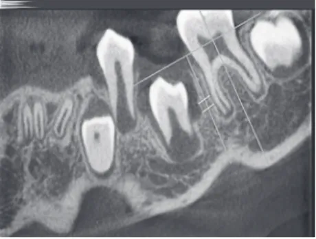
The images obtained were analyzed by Kodak den-tal imaging sotware (3D module V 2.2). At the time of measurements, scanned images were oriented in all three planes: sagittal, axial and coronal. In the posterior inter-radicular areas of the mandible, the sagittal slice was used to locate the inter-radicular area of interest for measure-ments (Fig 1). The vertical reference plane was made par-allel to the long axes of the roots, and the horizontal refer-ence plane was marked along the cementoenamel junc-tion (CEJ) of permanent mandibular irst molar10 (Fig 2).
Measurements were carried out at 6 and 8 mm apical to the cementoenamel junction. Mesiodistal bone width in the mandibular irst molar region was measured in sagit-tal slice (Fig 3) whereas the thickness of the buccal corti-cal plate (Fig 4) and buccolingual bone thickness or depth was measured in the areas between the second premolar and irst molar in the coronal slice (Fig 5).
STATISTICAL ANALYSIS
RESULTS
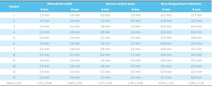
Of the 15 images obtained, three were discarded due to poor image quality. Mean and standard deviation for each of the variables were calculated. Mesiodistal bone width measurements at 6 mm and 8 mm ranged from 1.3 to 2.9 mm. Results for buccal cortical plate thick-ness and buccolingual bone depth ranged between 1.5 - 2.9 mm and 11.9 - 15.4 mm, respectively. Mean val-ues for mesiodistal bone width, buccal cortical plate thick-ness and buccolingual bone depth at 8 mm were found to be suicient for miniscrews placement with a diameter of 1.2 - 1.4 mm and length of 10 - 14 mm (Table 1).
Diferences in measurement at 6 and 8 mm for buccal cortical plate thickness (P < 0.05) and bucco-lingual bone thickness (P < 0.05) were found to be
signiicant, whereas for mesiodistal width it was insig-niicant (P > 0.05) (Table 2). The error of the method (EM) for mesiodistal bone width, buccal and palatal cortical plate thickness and buccopalatal bone depth measurements were found to be 0.40, 0.58 and 0.48, respectively (Table 3).
DISCUSSION
Miniscrews13-17 are now frequently used for
estab-lishing absolute anchorage for orthodontic tooth move-ment. They are easily inserted and removed without a mucoperiosteal lap, and can be loaded immediately ater insertion.18 Their potential applications include
improving anchorage, increasing the horizontal com-ponent of force applied during space closure, posterior
Figure 1 - Sagittal, coronal and axial slices. Figure 2 - Vertical and horizontal reference plane.
Figure 4 - Buccal cortical bone thickness at
6 mm and 8 mm.
Figure 3 - Mesiodistal bone width at 6 and 8 mm.
Figure 5 - Buccolingual bone depth at
Table 1 - Mean and standard deviation for mesiodistal bone width, buccal cortical plate thickness and buccolingual bone depth measurements.
Table 2 - Paired t-test for mesiodistal bone width, buccal and palatal cortical plate thickness and buccolingual bone depth measurements.
Table 3 - Error of the method for mesiodistal bone width, buccal and palatal cortical plate thickness and buccolingual bone depth measurements at 6 and 8 mm.
Patient Mesiodistal width Buccal cortical plate Buccolingual bone thickness
6 mm 8 mm 6 mm 8 mm 6 mm 8 mm
1 2.3 mm 2.6 mm 2.0 mm 2.9 mm 13.1 mm 13.7 mm
2 2.7 mm 2.6 mm 1.5 mm 2.6 mm 11.9 mm 12.1 mm
3 1.3 mm 2.2 mm 1.9 mm 1.9 mm 15.4 mm 15.4 mm
4 2.3 mm 2.8 mm 1.8 mm 2.4 mm 12.4 mm 12.8 mm
5 2.4 mm 2.9 mm 2.1 mm 2.5 mm 13.5 mm 14.8 mm
6 1.9 mm 2.6 mm 1.9 mm 2.2 mm 12.8 mm 13.1 mm
7 2.0 mm 2.8 mm 1.8 mm 2.6 mm 13.4 mm 15.1 mm
8 2.1 mm 2.7 mm 2.2 mm 2.7 mm 13.6 mm 14.3 mm
9 1.9 mm 2.4 mm 1.9 mm 2.5 mm 13.4 mm 13.7 mm
10 2.4 mm 2.8 mm 1.8 mm 2.3 mm 13.1 mm 13.5 mm
11 2.3 mm 2.5 mm 2.0 mm 2.0 mm 12.9 mm 12.5 mm
12 2.2 mm 2.5 mm 1.5 mm 2.4 mm 12.7 mm 12.8 mm
Mean ± SD 2.15 ± 0.49 2.48 ± 0.33 1.73 ± 0.41 2.42 ± 0.34 13.18 ± 1.22 13.65 ± 1.25
Mesiodistal width Buccal cortical plate thickness Buccolingual depth
T-test value 1.76 3.37 2.51
P value 0.13 (N.S., P > 0.05) 0.021 (Sig, P < 0.05) 0.044 (Sig, P < 0.05)
Mesiodistal width Buccal cortical plate thickness Buccolingual depth
Error of the method 0.40 0.58 0.48
intrusion in open-bite cases, distalization of molars, ex-trusion of impacted teeth, molar uprighting and correc-tion of midline diastema.7,8,18
The mandibular buccal region had the thickest cortical bone of all evaluated regions. Thicker cor-tical bone has been previously reported in the man-dible than in the maxilla.12,19 Increased cortical bone
thickness and higher bone mineral density have been shown in the mandibular buccal region when com-pared to the maxillary buccal and lingual regions,20-23
as the mandible is found to be always under torsional and bending strains or forces, whereas the maxilla is generally subjected to more compressive forces.24 Also,
in animal experiments, it has been demonstrated that regions which experience higher strain during func-tion develop thicker cortical bones.25
Thus, in humans, cortical bone in the mandibu-lar buccal region was found to be thicker posteriorly, and it becomes progressively thinner anteriorly.12,26
This pattern might also be explained by the higher
functional demands placed on posterior teeth. Van Ei-jden24 reported an increase in the longitudinal elastic
modulus (increase in stress per unit of strain) between the molar region and the symphysis. Stress and strain diferences could give rise to the diferences in cortical thickness in this region.
Age-related differences between younger, ado-lescent and older patients in cortical bone thickness might be explained by changes in functional capac-ity, because maximum bite forces, masticatory mus-cle size, and musmus-cle activity have the tendency to in-crease with age. Changes in the functional capacity, which alter biomechanical stresses and strains, have shown to manipulate cortical bone thickness and bone density because increased strains and stresses within a certain limit increase cortical bone thickness and bone mineral density.10
molar and first premolar and first premolar and ca-nine. These sites provide moderate inter-radicular space and sufficient cortical plate thickness. However, due to root proximity, the area suitable for miniscrew insertion is over 8 mm from the alveolar crest.6
In this study, the CEJ was selected as the start-ing point for measurements, as compared to other studies in which alveolar crest was used, which could be affected by different periodontal problems.27,28
The maximum level of measurement in this study was selected to be 6 and 8 mm from CEJ because miniscrew placement is most commonly advised in the area of attached gingiva.29
The selection of proper miniscrew diameter and length is important as it may hamper eruption or de-lect erupting premolars during mixed dentition. Hence, selection will depend upon inter-radicular mesiodistal bone width, buccal cortical plate thickness and bucco-lingual bone depth.28 Currently, most miniscrews have
diameters ranging from 1.2 to 2 mm. Presently, there are no relative data available on the amount of bone that is to be present between miniscrews and dental roots for both periodontal health and miniscrew stability. Considering that the width of the periodontal ligament is approxi-mately 0.25 mm, it is assumed that a minimum clear-ance of 1 mm of alveolar bone around the screw could be suicient for periodontal health.6,28 Combining this value
with our data, the safe zone for a miniscrew 1.2 mm in diameter, placed in the inter-radicular spaces have been identiied to be at 8 mm.
Radiographic analysis is a pre-requirement in determining anatomic sites for implant placement. Three-dimensional imaging techniques, such as CT or MRI imaging, have turned into important diag-nostic imaging in the head and neck.30 CT involves
a considerably higher radiation dose31 in comparison
to conventional radiography, as well as high working costs and considerable investment in equipment.32
Digital volume tomograph (DVT) is a new imaging technique which produces three-dimensional im-ages similar to CT, but at a low radiation dose which is comparable with panoramic radiograph, and at a lesser cost. DVT technology in clinical practice has numerous advantages, such as image accuracy, rap-id scan time and display modes which are unique to maxillofacial imaging. Three-dimensional volu-metric tomograph is also well suited for imaging the craniofacial area because it provides clear images of highly contrasted structures which are extremely useful for evaluating bone.33,34 Hence, in this study,
three-dimensional digital volumetric tomograph (DVT) was used to assess mesiodistal bone width, cortical bone thickness and buccolingual bone depth.
In the mandibular molar region, mini-implants placement between premolars is not recommended due to the presence of mental foramen.29 Hence, the
prox-imity of the mental foramina and bone density in the posterior region needs to be assessed in mixed denti-tion in order to provide a three-dimensional analysis for miniscrew placement. However, the results of the present study need to be correlated with clinical assess-ment so as to maintain optimum periodontal health and miniscrew stability.
CONCLUSION
Ater evaluating the amount of bone thickness in the inter-radicular spaces of the mandibular posterior region, the results of the present study show promising evidence for safe miniscrews placement in the mixed dentition period. This results need to be reevaluated in a larger scale.
1. Guidelines on the use of space maintainers following premature loss of primary teeth. J Can Dent Assoc. 1997;63(10):753-66.
2. Laing E, Ashley P, Naini FB, Gill DS. Space maintenance. Int J Paediatr Dent. 2009;19(3):155-62.
3. Ngan P, Randy GA, Fields JRH. Management of space problems in the primary and mixed dentitions. J Am Dent Assoc, 1999;130:1330-9.
4. Dincer M, Haydar S, Unsal B, Turk TS. Space maintainer efects on intercanine arch width and length. J Clin Pediatr Dent. 1996;21(1):47-50.
5. Cuoghi OA, Bertoz FA, de Mendonca MR, Santos EC. Loss of space and dental arch length after the loss of the lower irst primary molar: A longitudinal study. J Clin Pediatr Dent 1998;22(2):117-20.
6. Paola MP, Cristina I, Sefano V, Aldo C. Safe Zones: a guide for miniscrew positioning in the maxillary and mandibular arch. Angle Orthod. 2006;76(2):191-7. 7. Carano A, Velo S, Leone P, Siciliani G. Clinical applications of the Miniscrew
Anchorage System. J Clin Orthod. 2005;39(1):9-24.
8. Mark R, Yanosky, Holmes JD. Mini-implant temporary anchorage devices: orthodontic applications. Compend. 2008;29(1):12-20.
9. Kim SH, Yoon HG. Evaluation of interdental space of the maxillary posterior area for orthodontic mini-implants with cone-beam computed tomography. Am J Orthod Dentofacial Orthop. 2009;135(5):635-41.
10. Farnsworth D, Rossouw PE, Ceen RF, Buschangd PH. Cortical bone thickness at common miniscrew implant placement sites. Am J Orthod Dentofacial Orthop. 2011;139(4):495-503.
11. Kang S, Lee SJ, Ahn SJ, Heo MS, Kim TW. Bone thickness of the palate for orthodontic mini-implant anchorage in adults. Am J Orthod Dentofacial Orthop. 2007;131(4 Suppl):S74-81.
12. Schwartz CL, Dechow PC. Variations in cortical material properties throughout the human dentate mandible. Am J Phys Anthropol. 2003;120(3):252-77. 13. Costa A, Rafainl M, Melsen B. Miniscrews as orthodontic anchorage: a
preliminary report. Int J Adult Orthodon Orthognath Surg. 1998;13(3):201-9. 14. Kanomi R. Mini-implant for orthodontic anchorage. J Clin Orthod. 1997;31:763-7. 15. Park HS, Bae SM, Kyung HM, Sung JH. Micro-implant anchorage for treatment of
skeletal Class I bialveolar protrusion. J Clin Orthod. 2001;35(7):417-22. 16. Paik CH, Woo YJ, Boyd R. Treatment of an adult patient with vertical maxillary
excess using miniscrew ixation. J Clin Orthod. 2003;37(8):423-8. 17. Xun CL, Zeng XL, Wang X. Clinical application of miniscrew implant for
maximum anchorage treatment. Chin J Stomatol. 2004;39:505-8.
18. Fortini A, Cacciafesta V, Sfondrini MF, Cambi S, Lupoli M. Clinical applications and eiciency of miniscrews for extradental anchorage. Orthodontics. 2004;1(2):1-12. 19. Peterson J, Wang Q, Dechow PC. Material properties of the dentate maxilla. Anat
Rec A Discov Mol Cell Evol Biol. 2006;288(9):962-72.
REFERENCES
20. Ono A, Motoyoshi M, Shimizu N. Cortical bone thickness in the buccal posterior region for orthodontic mini-implants. Int J Oral Maxillofac Surg. 2008;37(4):334-40
21. Deguchi T, Nasu M, Murakami K, Yabuuchi T, Kamioka H, Takano-Yamamoto T. Quantitative evaluation of cortical bone thickness with computed tomographic scanning for orthodontic implants. Am J Orthod Dentofacial Orthop. 2006;129(6):721.e7-12.
22. Park HS, Lee YJ, Jeong SH, Kwon TG. Density of the alveolar and basal bones of the maxilla and the mandible. Am J Orthod Dentofacial Orthop. 2008;133(1):30-7.
23. Mitsuru M. Clinical indices for orthodontic mini-implants. J Oral Sci. 2011;3(4):407-12.
24. Van Eijden TM. Biomechanics of the mandible. Crit Rev Oral Biol Med. 2000;11(1):123-36.
25. Daegling DJ, Hylander WL. Experimental observation, theoretical models, and biomechanical inference in the study of mandibular form. Am J Phys Anthropol. 2000;112(4):541-51.
26. Katranji A, Misch K, Wang HL. Cortical bone thickness in dentate and edentulous human cadavers. J Periodontol. 2007;78(5):874-8.
27. Fayed MM, Pazera P, Katsaros C. Optimal sites for orthodontic mini-implant placement assessed by cone beam computed tomography. Angle Orthod. 2010;80(5):939-51.
28. Monnerat C, Restle L, Mucha JN. Tomographic mapping of mandibular interradicular spaces for placement of orthodontic mini-implants. Am J Orthod Dentofacial Orthop. 2009;135(4):428.e1-9; discussion 428-9.
29. Melsen B. Mini-implants: where are we? J Clin Orthod 2005;39:539-47. 30. Fuhrmann R, Klein HM, Wehrbein H, GuÈ nther RW, Dietrich P. Hochauo È
sende computertomographie fazialer und oraler knochendehiszenzen. Dtsch ZahnaÈrztl Z. 1993;48:242-6.
31. Hassfeld S, Streib S, Stahl H, Stratmann U, Fehrentz D, ZoÈ ller J. Low-dose-computertomographie des kieferknochens in derpra È implantologischen Diagnostik. Mund Kiefer Gesichts Chir. 1998;2:188-93.
32. Arai Y, Tammisalo E, Iwai K, Hashimoto K, Shinoda K. Development of a compact computed tomographic apparatus for dental use. Dentomaxillofac Radiol. 1999;28(4):245-8.
33. Ziegler CM, Woertche R, Brief J, Hassfeld S. Clinical indications for digital volume tomography in oral and maxillofacial surgery. Dentomaxillofac Radiol. 2002;31(2):126-30.