Reproducibility of bone plate thickness
measurements with Cone-Beam Computed
Tomography using different image
acquisition protocols
Carolina Carmo de Menezes*, Guilherme Janson**, Camila da Silveira Massaro***, Lucas Cambiaghi***, Daniela G. Garib****
Introduction: A smaller voxel dimension leads to greater resolution of Cone-Beam Computed Tomography (CBCT), but a greater dosage of radiation is emitted. Objec-tive: Assess and compare the reproducibility of buccal and lingual bone plate thickness measurements in CBCT images using different image acquisition protocols, with varia-tions in the voxel dimension. Methods: CBCT exams were taken of 12 dried human mandibles with voxel dimensions of 0.2, 0.3 and 0.4 mm using the i-CAT Cone-Beam 3-D Dental Imaging System. The thickness of the buccal and lingual bone plates was measured, with the i-CAT Vision software, on an axial section passing 12 mm above the right mental foramen. Intra-examiner and inter-examiner reproducibility was assessed using the paired t-test and independent t-test, respectively, with the level of significance set at 5%. Results: Excellent inter-examiner reproducibility was observed for the three protocols analyzed. Intra-examiner reproducibility was very good, with the exception of some regions of the anterior teeth, which exhibited statistically significant differences regardless of the voxel dimensions. Conclusion: The measurement of buccal and lingual bone plate thickness on CBCT images demonstrated good precision for voxel dimen-sions of 0.2, 0.3 and 0.4 mm. The reproducibility of the measurements of the anterior region of the mandible was more critical than that of the posterior region.
Abstract
Keywords: Cone-Beam Computed Tomography. Alveolar bone. Reproducibility.
* Master’s Student, Program of Applied Oral Science, Major in Orthodontics, Bauru Dental School, University of São Paulo, Brazil. ** Undergraduate Student, Bauru Dental School, University of São Paulo, Brazil.
*** Professor of Orthodontics and Head of the Department of Pediatric Dentistry, Orthodontics and Community Dentistry, Bauru Dental School, University of São Paulo, Brazil.
IntROduCtIOn
A correct and precise diagnosis and treatment plan are fundamental for the success of orthodon-tic treatment. With the advent of Cone-Beam Computed Tomography (CBCT), orthodontists are able to obtain all the two-dimensional im-ages (2D) that compose the orthodontic docu-mentation during a single exam with the same precision of conventional radiographs, along with a detailed view of dentofacial structures.1,8,9
CBCT offers images of the labial/buccal and lingual bone plates, which are not apparent in conventional two-dimensional x-rays due to im-age superimposition.4 Tooth movements in the
buccolingual direction may cause bone dehis-cence, as documented in studies involving ani-mals and humans.17,18 That constitutes a concern
regarding the long-term periodontal integrity. Moreover, many patients, especially adults, may exhibit bone dehiscence prior to orthodontic treatment, which requires the orthodontist to plan more parsimonious dental movements.6,19
Facial type has an effect on the thickness of the alveolar bone. Patients with a horizontal growth pattern have a greater buccolingual dimension of the alveolar ridge in comparison to hyperdi-vergent patients.6 Thus, the morphology of the
alveolar bone is one of the limiting factors of orthodontic movements.6
Previous studies have validated CBCT for quantitative analyses, demonstrating its highly precise measurements.2 Measurement precision
is related to the resolution of the image.11 The
spatial resolution of CBCT, in turn, depends upon the voxel dimension, which is the low-est image unit. A smaller voxel dimension leads to greater image resolution,14 but also a higher
dose of radiation.3
A number of studies have demonstrated the precision of linear measurements performed on CBCT images.7,10,11,12,15 However, the influence
of the voxel dimension on measurement preci-sion of delicate structures, such as the buccal
and lingual bone plates, has yet to be demon-strated. Thus, the aim of the present study was to assess and compare the reproducibility of buccal and lingual bone plate thickness mea-surements in CBCT images using different im-age acquisition protocols with variations in the voxel dimension.
MAtERIALS And MEtHOdS
Twelve dried human mandibles with perma-nent dentition were selected from the Anatomy Department of the Bauru Dental School, Uni-versidade de São Paulo, Brazil. CBCT scans were performed on each specimen using the i-CAT Cone-Beam 3-D Dental Imaging System (USA). Each mandible was embedded in a cube of no. 7 dental wax with water and detergent in order to simulate the density of the soft tissue. The base of the mandible was directly supported on the floor of the box and parallel to the ground. The following image acquisition protocols were used for each specimen:
1. Protocol 1: Field of view (FOV) of 8 cm, 120 kVp, 36.12 mAs, 0.2-mm voxel, 40-second scan time
2. Protocol 2: FOV of 8 cm, 120 kVp, 18.45 ma, 0.3-mm voxel, 20-second scan time 3. Protocol 3: FOV of 8 cm, 120 kVp, 18.45
ma, 0.4-mm voxel, 20-second scan time The difference between protocols was essen-tially the voxel dimension, which is the small-est unit of the tomographic image. Thirty-six CBCT scans were performed, composing the overall sample.
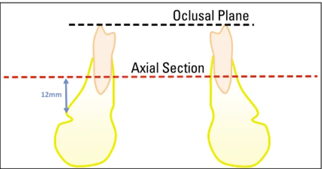
12mm
Due to the variation in the morphology of the mandibles analyzed, the cursor was moved more or less than 12 mm on some specimens in order to reach the region between the middle and api-cal thirds of the tooth roots.
On the axial section, the thickness of the labial/buccal and lingual bone plates was sured on all permanent teeth (Fig 2). The mea-surement extended from the external limit of the root to the external limit of the cortical bone, perpendicular to the contour of the dental arch on both sides (Fig 3).
The measurements were performed by two previously calibrated examiners. The first exam-iner repeated the measurements after an interval of at least 15 days. Statistical analysis involved the calculation of mean and standard deviation values of the labial/buccal and lingual bone plate thickness measurements for each tooth group (incisors, canines, premolars and molars). Paired t-tests were used for the intra-examiner comparison and the independent t-tests were used for the inter-examiner comparison, with the significance level of 5%.
RESuLtS
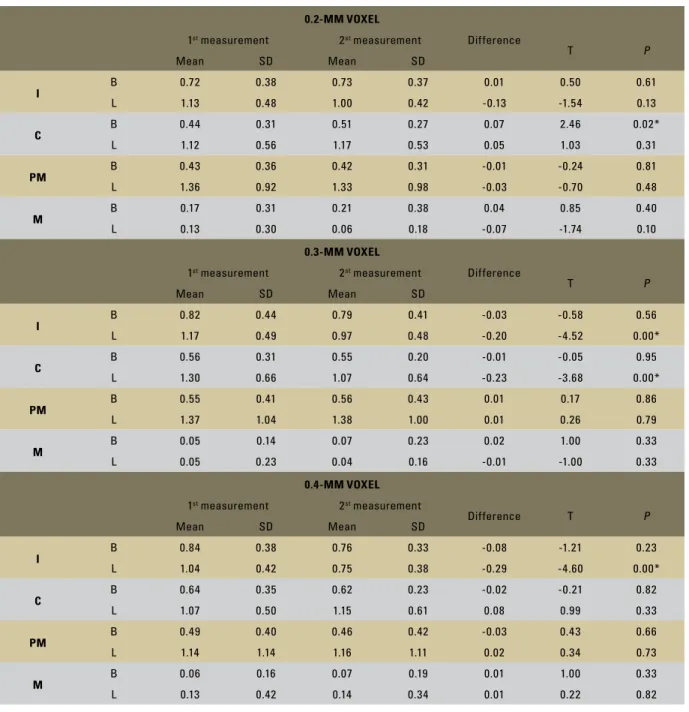
Table 1 displays the mean and standard devia-tion values for the measurements of labial/buccal and lingual bone plate thickness, along with the results of the intra-examiner comparison. There were statistically significant differences between the first and the second measurements for a sin-gle area using the 0.2-mm voxel protocol (buccal canine surface), for two areas using the 0.3-mm voxel protocol (lingual surface of incisors and canines) and for a single area using the 0.4-mm voxel protocol (lingual surface of incisors).
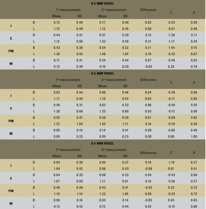
Table 2 shows the mean and standard devia-tion values for the measurements of buccal and lingual bone plate thickness, along with the re-sults of the inter-examiner comparison. No sta-tistically significant differences were found be-tween the measurements of the two examiners.
FIGURE 1 - Frontal reconstruction showing the right mental foramen used as reference to define the axial section for taking the measurements. The axial section passing an average of 12 mm above the superior border of the right mental foramen was used.
FIGURE 2 - Schematic representation of buccal and lingual bone plate thickness measurements in the selected axial section.
FIGURE 3 - Buccal and lingual bone plate thickness measurements in the axial section of one specimen (0.2-mm voxel).
Oclusal Plane
TABLE 1 - Intra-examiner comparison for buccal and lingual bone plate thickness measurements (in millimeters) on CBCT images with voxel dimensions of 0.2, 0.3 and 0.4 mm.
I: incisors; C: canines; PM: premolars; M: molars; B: buccal bone plate; L: lingual bone plate; * p < 0.05.
0.2-mm Voxel
1st measurement 2st measurement Difference
T P
Mean SD Mean SD
I
B 0.72 0.38 0.73 0.37 0.01 0.50 0.61
L 1.13 0.48 1.00 0.42 -0.13 -1.54 0.13
C
B 0.44 0.31 0.51 0.27 0.07 2.46 0.02*
L 1.12 0.56 1.17 0.53 0.05 1.03 0.31
Pm
B 0.43 0.36 0.42 0.31 -0.01 -0.24 0.81
L 1.36 0.92 1.33 0.98 -0.03 -0.70 0.48
m
B 0.17 0.31 0.21 0.38 0.04 0.85 0.40
L 0.13 0.30 0.06 0.18 -0.07 -1.74 0.10
0.3-mm Voxel
1st measurement 2st measurement Difference
T P
Mean SD Mean SD
I
B 0.82 0.44 0.79 0.41 -0.03 -0.58 0.56
L 1.17 0.49 0.97 0.48 -0.20 -4.52 0.00*
C
B 0.56 0.31 0.55 0.20 -0.01 -0.05 0.95
L 1.30 0.66 1.07 0.64 -0.23 -3.68 0.00*
Pm
B 0.55 0.41 0.56 0.43 0.01 0.17 0.86
L 1.37 1.04 1.38 1.00 0.01 0.26 0.79
m
B 0.05 0.14 0.07 0.23 0.02 1.00 0.33
L 0.05 0.23 0.04 0.16 -0.01 -1.00 0.33
0.4-mm Voxel
1st measurement 2st measurement
Difference T P
Mean SD Mean SD
I B 0.84 0.38 0.76 0.33 -0.08 -1.21 0.23
L 1.04 0.42 0.75 0.38 -0.29 -4.60 0.00*
C B 0.64 0.35 0.62 0.23 -0.02 -0.21 0.82
L 1.07 0.50 1.15 0.61 0.08 0.99 0.33
Pm B 0.49 0.40 0.46 0.42 -0.03 0.43 0.66
L 1.14 1.14 1.16 1.11 0.02 0.34 0.73
m B 0.06 0.16 0.07 0.19 0.01 1.00 0.33
L 0.13 0.42 0.14 0.34 0.01 0.22 0.82
dISCuSSIOn
Considering the increasing applicability of CBCT in Dentistry, it is very important to de-termine an image acquisition protocol capable of providing a three-dimensional view with the ap-propriate resolution to measure small structures,
such as buccal and lingual bone plates. A smaller voxel dimension leads to greater spatial reso-lution of the image, but also emits a greater amount of radiation.3 In other words, the voxel
TABLE 2 - Inter-examiner comparison for buccal and lingual bone plate thickness measurements (in millimeters) on CBCT images with voxel dimensions of 0.2, 0.3 and 0.4 mm.
I: incisors; C: canines; PM: premolars; M: molars; B: buccal bone plate; L: lingual bone plate; * p < 0.05.
0.2-mm Voxel
1st measurement 2st measurement Difference
T P
Mean SD Mean SD
I
B 0.72 0.40 0.77 0.40 0.05 -0.53 0.59
L 1.13 0.48 1.13 0.45 0.00 -0.01 0.98
C
B 0.44 0.31 0.57 0.29 0.13 -1.38 0.17
L 1.12 0.56 1.33 0.59 0.21 -1.17 0.24
Pm
B 0.43 0.36 0.54 0.32 0.11 -1.44 0.15
L 1.36 0.92 1.46 1.04 0.10 -0.42 0.67
m
B 0.17 0.31 0.24 0.44 0.07 -0.48 0.62
L 0.13 0.30 0.10 0.29 -0.03 0.26 0.79
0.3-mm Voxel
1st measurement 2st measurement Difference
T P
Mean SD Mean SD
I
B 0.82 0.44 0.86 0.46 0.04 -0.39 0.69
L 1.17 0.49 1.19 0.54 0.02 -0.17 0.85
C
B 0.56 0.31 0.62 0.33 0.06 -0.59 0.55
L 1.30 0.66 1.33 0.60 0.03 -0.13 0.89
Pm
B 0.55 0.41 0.56 0.39 0.01 -0.09 0.92
L 1.37 1.04 1.55 1.11 0.18 -0.70 0.48
m
B 0.05 0.14 0.14 0.41 0.09 -0.85 0.40
L 0.05 0.23 0.05 0.23 0.00 0.00 1.00
0.4-mm Voxel
1st measurement 2st measurement
Difference T P
Mean SD Mean SD
I B 0.84 0.38 0.94 0.37 0.10 -1.10 0.27
L 1.04 0.42 0.96 0.43 -0.08 0.81 0.41
C B 0.64 0.35 0.68 0.33 0.04 -0.43 0.66
L 1.07 0.50 1.17 0.61 0.10 -0.56 0.57
Pm B 0.46 0.40 0.43 0.41 -0.03 0.33 0.73
L 1.14 1.14 1.23 1.65 0.09 -0.33 0.73
m B 0.06 0.16 0.03 0.14 -0.03 0.45 0.65
L 0.13 0.42 0.15 0.44 0.02 -0.14 0.88
submitted during the procedure. Thus, before se-lecting the image acquisition protocol, it is nec-essary to determine its cost-benefit ratio based on the ALARA principle (as low as reasonably achievable dose of radiation), in which the pro-fessional chooses the scanning protocol with the
lowest possible radiation dose, but with sufficient resolution for the identification of the structures to be assessed.
acquisition protocol. Thus, the aim of the pres-ent study was to compare the reproducibility of thickness measurements of the buccal and lin-gual bone plates of permanent teeth in CBCT images with different voxel dimensions (0.2, 0.3 and 0.4 mm). The results revealed statisti-cally significant differences in the intra-examin-er comparison in some regions of the antintra-examin-erior teeth (Table 1). This corroborates the findings of previous studies. Tsunori et al16 have
mea-sured the buccal, lingual and basal cortical bone thickness as well as the buccolingual width and height of the alveolar ridge using CBCT of 39 dry skulls and found few significant differences between the first and second measurements by a single examiner.16
Mol and Balasundaram13 analyzed the
preci-sion of measurements of bone dehiscence using CBCT on five dry skulls. The authors compared measurements performed by six examiners us-ing CBCT, conventional radiographs and the anatomic specimens and concluded that CBCT achieved the greatest diagnostic precision of the three methods. However, the authors found that the region of the mandibular anterior teeth showed less precision in comparison to other ar-eas and concluded that the mar-easurement of bone dehiscence in the anterior region is more limited with the NewTom 9000 scanner.13
In the present study, significant intra-exam-iner differences were found in the region of the anterior teeth (incisors and canines) although the differences between the first and second measurements did not surpass 0.30 mm (Table 1). The measurements of the bone plates in the posterior region were highly precise. It is likely that the difference in the reproducibility of the measurements between anterior and posterior teeth is due to the fact that the thickness of the bone plates is thinner in the anterior region compared with the posterior region. A thinner bone plate has less image resolution, decreas-ing the precision of linear measurements.14
This limitation of computed tomography may be due to the property denominated “partial volume averaging”; when the limit between two tissues is in the middle of a voxel, its density corresponds to the average density of the two structures it en-compasses.14 These results are in agreement with
those described by Mol and Balasundaram13,
who found less accuracy in the measurement of buccal bone dehiscence in the anterior region of the mandible in comparison with the posterior region on images generated with the NewTom 9000 scanner.Using helical computed tomogra-phy, Fuhrman found that only bone plates with a thickness of less than 0.2 mm were not apparent on the exam.5 To date, no studies have indicated
the least bone plate thickness that can be identi-fied on CBCT images.
In 2008, Loubele et al10 performed linear
measurements of the buccolingual diameter of the alveolar ridge at previously marked points on an human maxilla comparing CBCT with heli-cal CT and found no significant inter-examiner differences.The present study corroborates this finding, as inter-examiner reproducibility was ex-cellent (Table 2).
Based on the results of the present study, the measurement of bone plate thickness proved to have similar reproducibility in the different im-age acquisition protocols, although the 0.2 mm voxel protocol has produced sharper images than the 0.3 and 0.4 mm voxel protocols. Further studies should be carried out to determine the accuracy of bone plate thickness measurements using CBCT images.
COnCLuSIOn
Contact address
Daniela G. Garib
Av. José Affonso Aiello 6-100 CEP: 17.018-520 – Bauru / SP, Brazil E-mail: [email protected]
1. Baumgaertel S, Hans MG. Buccal cortical bone thickness for mini-implant placement. Am J Orthod Dentofacial Orthop. 2009 Aug;136(2):230-5.
2. Cevidanes LH, Franco AA, Scanavini MA, Vigorito JW, Enlow
DH, Profit WR. Clinical outcomes of Fränkel appliance therapy
assessed with a counterpart analysis. Am J Orthod Dentofacial Orthop. 2003 Apr;123(4):379-87.
3. Farman AG, Scarfe WC. Development of imaging selection criteria and procedures should precede cephalometric assessment with cone-beam computed tomography. Am J Orthod Dentofacial Orthop. 2006 Aug;130(2):257-65. 4. Fuhrmann RA, Bücker A, Diedrich PR. Furcation involvement:
comparison of dental radiographs and HR-CT-slices in human specimens. J Periodontal Res. 1997 Jul;32(5):409-18. 5. Fuhrmann RA, Wehrbein H, Langen HJ, Diedrich PR.
Assessment of the dentate alveolar process with high resolution computed tomography. Dentomaxillofac Radiol. 1995 Feb;24(1):50-4.
6. Gracco A, Lombardo L, Mancuso G, Gravina V, Siciliani G. Upper incisor position and bony support in untreated patients as seen on CBCT. Angle Orthod. 2009 Jul;79(4):692-702. 7. Howerton WB Jr, Mora MA. Advancements in digital imaging:
What is new and on the horizon? J Am Dent Assoc. 2008 Jun;139 Suppl:20S-24S.
8. Lamichane M, Anderson NK, Rigali PH, Seldin EB, Will LA. Accuracy of reconstructed images from cone-beam computed tomography scans. Am J Orthod Dentofacial Orthop. 2009 Aug;136(2):156.e1-6.
9. Loubele M, Maes F, Schutyser F, Marchal G, Jacobs R, Suetens P. Assessment of bone segmentation quality of cone-beam CT versus multislice spiral CT: a pilot study. Oral Surg Oral Med Oral Pathol Oral Radiol Endod. 2006 Aug;102(2):225-34. 10. Loubele M, Van Assche N, Carpentier K, Maes F, Jacobs R, van
Steenberghe D, et al. Comparative localized linear accuracy of
small-ield cone-beam CT and multislice CT for alveolar bone
measurements. Oral Surg Oral Med Oral Pathol Oral Radiol Endod. 2008 Apr;105(4):512-8.
REfEREnCES
11. Ludlow JB, Laster WS, See M, Bailey LJ, Hershey HG. Accuracy of measurements of mandibular anatomy in cone beam computed tomography images. Oral Surg Oral Med Oral Pathol Oral Radiol Endod. 2007 Apr;103(4):534-42. 12. Misch KA, Yi ES, Sarment DP. Accuracy of Cone Beam
Computed Tomography for periodontal defect measurements. J Periodontol. 2006 Jul;77(7):1261-6.
13. Mol A, Balasundaram A. In vitro cone beam computed tomography imaging of periodontal bone. Dentomaxillofac Radiol. 2008 Sep;37(6):319-24.
14. Molen AD. Considerations in the use of cone-beam computed tomography for buccal bone measurements. Am J Orthod Dentofacial Orthop. 2010 Apr;137(4 Suppl):S130-5. 15. Stavropoulos A, Wenzel A. Accuracy of cone beam dental
CT, intraoral digital and conventional ilm radiography for the
detection of periapical lesions. An ex vivo study in pig jaws. Clin Oral Investig. 2007 Mar;11(1):101-6.
16. Tsunori M, Mashita M, Kasai K. Relationship between facial types and tooth and bone characteristics of the mandible obtained by CT scanning. Angle Orthod. 1998 Dec;68(6):557-62.
17. Wehrbein H, Bauer W, Diedrich P. Mandibular incisors, alveolar bone, and symphysis after orthodontic treatment. A retrospective study. Am J Orthod Dentofacial Orthop. 1996 Sep;110(3):239-46.
18. Wennström JL, Lindhe J, Sinclair F, Thilander B. Some periodontal tissue reactions to orthodontic tooth movement in monkeys. J Clin Periodontol. 1987 Mar;14(3):121-9.
19. Yamada C, Kitai N, Kakimoto N, Murakami S, Furukawa S, Takada K. Spatial relationships between the mandibular central incisor and associated alveolar bone in adults with mandibular prognathism. Angle Orthod. 2007 Sep;77(5):766-72.