Immediate periodontal bone plate changes induced by rapid
maxillary expansion in the early mixed dentition: CT findings
Daniela Gamba Garib1, Maria Helena Ocké Menezes2, Omar Gabriel da Silva Filho3, Patricia Bittencourt Dutra dos Santos4
Objective:This study aimed at evaluating buccal and lingual bone plate changes caused by rapid maxillary expansion (RME) in the mixed dentition by means of computed tomography (CT). Methods: The sample comprised spiral CT exams taken from 22 mixed dentition patients from 6 to 9 years of age (mean age of 8.1 years) presenting constricted maxillary arch treated with Haas-type expanders. Patients were submitted to spiral CT scan before expansion and after the screw activation period with a 30-day interval between T1 and T2. Multiplanar reconstruction was used to measure buccal and lingual bone plate thickness and buccal bone crest level of maxillary posterior deciduous and permanent teeth. Changes induced by expansion were evaluated using paired t test (p < 0.05). Results: Thickness of buccal and lingual bone plates of posterior teeth remained unchanged during the expansion period, except for decidu-ous second molars which showed a slight reduction in bone thickness at the distal region of its buccal aspect. Buccal bone dehiscences were not observed in the supporting teeth after expansion. Conclusion: RME performed in mixed dentition did not produce immediate undesirable effects on periodontal bone tissues.
Keywords:Palatal expansion technique. Periodontium. Spiral computed tomography.
How to cite this article: Garib DG, Menezes MHO, Silva Filho OG, Santos PBD. Immediate periodontal bone plate changes induced by rapid maxillary ex-pansion in the early mixed dentition: CT findings. Dental Press J Orthod. 2014 May-June;19(3):36-43. DOI: http://dx.doi.org/10.1590/2176-9451.19.3.036-043.oar
Submitted: April 10, 2012 - Revised and accepted: August 30, 2012 » The authors report no commercial, proprietary or financial interest in the prod-ucts or companies described in this article.
Contact address: Daniela Gamba Garib
Faculdade de Odontologia de Bauru – Al. Octávio Pinheiro de Brisola 9-75 CEP: 17.012-901 – Bauru/SP – Brazil — E-mail: [email protected] » Patients displayed in this article previously approved the use of their facial and
intraoral photographs.
1 Full professor, School of Dentistry — University of São Paulo/Bauru.(FOB-USP).
2 MSc in Orthodontics, University São Paulo, UNICID. 3 MSc in Orthodontics, São Paulo State University (UNESP). 4 PhD resident in Applied Dental Sciences (FOB-USP).
DOI: http://dx.doi.org/10.1590/2176-9451.19.3.036-043.oar
Objetivo:o presente estudo teve como objetivo avaliar alterações das tábuas ósseas vestibulares e linguais decorrentes da expansão rápida da maxila (ERM), em pacientes na dentição mista, por meio de tomografia computadorizada (TC).
Métodos: a amostra foi constituída por exames de TC helicoidal, realizados de 22 pacientes com dentição mista, dos 6 aos 9 anos de idade (média de 8,1 anos), com atresia maxilar, tratados com expansores do tipo Haas. Os pacientes foram submetidos a tomografia computadorizada helicoidal antes da expansão e após o período de ativação de parafuso expansor, com 30 dias de intervalo entre as fases T1 e T2. A reconstrução multiplanar foi usada para medir a espes-sura da tábua óssea vestibular e lingual e a altura da crista óssea alveolar dos dentes posteriores decíduos e dos dentes permanentes. As alterações induzidas pela expansão foram avaliadas usando o teste t pareado (p < 0,05). Resultados:
a espessura das tábuas ósseas vestibular e lingual dos dentes posteriores permaneceu inalterada durante o período de expansão, com exceção dos segundos molares decíduos, que mostraram uma ligeira redução da espessura do osso na região distal. Deiscências ósseas vestibulares não foram observadas nos dentes de suporte após a expansão. Conclusão:
a ERM, realizada na dentição mista, não produziu efeitos imediatos indesejáveis sobre os tecidos ósseos periodontais.
INTRODUCTION
During rapid maxillary expansion, orthopedic efect is produced by midpalatal suture splitting. Addition-ally, a dental efect characterized by buccal movement
of supporting teeth is also produced.19-22,27,33 As a result,
maxillary posterior teeth are buccally displaced by an association of inclination and translation. The litera-ture evinces that buccal tooth movement is associated with the occurrence of bone dehiscences. Engelking
and Zachrisson,15 Steiner et al,28 Thilander et al29 and
Wennströn et al32 conducted animal investigations and
demonstrated that buccal tooth movement with mild forces increases the distance between the
cementoenam-el junction and the buccal alveolar crest. Wehrbein et al31
reached a similar conclusion when conducting a cadav-er study. Buccolingual tooth movement seems to oc-cur through the alveolar bone and not along the bone.
It leads to bone dehiscences in the short-term15,28,29,31,32
and to gingival recession in the long-term.1-4
Recent studies conducted with CT have shown api-cal migration of bucapi-cal alveolar crest of posterior teeth ater RME is performed in permanent dentition
pa-tients.18,25 By means of CT methodology, Garib et al18
assessed a sample of eight female adolescents before RME and ater the removal of the expander follow-ing a 3-month retention period. The authors con-cluded that RME induced bone dehiscences on the buccal aspect of supporting teeth (irst premolars and irst molars), especially in subjects who initially pre-sented thinner buccal bone plate. Rungcharassaeng
et al25 found similar results in a sample of thirty
con-secutive RME patients with a mean age of 13.8 years. Their indings included buccal bone loss in both hori-zontal and vertical dimensions for all posterior teeth ater expansion, and displayed a signiicant correlation with age, amount of expansion and initial buccal bone
thickness. Ballanti et al8 observed, at the end of the
ac-tive expansion phase, a signiicant decrease of buccal bone plate thickness of permanent maxillary irst mo-lars in a sample of 17 prepubertal patients with a mean age of 11.2 years. However, a tendency for partial re-covery was found six months ater expansion.
Buccal bone changes produced by rapid maxillary expansion in the permanent dentition raise the ques-tion about the periodontal efects of RME performed in the early phases of mixed dentition. During decidu-ous and early mixed dentitions, RME produces greater
orthopedic efects14,23 and transfers anchorage to
decidu-ous molars and canines. A classic implant study showed that, in adolescents, skeletal efects corresponded to 35% of expansion, whereas dental efects accounted
for 65%.23 On the other hand, in young children, the
proportion between skeletal and dental efects was 1:1.
Baccetti et al5 also observed that RME performed before
the peak of skeletal maturation produced more skeletal efects than RME performed ater the peak. Thus, the periodontal changes related to the orthodontic efect of RME in the early phases of mixed dentition deserve to be diferentiated from those observed in late mixed den-tition or even in permanent denden-tition. Therefore, the aim of this study was to investigate, by means of spi-ral CT, the periodontal bone changes of RME in early mixed dentition using deciduous teeth as anchorage.
MATERIAL AND METHODS
This study was approved by the School of Dentistry — University of São Paulo/Bauru Institutional Review Board. The sample comprised spiral CT exams taken from 22 orthodontic patients (10 males and 12 females) with mean initial age of 8.1 years (ranging from 6 to 9
years). The exams had been taken for a previous study,13
in 2002, before CBCT was introduced in Brazil. In
se-lecting the sample, the following inclusioncriteria were
applied: patients in early mixed dentition, and the pres-ence of constricted maxillary arch with or without pos-terior crossbites. In the examined sample, all subjects were in the early transitional phase, as described by Van
der Linden,30 and either at stage CS1 or CS2 at the time
of treatment.6In other words, patients were treated
Otawara-Shi, Japan) was used at 120 kV and 100 mA, with a scanning time of one second per section. A FC 30 scanning ilter, ield of view (FOV) of 12.6 x 12.6 cm and matrix of 512 x 512 pixels was used. Window width was 2400 HU with a center of 1300 HU.
For standardization of head positioning in all three planes, the perpendicular light beam resource provided by the machine was used; thereby, allowing
compari-son of images obtained before and ater expansion.18, 19
For that purpose, patients were positioned with Camper’s plane perpendicular to the ground, while the longitudinal light beam passed through the center of the glabella and the iltrum, and the transverse light beam passed through the lateral eye canthus. Teeth were kept apart in order
to avoid imaging the mandibular dental arch. One-milli-meter thick axial sections were performed parallel to the palatal plane, including the dentoalveolar and basal areas of the maxilla, up to the lower third of the nasal cavity. The imaged area encompassed 36 to 40 mm, totalizing 36 to 40 sections. This protocol results in images with a
spacial resolution that ranges from 0.2 to 0.5 mm.17
Data were transferred to a network computer work-station (Silicon Graphics, Toshiba Corporation Medical Systems Company, Otawara-Shi, Japan), using Alato-view sotware (Toshiba Corporation Medical Systems Company, Otawara-Shi, Japan) on which 2D reformat-ted images were generareformat-ted and measured by the comput-erized method. The Allatoview sotware generates a very small ball size pointer for linear measurements.
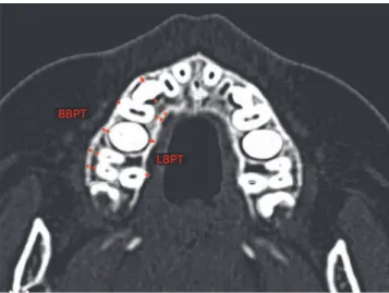
Measurement of alveolar bone plate thickness of max-illary posterior teeth at the buccal and lingual aspects was conducted on two axial sections parallel to the palatal plane, one at the level of right maxillary permanent irst molar furcation and another at the level of right maxil-lary deciduous second molar furcation (Figs 2 and 3). Measurements of alveolar bone thickness were performed on magniied images (4x), before and ater expansion. Whenever tooth rotations were present, bone plate thick-ness was measured at the area where the root was closer to the external contour of the alveolar ridge.
Evaluation of the buccal alveolar bone crest level of maxillary posterior teeth was conducted by means
Figure 1 - Haas-type expander used in this study.
Figure 2 - Measurements of buccal and lingual bone plate thickness (BBPT and LBPT) of permanent erupted and non-erupted posterior teeth were performed in the axial section, parallel to the palatal plane, passing at the level of right maxillary permanent first molar furcation.
Figure 3 - Measurements of buccal and lingual bone plate thickness (BBPT and LBPT) of deciduous posterior teeth were performed in the axial section, parallel to the palatal plane, passing at the level of right maxillary deciduous second molar furcation.
BBPT
BBPT
LBPT
RESULTS
The results of the error of the method are shown in Table 1. Two out of 22 variables had a statistically signii-cant systematic error (LBPT of the permanent irst molars and BABCL at the center of deciduous second molars — as shown in Figures 2 and 4). Casual errors ranged from 0.11 (BBPT at the distal region of permanent irst molars) to 0.52 (BBPT at the mesial region of permanent irst molars).
Buccal and lingual bone plate thickness (BBPT and LBPT)
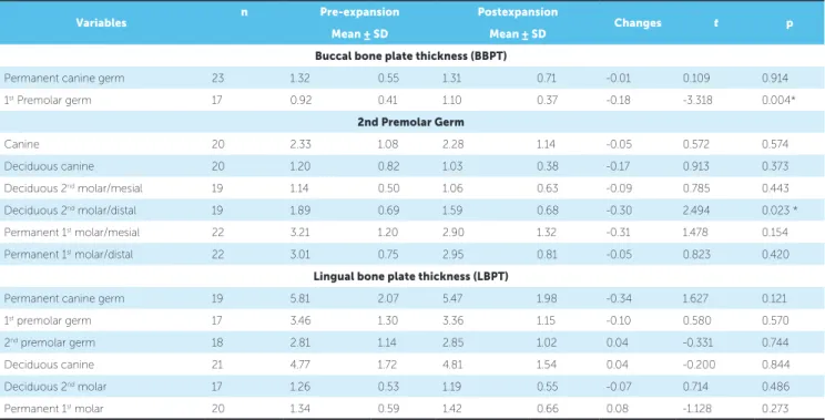
Table 2 shows that buccal bone plate thickness of sup-porting teeth was not signiicantly changed by RME. Additionally, no changes were observed in the buccal bone plate thickness of tooth germs and permanent irst molars (Table 2). The only exceptions were the buccal bone plate of irst premolar germ which showed a sta-tistically signiicant decrease (mean of 0.18 mm), and the buccal bone plate of deciduous second molar which showed a statistically signiicant decrease at the distal re-gion (mean of 0.3 mm).
No changes were observed for the lingual bone plate thickness of deciduous and permanent teeth (Table 2).
Buccal alveolar bone crest level (BABCL)
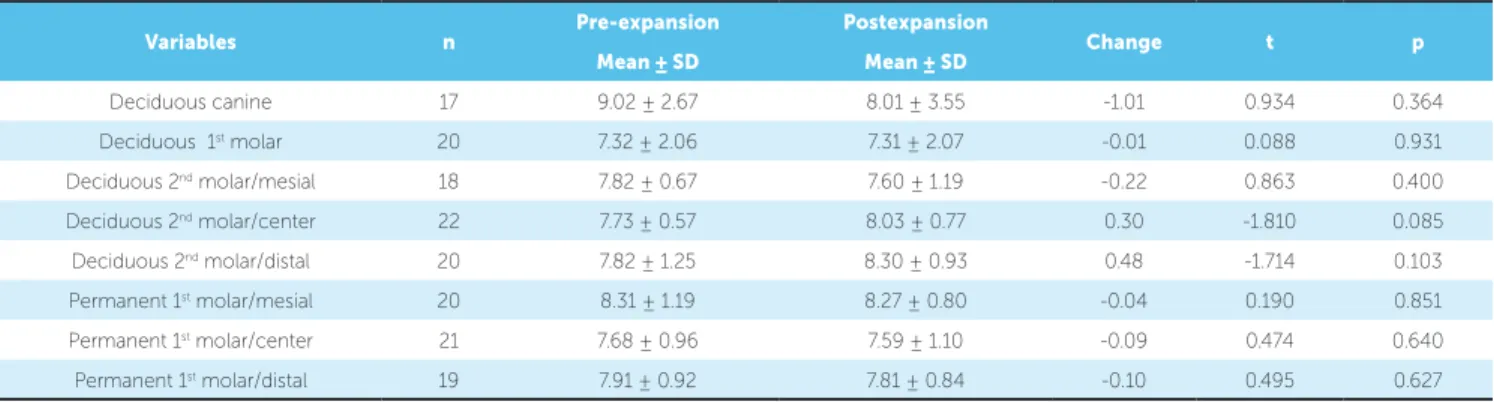
No statistically signiicant changes were observed in the level of buccal alveolar bone crest of supporting teeth (deciduous canines and deciduous second molars) and permanent irst molars (Table 3).
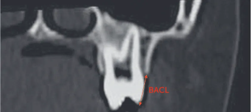
of orthoradially reformatted images perpendicular to the contour of the dental arch (cross sections), passing through the center of the buccal aspect of the deciduous canines, deciduous irst molar and through the center, mesial and distal areas of the buccal aspect of decidu-ous second molars and permanent irst molars. Figure 4 illustrates the linear variable obtained on each of these eight images both before and ater expansion.
Statistical analyses
All measurements were performed twice within a monthly interval by the same calibrated examiner. Statistical analysis was performed taking into account the mean of these two measurements. Each tooth category corresponded to the mean of right and let side teeth. Mean and standard deviation of each variable were calcu-lated before and ater expansion, and so were the changes between these stages. An exploratory test revealed normal distribution of data. Therefore, dependent t-tests were used to compare inter-phase changes. Sample size cal-culation (with an alfa value of 0.05 and statistical power of 90%) revealed that a sample size of 23 was more than enough. Results were regarded as signiicant for p < 0.05.
Systemic and casual errors
Casual and systematic errors were calculated by comparing the irst and second measurements with Dahlberg’s formula and dependent t-test respectively, at a signiicance level of 5%.
Figure 4 - Measurement of maxillary posterior teeth buccal alveo-lar crest level. BACL: buccal alveoalveo-lar crest level measured from the buccal cusp tip to the buccal alveolar crest.
Table 1 - Systematic and casual errors (dependent t-tests and Dalhberg’s formula).
Table 2 - Buccal and lingual bone plate thickness expansion changes (Paired t test). *Statistically significant at P < 0.05
*Statistically significant at P < 0.05
Variables First Second t p Dalhberg
Mean ± SD Mean ± SD
Buccal bone plate thickness (BBPT)
Permanent canine germ 1.54 ± 0.60 1.52 ± 0.59 0.260 0.798 0.21
1st premolar germ 0.97 ± 0.46 0.95 ± 0.42 0.356 0.727 0.12
2nd premolar germ 2.10 ± 1.01 2.15 ± 1.17 -0.612 0.549 0.25
Deciduous canine 1.13 ± 0.64 1.15 ± 0.68 0.74 0.463 0.12
Deciduous 2nd molar/mesial 1.11 ± 0.58 1.14 ± 0.59 0.91 0.367 0.12
Deciduous 2nd molar/distal 1.72 ± 0.70 1.75 ± 0.67 0.95 0.349 0.15
Permanent 1st molar/mesial 2.96 ± 1.21 2.77 ± 1.21 1.121 0.277 0.52
Permanent 1st molar/distal 2.92 ± 0.88 2.87 ± 0.90 1.334 0.200 0.11
Lingual bone plate thickness (LBPT)
Permanent canine germ 5.60 ± 2.03 5.72 ± 2.07 -1.170 0.259 0.30
1st premolar germ 3.09 ± 0.97 3.03 ± 1.05 0.637 0.534 0.29
2nd premolar germ 2.71 ± 1.05 2.74 ± 1.02 -0.421 0.680 0.16
Deciduous canine 4.73 ± 1.58 4.79 ± 1.61 1.955 0.057 0.16
Deciduous 2nd molar 1.16 ± 0.55 1.18 ± 0.55 0.64 0.527 0.11
Permanent 1st molar 1.41 ± 0.57 1.30 ± 0.61 3.082 0.007* 0.13
Buccal alveolar crest level (BACL)
Deciduous canine 9.14 ± 2.02 9.28 ± 2.12 -0.690 0.512 0.39
Deciduous 1st molar 7.66 ± 1.16 7.67 ± 1.18 -0.031 0.976 0.27
Deciduous 2nd molar/mesial 7.72 ± 1.40 7.72 ± 1.44 -0.026 0.980 0.35
Deciduous 2nd molar/center 7.80 ± 0.45 7.98 ± 0.40 -2.545 0.020 * 0.24
Deciduous 2nd molar/distal 8.10 ± 0.58 8.03 ± 0.62 1.108 0.284 0.18
Permanent 1st molar/mesial 8.39 ± 1.24 8.50 ± 1.08 -1.130 0.275 0.29
Permanent 1st molar/center 7.64 ± 1.02 7.71 ± 1.21 -0.725 0.479 0.27
Permanent 1st molar/distal 7.79 ± 0.88 7.79 ± 0.90 0.000 1.000 0.22
Variables n Pre-expansion Postexpansion Changes t p
Mean ± SD Mean ± SD
Buccal bone plate thickness (BBPT)
Permanent canine germ 23 1.32 0.55 1.31 0.71 -0.01 0.109 0.914
1st Premolar germ 17 0.92 0.41 1.10 0.37 -0.18 -3.318 0.004*
2nd Premolar Germ
Canine 20 2.33 1.08 2.28 1.14 -0.05 0.572 0.574
Deciduous canine 20 1.20 0.82 1.03 0.38 -0.17 0.913 0.373
Deciduous 2nd molar/mesial 19 1.14 0.50 1.06 0.63 -0.09 0.785 0.443
Deciduous 2nd molar/distal 19 1.89 0.69 1.59 0.68 -0.30 2.494 0.023 *
Permanent 1st molar/mesial 22 3.21 1.20 2.90 1.32 -0.31 1.478 0.154
Permanent 1st molar/distal 22 3.01 0.75 2.95 0.81 -0.05 0.823 0.420
Lingual bone plate thickness (LBPT)
Permanent canine germ 19 5.81 2.07 5.47 1.98 -0.34 1.627 0.121
1st premolar germ 17 3.46 1.30 3.36 1.15 -0.10 0.580 0.570
2nd premolar germ 18 2.81 1.14 2.85 1.02 0.04 -0.331 0.744
Deciduous canine 21 4.77 1.72 4.81 1.54 0.04 -0.200 0.844
Deciduous 2nd molar 17 1.26 0.53 1.19 0.55 -0.07 0.714 0.486
Table 3 - Buccal alveolar crest level (BACL) expansion changes (Paired t test).
*Statistically significant at P < 0.05
Variables n Pre-expansion Postexpansion Change t p
Mean ± SD Mean ± SD
Deciduous canine 17 9.02 ± 2.67 8.01 ± 3.55 -1.01 0.934 0.364
Deciduous 1st molar 20 7.32 ± 2.06 7.31 ± 2.07 -0.01 0.088 0.931
Deciduous 2nd molar/mesial 18 7.82 ± 0.67 7.60 ± 1.19 -0.22 0.863 0.400
Deciduous 2nd molar/center 22 7.73 ± 0.57 8.03 ± 0.77 0.30 -1.810 0.085
Deciduous 2nd molar/distal 20 7.82 ± 1.25 8.30 ± 0.93 0.48 -1.714 0.103
Permanent 1st molar/mesial 20 8.31 ± 1.19 8.27 ± 0.80 -0.04 0.190 0.851
Permanent 1st molar/center 21 7.68 ± 0.96 7.59 ± 1.10 -0.09 0.474 0.640
Permanent 1st molar/distal 19 7.91 ± 0.92 7.81 ± 0.84 -0.10 0.495 0.627
DISCUSSION
Spiral CT proves valuable in assessing alveolar bone
thickness and level . Fuhrmann et al16 demonstrated that
buccal and lingual bone plates can be identiied in spiral CT images provided that they are at least 0.5 mm thick. On the other hand, when the periodontal ligament space is apparent, CT identiies even thinner buccal and lingual bone plates (0.2 mm). Measurements taken with spiral
CT showed high accuracy and precision.10,11 While
peri-apical radiographs underestimate horizontal alveolar bone defects in 0.6 to 2.2 mm, spiral CT overestimate them in
0.2 mm.17 Moreover, previous studies measuring alveolar
bone plate thickness and level in spiral CT showed high
reproducibility and no-signiicant errors.8,18,24
This study assessed the changes in alveolar bone thickness and level at the region of maxillary posterior teeth ater RME was performed during early mixed dentition. The comparison between initial and post ex-pansion CT images was possible due to standardization of head positioning during the exam associated with selection of standardized image sections for measure-ments. Molar furcation was used as reference to obtain standardized axial sections before and ater expansion. This region is relatively stable, since — in reference to the palatal plane — posterior tooth extrusion is very small during RME and may be compensated by tooth
buccal tipping.9
With regard to changes in buccal bone plate thickness of supporting teeth, deciduous canines, which received a bonded C wire extension, did not reveal reduction in BBPT ater expansion (Table 2). Conversely, deciduous second molars, which received bands, showed statisti-cally signiicant reduction in buccal bone plate thick-ness at the distal region ater expansion, while the mesial aspect of buccal bone plate remained stable (Table 2).
The mean decrease in thickness of the distal aspect of buccal bone plate of deciduous second molars was 0.3 mm. Although statistically signiicant, this reduction was of lesser magnitude than the reduction in BBPT observed for permanent irst molars when RME is
per-formed in permanent dentition.18,25
RME performed in mixed dentition and anchored exclusively in deciduous teeth produces permanent irst molar expansion of one-half the amount of screw
ex-pansion, following the orthopedic efect of basal bones.12
In this study, palatal wire extension was used at perma-nent irst molars and probably produced further orth-odontic efect in this region. However, results revealed that buccal bone plate of permanent irst molars did not undergo any changes (Table 2). Previous studies assess-ing changes in buccal bone plate thickness of posterior teeth ater rapid maxillary expansion were performed in
permanent dentition18,25 or in late mixed and permanent
dentitions.8 In these previous studies, only permanent
teeth were analyzed.8 Garib et al18 reported a
signii-cant decrease in buccal bone plate thickness of banded supporting teeth (irst premolars and permanent irst molars) that ranged from 0.6 to 0.9 mm, three months
ater expansion. Rungcharassaeng et al25 corroborated
the indings by Garib et al18 and showed a signiicant
decrease in buccal bone plate thickness of irst premo-lars, second premolars and permanent irst mopremo-lars, with an average of 1.1 mm, 0.8 mm and 1.2 mm,
respec-tively, three months ater expansion. Ballanti et al8
The efects of RME on mixed dentition are similar to the efects observed in permanent dentition, including an orthopedic efect represented by midpalatal suture split and an orthodontic efect represented by buccal movement of
posterior teeth.13,14 The V shaped maxillary split observed
ater RME, in both occlusal and frontal planes during per-manent dentition, is also observed during mixed
denti-tion.13,14 However, maxillary halves separation is greater in
mixed dentition and corresponds to 50% of screw activa-tion, while in permanent dentition it corresponds to
ap-proximately 30% of screw activation.23 Consequently, the
amount of orthodontic efect decreases in mixed dentition in comparison to permanent dentition. Considering that periodontal bone changes are related to tooth movement in the alveolar ridge, it would be expected that RME dur-ing the mixed dentition causes less changes in buccal bone plate thickness, as conirmed in this study.
Lingual bone plate thickness (LBPT) of support-ing teeth and permanent irst molars did not change ater RME (Table 2). A previous study performed in permanent dentition reported an increase in lingual bone thickness of posterior teeth ater RME,
there-by relecting the buccal movement of these teeth.18
This increase had a mean value of 0.7 to 1.4 mm, three
months ater expansion.18 The absence of changes in
lingual bone plate thickness in mixed dentition ob-served in this study can be explained by the smaller amount of orthodontic efects caused by RME
dur-ing childhood.5,23 Furtheremore, the relatively short
interval between the irst and second CT exam might have inluenced the results. A previous study assessing patients in permanent and late mixed dentitions re-ported that lingual bone plate thickness of permanent irst molars did not immediately change ater the active phase of expansion; however, an increase was observed
6 months ater RME.8
All patients comprising the sample had maxillary per-manent canines and premolars unerupted when RME was performed. Thus, one of the goals of the present study was to observe the behavior of posterior tooth germs during expansion. Results showed that buccolingual position of posterior tooth germs is not afected by RME. Thick-ness of buccal and lingual bone plates of posterior tooth germs remained unchanged from T1 to T2 (Table 2).
Only the buccal bone plate of irst premolars germs demonstrated a small decrease. Such decrease might be related to tooth eruption, although the mean decrease of 0.18 mm is not clinically relevant. It is interesting to note that a favorable side efect of RME in early mixed dentition consists in facilitating eruption of palatally
displaced maxillary permanent canines.7 This aspect can
be regarded as a “bonus” of early expansion therapy at the maxillary arch.
The level of buccal alveolar crest of posterior teeth showed a slight reduction in the distal region of de-ciduous second molars, only; although changes were not statistically signiicant (Table 3). Results indicated that bone dehiscences did not occur immediately af-ter RME in the early mixed dentition. In permanent
dentition, Garib et al18 found that banded teeth showed
a signiicant reduction in buccal alveolar crest level, with a mean loss of 7 mm in the irst premolar and 3.5 mm at the mesial aspect of irst molars.
Rungcharas-saeng et al25 observed signiicant vertical bone losses at
the buccal aspect of all supporting teeth ater RME in permanent dentition. The mean change in buccal crest level of irst premolars, second premolars and irst
mo-lars was 4.4, 1.3 and 2.9 mm, respectively.25 Therefore,
RME performed in early mixed dentition seems to preserve the integrity of buccal bone plate more than RME performed in permanent dentition. The possible explanation is that the reduced orthodontic efect of RME on mixed dentition in comparison to permanent
dentition5,23 is not enough for moving posterior teeth
throughout the alveolar bone.
Evidence provided by current investigation suggests that early mixed dentition proves adequate to accomplish
orthopedic expansion. In addition to good stability26 and
greater orthopedic efect,5,23 RME performed in early
mixed dentition may avoid collateral buccal bone changes that predispose gingival recession in the long-term.
CONCLUSION
1. Andlin-Sobocki A, Bodin L. Dimensional alterations of the gingiva related to changes of facial/lingual tooth position in permanent anterior teeth of children. A 2-year longitudinal study. J Clin Periodontol. 1993;20(3):219-24.
2. Andlin-Sobocki A, Persson M. The association between spontaneous reversal of gingival recession in mandibular incisors and dentofacial changes in children. A 3-year longitudinal study. Eur J Orthod. 1994;16(3):229-39.
3. Artun J, Grobety D. Periodontal status of mandibular incisors after pronounced orthodontic advancement during adolescence: a follow-up evaluation. Am J Orthod Dentofacial Orthop. 2001;119(1):2-10. 4. Artun J, Krogstad O. Periodontal status of mandibular incisors following excessive proclination. A study in adults with surgically treated mandibular prognathism. Am J Orthod Dentofacial Orthop. 1987;91(3):225-32.
5. Baccetti T, Franchi L, Cameron CG, McNamara JA Jr. Treatment timing for rapid maxillary expansion. Angle Orthod. 2001;71(5):343-50. 6. Baccetti T, Franchi L, McNamara JA Jr. An improved version of the
cervical vertebral maturation (CVM) method for the assessment of mandibular growth. Angle Orthod. 2002;72(4):316-23.
7. Baccetti T, Mucedero M, Leonardi M, Cozza P. Interceptive treatment of palatal impaction of maxillary canines with rapid maxillary expansion: a randomized clinical trial. Am J Orthod Dentofacial Orthop. 2009;136(5):657-61.
8. Ballanti F, Lione R, Fanucci E, Franchi L, Baccetti T, Cozza P. Immediate and post-retention effects of rapid maxillary expansion investigated by computed tomography in growing patients. Angle Orthod. 2009;79(1):24-9.
9. Byrum AG Jr. Evaluation of anterior-posterior and vertical skeletal change vs. dental change in rapid palatal expansion cases as studied by lateral cephalograms. Am J Orthod. 1971;60(4):419.
10. Cavalcanti MG, Yang J, Ruprecht A, Vannier MW. Accurate linear measurements in the anterior maxilla using orthoradially reformatted spiral computed tomography. Dentomaxillofac Radiol. 1999;28(3):137-40. 11. Cavalcanti MG, Yang J, Ruprecht A, Vannier MW. Validation of spiral
computed tomography for dental implants. Dentomaxillofac Radiol. 1998;27(6):329-33.
12. Cozzani M, Rosa M, Cozzani P, Siciliani G. Deciduous dentition-anchored rapid maxillary expansion in crossbite and non-crossbite mixed dentition patients: reaction of the permanent first molar. Prog Orthod. 2003;4:15-22.
13. Silva Filho OG, Lara TS, Almeida AM, Silva HC. Evaluation of the midpalatal suture during rapid palatal expansion in children: a CT study. J Clin Pediatr Dent. 2005;29(3):231-8.
14. Silva Filho OG, Montes LA, Torelly LF. Rapid maxillary expansion in the deciduous and mixed dentition evaluated through posteroanterior cephalometric analysis. Am J Orthod Dentofacial Orthop. 1995;107(3):268-75.
15. Engelking G, Zachrisson BU. Efects of incisor repositioning on monkey periodontium after expansion through the cortical plate. Am J Orthod. 1982;82(1):23-32.
REFERENCES
16. Fuhrmann RA, Bucker A, Diedrich PR. Assessment of alveolar bone loss with high resolution computed tomography. J Periodontal Res. 1995;30(4):258-63.
17. Fuhrmann RA, Wehrbein H, Langen HJ, Diedrich PR. Assessment of the dentate alveolar process with high resolution computed tomography. Dentomaxillofac Radiol. 1995;24(1):50-4.
18. Garib DG, Henriques JF, Janson G, Freitas MR, Fernandes AY. Periodontal efects of rapid maxillary expansion with tissue-borne and tooth-borne expanders: a computed tomography evaluation. Am J Orthod Dentofacial Orthop. 2006;129(6):749-58.
19. Garib DG, Henriques JF, Janson G, Freitas MR, Coelho RA. Rapid maxillary expansion--tooth tissue-borne versus tooth-borne expanders: a computed tomography evaluation of dentoskeletal efects. Angle Orthod. 2005;75(4):548-57.
20. Haas AJ. Palatal expansion: just the beginning of dentofacial orthopedics. Am J Orthod. 1970;57(3):219-55.
21. Haas AJ. Rapid expansion of the maxillary dental arch and nasal cavity by opening the midpalatal suture. Angle Orthod. 1963;31(2):73-90. 22. Haas AJ. The treatment of maxillary deiciency by opening the midpalatal
suture. Angle Orthod. 1965;35(3):200-17.
23. Krebs A. Midpalatal suture expansion studies by the implant method over a seven-year period. Rep Congr Eur Orthod Soc. 1964;40:131-42. 24. Loubele M, Van Assche N, Carpentier K, Maes F, Jacobs R, van
Steenberghe D, et al. Comparative localized linear accuracy of small-ield cone-beam CT and multislice CT for alveolar bone measurements. Oral Surg Oral Med Oral Pathol Oral Radiol Endod. 2008;105(4):512-8. 25. Rungcharassaeng K, Caruso JM, Kan JY, Kim J, Taylor G. Factors afecting
buccal bone changes of maxillary posterior teeth after rapid maxillary expansion. Am J Orthod Dentofacial Orthop. 2007;132(4):428.e1-8. 26. Spillane LM, McNamara JA Jr. Maxillary adaptation to expansion in the
mixed dentition. Semin Orthod. 1995;1(3):176-87.
27. Starnbach H, Bayne D, Cleall J, Subtelny JD. Facioskeletal and dental changes resulting from rapid maxillary expansion. Angle Orthod. 1966;36(2):152-64.
28. Steiner GG, Pearson JK, Ainamo J. Changes of the marginal periodontium as a result of labial tooth movement in monkeys. J Periodontol. 1981;52(6):314-20.
29. Thilander B, Nyman S, Karring T, Magnusson I. Bone regeneration in alveolar bone dehiscences related to orthodontic tooth movements. Eur J Orthod. 1983;5(2):105-14.
30. Van der Linden FPGM. Development of the dentition. Chicago: Quintessence; 1983.
31. Wehrbein H, Fuhrmann RA, Diedrich PR. Periodontal conditions after facial root tipping and palatal root torque of incisors. Am J Orthod Dentofacial Orthop. 1994;106(5):455-62.
32. Wennstrom JL, Lindhe J, Sinclair F, Thilander B. Some periodontal tissue reactions to orthodontic tooth movement in monkeys. J Clin Periodontol. 1987;14(3):121-9.