Instituto Dante Pazzanese de Cardiologia
Mailing address: Silvia Judith Fortunato de Cano – Rua Sampaio Viana, 203/81 04004-000 – São Paulo, SP, Brazil – E-mail: [email protected]
English version by Stela Maris C. e Gandour
Objective - Analyze the dromotropic disturbances (vector-electrocardiographic), and the possible anatomic causes, provoked by selective alcohol injection in the septal branch, for percutaneous treatment, of obstructive hypertrophic cardiomyopathy.
Methods - Ten patients with a mean age of 52.7 years underwent percutaneous septal ablation (PTSA) from oc-tober 1998; all in functional class III/IV). Twelve-lead electrocardiogram was performed prior to and during PTSA, and later electrocardiogram and vectorcardio-gram according to Frank’s method. The patients were fol-lowed up for 32 months.
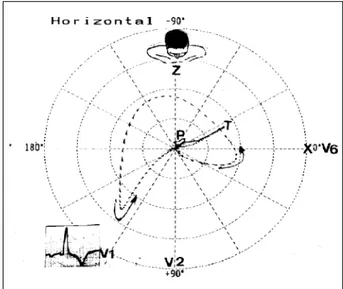
Results - On electrocardiogram (ECG) prior to PTSA all patients had sinus rhythm and left atrial enlar-gement, 8 left ventricular hypertrophy of systolic pattern. On ECG immediately after PTSA, 8 had complete right bundle-branch block; 1 transient total atrioventricular block; 1 alternating transient bundle-branch block either right or hemiblock. On late ECG 8 had complete right bundle-branch block confirmed by vectorcardiogram, type 1 or Grishman.
Conclusion - Septal fibrosis following alcohol injec-tion caused a predominance of complete right bundle-branch block, different from surgery of myotomy/myectomy.
Key words: percutaneous septal ablation, obstructive hypertrophic cardiomyopathy, vector elec-trocardiographic alterations
Arq Bras Cardiol, volume 79 (nº 5), 471-5, 2002
Andrés Ricardo Pérez Riera, Silvia Judith Fortunato de Cano, Manuel Nicolás Cano, Vera Márcia L. Gimenez, Luciano Augusto de Padua Fleury Neto,
José Eduardo M. R. Sousa
São Paulo, SP - Brazil
Vector Electrocardiographic Alterations After Percutaneous
Septal Ablation in Obstructive Hypertrophic
Cardiomyopathy. Possible Anatomic Causes
The appearance of decreasing exercise capacity in patients with obstructive hypertrophic cardiomyopathy under optimal medical treatment has spawned the intro-duction of different techniques to reduce left ventricular outflow tract gradient and to relieve symptoms. Percuta-neous septal ablation is a promising alternative to myoto-my/myectomy surgery and to DDD pacemaker implantation for the treatment of obstructive hypertrophic cardiomyopa-thy with significant septal thickness and gradient (thickness always > 18 mm and gradient > 50 mmHg at rest) in the ab-sence of intrinsic mitral disease or significant systolic dys-function 1 in patients with limiting symptoms refractory to
drug treatment, who refuse to undergo surgery or who have a high surgical risk.
The procedure consisted of causing a septal infarc-tion by injecting absolute alcohol into the first great perfo-rating septal branch of the anterior descending artery, redu-cing septal thickness with fibrosis and relieving the obs-tructive component 2.
Although promising, this is still an experimental method, because less than 1,000 cases have been reported worldwide, and, in Brazil, only 5. Randomized comparative studies with the widely accepted transaortic invasive surgi-cal technique are still lacking. The transaortic invasive sur-gical technique has an acceptable estimated mortality lower than 5%, ranging from 3% (large centers) to 15% 3,4, and a
mean complication rate of 6% of the patients operated on. The complications include significant residual aortic insuffi-ciency, which is present in almost 4% of surgical patients 5,
endocarditis, complete left bundle-branch block, and total atrioventricular block.
Methods
The protocol used was that proposed by Herrmann 6 in
a recent publication. Patients with the following characteris-tics were excluded from the study: diastolic septal thickness < 18 mm; hypertrophy located out of the left ventricular outflow tract (for example: in the medioseptal region); absence of a significant resting or provoked gradient (< 50 mmHg); presence of systolic dysfunction; presence of intrinsic mitral valve disorder; good response to treatment with negative inotropic medication; NYHA functional class I and II; and those who spontaneously chose to undergo surgery. Previous right bundle-branch block was not an exclusion criterion, as recommended in the literature 7, because we
found no rationale for its contraindication, as none of the patients had this dromotropic disorder prior to the procedure. Our case series consisted of 10 consecutive patients from October 1998 to July 2001 patients (5 males) with obs-tructive hypertrophic cardiomyopathy, whose ages ranged from 34 to 75 years. All of them were symptomatic, 9 were refractory to clinical treatment, and 1 had relapse of the obs-truction after myectomy. The most frequent symptoms were progressive dyspnea, which occurred on minimum exertion at the time of intervention, shortness of breath, angina pectoris, and angina equivalent. All patients were in NYHA functional classes III or IV. Conventional 12-lead elec-trocardiography, Doppler echocardiography, exercise testing, and coronary angiography were performed before and right after the procedure, and, 3 months later, vector car-diography was performed in 8 patients according to Frank’s method using the 7100 model, 1992, of the Body Surface Po-tential Mapping Fukuda-Denshi device.
The patients signed a formal written consent and were aware of the existence of another widely accepted therapeuti-cal surgitherapeuti-cal option. The technique of alcohol injection was re-commended for the patients who refused to undergo surgery or who had a high surgical risk, as long as they agreed to it.
The patients were sedated with a solution of midazo-lam (5 mg in 9 mL of saline solution) slowly applied
accor-ding to each patient’s clinical response. After the proce-dure, the patients were maintained in the coronary unit where they were continuously monitored for 48 hours with serial sampling of the cardiac enzymes and daily electrocar-diographic recording for clinical control of the septal infarction. The functional septal alteration was followed up with daily echocardiography. Hospital discharge occurred in an average of 2 days.
Results
The technique of percutaneous septal ablation was successful in all patients, resulting in a reduction in the mean left ventricular outflow tract gradient from 88.7 mmHg (prior to the procedure) to 12.8 mmHg (after the procedure), which correlated with an increase in CK-MB, confirming the septal infarction. Symptom relief was immediate.
On the electrocardiography prior to percutaneous septal ablation, all patients had sinus rhythm, 8 patients
Table I - Results
Patient Sex Age NYHA class ECG (pre) ECG (post) Late ECG and VCG
1 M 69 IV LVH-1 EISA-CRBBB CRBBB, FA
2 F 65 IV LVH-1 EISA-CRBBB CRBBB
3 F 47 III/1V LVH-1 Alternating R/LBBB SR-CRBBB
EISA-TAVB
4 F 63 III SR –LVH-0 EISA CRBBB
Mild LVH/LAH
5 M 75 IV LVH-1 EISA-CRBBB CRBBB
6 M 38 III/IV LVH-1 EISA-CRBBB/LPIDB CRBBB
7 F 34 III/IV LVH-0 EISA - mild LVH CRBBB
8 F 59 IV LVH-1 EISA-CRBBB CRBBB
9 M 48 III/IV SR –LVH-0 EISA Mild LVH/LAH
Mild LVH/LAH
10 M 56 IV SR –LVH-0 EISA Mild LVH/LAH
Mild LVH/LAH
LAH - left atrial hypertrophy; LVH-0- left ventricular hypertrophy with no systolic pattern of ventricular repolarization; LVH-1- left ventricular hypertrophy with systolic pattern of ventricular repolarization; CRBBB – complete right bundle-branch block; LBBB – left bundle-branch block; EISA - electrically inactive septal area; ASEISA - anteroseptal electrically inactive septal area; SR – sinus rhythm; LPIDB – left posteroinferior divisional block.
had left atrial hypertrophy, 7 had left ventricular hypertro-phy with a repolarization pattern of the Cabrera systolic type (strain pattern), 2 had no systolic pattern, and 1 had an almost normal pattern (tab. I and fig. 1).
On the electrocardiogram performed immediately after percutaneous septal ablation, complete right bundle-branch block appeared isolated or associated with left divi-sional block in 80% of the cases. Our result was better than that reported in the literature with a predominance of com-plete right bundle-branch block (approximately 55% to 60% of the cases) (fig. 2).
Only 1 bifascicular block of the complete right bundle-branch block type was observed associated with left poste-roinferior divisional block.
One patient, who had transient total atrioventricular block treated with a pacemaker, recovered sinus rhythm in 24 hours. Another patient had alternating transient left and right block, anterosuperior divisional left bundle-branch block, and posteroinferior divisional block after alcohol jection. Two patients had ventricular tachycardia in the in-tensive care unit, which was cardioverted to sinus rhythm. Neither definitive total atrioventricular block nor complete left bundle-branch block was observed.
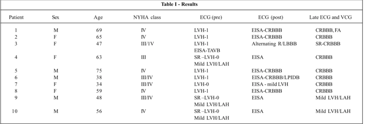
On electrocardiography and vectorcardiography per-formed during late follow-up, sinus rhythm was present in all patients except 1 who had atrial fibrillation 32 months after the procedure. Septal or anteroseptal infarction was present in all patients, and complete right bundle-branch block was observed in 8 patients. The predominating vector cardiographic pattern was type I or “Grishman” complete right bundle-branch block characterized, in the horizontal plane, by the afferent branch of the QRS loop located behind the X line and a final delay, similar to a glove finger, forward and to the right (fig. 3).
Discussion
In 1995, Ulrich Sigwart 8 successfully introduced the
percutaneous septal ablation technique for treating obstruc-tive hypertrophic cardiomyopathy in 3 patients at the Royal Bromptom Hospital in London. Gietzen and colleagues 9
achieved relief of obstruction by using transient occlusion
with an inflated balloon in the first perforating septal branch. Successive series have shown promising results, and, so far, almost 1,000 cases have already been reported in the worldwide medical literature, most of them by the German school 10-12.
The immediate appearance of the complete right bundle-branch block pattern after alcohol injection in most cases caught our attention, leading us to consider the parti-cipation of the first perforating septal branch in the irrigation of the right His’ system, which can be divided into 6 succe-ssive segments 13, according to figure 4.
Irrigation of the right His’ system is as follows: its trunk segment is irrigated by the first septal perforating branch of the anterior descending artery; its branching or perforating segment, or both of these, is irrigated by the perforating
Fig. 2 – Electrocardiogram after percutaneous septal ablation (same patient) – Sinus rhythm, anteroseptal current of injury, complete right bundle-branch block.
Fig. 3 – Horizontal vector cardiogram – HP loop VCG with typical type I or “Grishman” complete right bundle-branch block. Note the afferent branch of the QRS loop located behind the X line and the final delay similar to a glove finger for-ward and to the right.
Fig. 4 – The right His’ system: right penetrating (1) and branching (2) portions. The actual right branch formed by 3 portions: (3) trunk, proximal or membranous; (4) middle, intramyocardial or mimetic; and (5) inferior, distal or moderator band.
Divisional segment of the right branch (6): formed by 3 fascicles at which the parietal Purkinje of the right ventricle originates: a) superior, anterosuperior, or sub-pulmonary division; b) middle or anteroinferior division; c) inferior, posterior, or posteroinferior division.
I - His system
Right his Branch
branches of the posterior descending artery, the 2nd septal
perforating branch of the anterior descending artery, and Kugel’s artery, which is a branch of the circumflex artery (fig. 4: 1 and 2).
One proof of the participation of the irrigation of the first perforating branch is the frequent appearance of conco-mitant anterosuperior divisional left bundle-branch block, creating, therefore, a bifascicular block: right bundle-branch block + anterosuperior divisional left bundle-bundle-branch block as a consequence of the close proximity existing bet-ween the anterosuperior division and the branching portion of the right His’ bundle.
The penetrating portion of the right His’ bundle ex-tends from the inferior limit of the AV node to the origin of the first fibers that form the posteroinferior division of the left bundle-branch. Its length ranges from 50 to 100 mm and the designation “penetrating” is used because the right His’ system, at that level, crosses the fibrous trigone.
Histologic studies of the His’ bundle show that its right half carries some fibers to the ipsilateral ventricle.
The branching portion of the right His’ bundle (fig. 4: 2) extends from the origin of the posteroinferior division of the left branch to the origin of the trunk of the right branch and the anterosuperior division of the left branch. It is closely related to the interventricular membranous septum and to the noncoronary and right coronary leaflets of the aortic valve. A lesion in this portion causes, almost invariably, complete right bundle-branch block and concomitant an-terosuperior divisional left bundle-branch block, showing the close relation between both. The trunk, proximal, or membranous portion of the right bundle-branch (fig. 4: 3) is irrigated from 2 sources: one originating in the artery of the AV node of the right coronary artery and another in the first septal perforating branch of the anterior descending artery. Irrigation of the middle portion of the right bundle-branch (fig. 4: 4) is provided by the 3 following sources: the septal branches of the posterior descending artery, the 2nd septal
perforating branch of the anterior descending artery, and Kugel’s artery, which is a branch of the circumflex artery.
Finally, the middle and distal portions of the right bundle-branch (fig. 4: 5) are irrigated by the ramus limbi dextri, which is a branch of the 2nd septal perforating branch
of the anterior descending artery.
The only regions of the right His’ system exclusively irrigated by the first perforating branch of the anterior des-cending artery are the perforating and branching portions of the right His’ bundle. This explains why complete right bundle-branch block is the most frequent dromotropic di-sorder after percutaneous septal ablation caused by abso-lute alcohol injection in the great first septal perforating branch. Recent studies performed in human hearts during autopsies with the injection of dyed gelatinous barium showed that the irrigation of the anterosuperior division depends, exclusively, on the first perforating branch only in 40% of cases. In 50% of cases, double irrigation is provided by the anterior descending artery and branches of the right coronary artery. In 10% of cases, the right coronary artery is
the exclusive source 14. This explains why, only
approxima-tely half of the cases of alcohol injection in the first perfora-ting branch develop complete right bundle-branch block associated with anterosuperior divisional left bundle-branch block.
Among recent publications, we found one by Heneim et al 15, who studied 20 patients with characteristics similar
to those of our sample. Complete right bundle-branch oc-curred in 80% of the patients in the acute phase; in 3 pa-tients, the incomplete left bundle-branch block that existed prior to percutaneous septal ablation coincided with our fin-dings in individuals with severe left ventricular overload. Those authors attributed the appearance of complete right bundle-branch block to an alteration in activation and con-sequent change in ventricular contractility, which would lead to a reduction in the left ventricular outflow tract gra-dient. Undoubtedly, ventricular remodeling contributed to improving these patients. However, it does not elucidate completely the process by which complete right bundle-branch is produced. Therefore, activation with a pacemaker in obstructive hypertrophic cardiomyopathy would be the most effective treatment; however, we found a random, in-constant, and short-lasting response in these patients, which led some authors to consider the possible benefit of the pacemaker as a placebo effect 16.
As far as we are concerned, interruption of irrigation in the interventricular septum due to alcohol injection cau-ses a localized infarction and mainly impairs the branching segment of the His’ bundle and the proximal segment of the right bundle-branch. An association with incomplete left bundle-branch block exists due to the anatomic proximity with the right segment.
In most cases of septal myotomy/myectomy or tran-saortic cardiomyectomy, left bundle-branch block is obser-ved 17. The procedure was introduced in 1961 18 and has
been maintained with slight modifications.
After extracorporeal circulation, cardioplegia, and hy-pothermia, the aorta is very carefully sectioned obliquely in the noncoronary sinus so as not to injure the aortic valve. Then, 2 deep and extensive incisions are performed parallel to the bulging septum, a few millimeters below the valve until reaching the left ventricular apex. The myocardium lo-cated between the 2 incisions is resected and removed, and palpation is performed to assess the depth of the resection in septal thickness.
The His’ left bundle runs between the incisions, and this is why in most cases, the patient has patent left branch block. It is worth remembering that the left bundle-branch is closely related to the noncoronary and right coronary aortic valve leaflets (exactly the section line in Morrow’s technique), to the aortic ring (left bundle-branch block is frequent in aortic valve diseases), membranous septum, subaortic septal endocardium, and apex of the mus-cular septum. These anatomic relations justify the predomi-nance of left bundle-branch dromotropic disorder in transa-ortic myotomy/myectomy.
te-References
1. Fananapazir L, Mc Arevery D. Therapeutic options in patients with obstructive hypertrophic cardiomiopaty and severe drug-refractory symptoms. J Am Coll Cardiol 1998; 31: 359-64.
2. Knight C, Kurbaan AS, Seggewiss H, et al. Nonsurgical septal reduction for hy-pertrophic obstructive cardiomyopathy outcome in the first series of patients. Circulation 1997; 95: 2075-81.
3. Seiler C, Hess OM, Schoenbenbeck M, et al. Long term follow-up of medical versus surgical theraphy for hypertrophic cardyomiopathy: a restrospective study. J Am Coll Cardiol 1996; 17: 634-42.
4. Schulte HD, Bircks MH, Loesse B, et al. Prognosis of patientes with hypertro-phic obstructive cardiomyopathy after transaortic myectomy: late results up to twenty-five years. J Thorac Cardiovasc Surg 1993; 106: 709-17.
5. Brown PS Jr, Roberts CS, McIntosh CL, et al. Aortic regurgitation after left ventri-cular myotomy and myectomy. Ann Thorac Surg 1991; 51: 585-92.
6. Herrmann JLV. Oclusão da artéria septal na cardiomiopatia hipertrófica. Rev Soc Cardiol Est SP 2000; 10: 499-508.
7. Fananapazir L, Mc Arevery D. Therapeutic Options in patients with obstructive hypertrophic cardiomiopaty and severe drug-refractory symptoms. J Am Coll Car-diol 1998; 31: 359-64.
8. Sigwart U. Non cirurgical myocardial reduction of hypertrophic obstructive car-diomiopathy. Lancet 1995; 346: 211-4.
9. Gietzen F, Leuner Ch, Gerenkamp T, Kuhn H. Relief of obstruction in hypertro-phic cardiomyopathy by transient oclusion of the first septal branch of the left co-ronary artery. Eur Heart J 1994; 15(Suppl): 125-30.
10. Seggewiss L, Faber L, Gleichman U. Percutaneous transluminal septal ablation in hypertrophic obstructive cardiomyopathy. Thorac Cardiovasc Surg 1999; 47: 94-100.
11. Knight C, Kurbaan AS, Seggewiss H, et al. Noncirugical septal rediction for hy-pertrophic obstructive cardiomyopathy: outcome in first series of patients. Cir-culation 1997; 95: 2075-81.
12. Gietzen FH, Leuner ChJ, Rauber-Kreunsen V, et al. Acute and long-term results after transcoronary ablation of septal hypertrophy (TASH) catheter interventio-nal treatment for hypertrophic obstructive cardiomyopathy. Eur Heart J 1999; 20: 1342-54.
13. Rosenbaum MB, Elizari MV, Lazzari JO. Los Hemibloqueos. Cap. II. Anatomía del Sistema de Conducción. Editorial Paidos S.A. I.C.F, 1967: 43-91. 14. Pileggi F, Moffa PJ, Sosa E. In: Rivera IC. Cardiologia Electrocardiografía y
Vec-tocardiografia. Editora Médica Panamericana, S.A. de C.V., 1993; Vol. 1, cap. 7: 270-365, 332.
15. Heneim MY, et al. Eletromechanical left ventricular behaviour after nonsurgical seotal reduction in patients with hypertrophic obstructive cardiomiopathy. J Am Coll Cardiol 1999; 34: 1117-22.
16. Nishimura RA, Trusty JM, Hayes DL. Dual chamber pacing for hypertrophic obstructive cardiomyopathy: a randomized, double blind, crossover study. J Am Coll Cardiol 1997; 29: 435-41.
17. McIntosh C, Maron C. Current operative treatment of obstructive hypertrophic cardiomyopathy. Circulation 1988; 78: 487.
18. Morrow AG, Brockenbrough EC. Surgical treatment of idiopatic hypertrophic subaortic stenosis: technique and hemodynamic results of subaortic ventriculomyotomy. Ann Surg 1961; 154: 181-9.
19. Lakkis NM, Sherif F, Kay Dumn J, et al. Non surgecil septal reduction therpy for hypertrophic obstructive cardiomiopathy: one year follow up. J Am Coll Cardiol 2000; 36: 852-5.
chnique that allows widening of the left ventricular outflow tract, a reduction in the gradient 19, and clinical improvement
in symptoms. We observe a marked percentage difference in the type of resulting bundle-branch block with these 2 invasive therapeutic options. On the one hand, the percuta-neous technique of alcohol injection causes mainly complete right bundle-branch block; on the other, the myectomy surgery produces left bundle-branch block in 75% of cases. According to our understanding, these alterations derive from anatomic relations specific to each therapeutic procedure.
No reference exists in the literature about the vector cardiographic type of right bundle-branch block resulting
from alcohol injection. We emphasize the predominance of type I or “Grishman” complete right bundle-branch block, in which the afferent branch of the QRS loop in the horizon-tal plane is located behind the X line, a fact that is in accor-dance with the association between right bundle-branch block and left ventricular hypertrophy.