Arq Neuropsiquiatr 2009;67(3-A):712-714
712
Clinical / Scientiic note
Bilateral FaCial PalSy in a young Patient
aFter meningoCoCCal meningitiS,
aSSoCiated to herPetiC inFeCtion
Edna Quintas¹, Antónia Silva², António Sarmento¹
ParaliSia FaCial Bilateral em JoVem aPÓS meningite meningoCÓCiCa aSSoCiada a inFeCÇÃo herPÉtiCa
1Department of Infectious Diseases, Medical School, University of Porto, Portugal; 2Department of Neurology, Medical School, University of Porto, Portugal.
Received 28 November 2008, received in inal form 20 May 2009. Accepted 15 June 2009.
Dra. Edna Quintas – Serviço de Doenças Infecciosas / Hospital de São João - Al. Prof. Hernâni Monteiro - 4200-319 Porto - Portugal. E-mail: edna19d@ hotmail.com
Bilateral facial palsy is a rare clinical entity, which is
often a special inding of a systemic disease, occurring in
0.3% to 2.0% of facial palsy cases
1,2. Many of them are
po-tentially life-threatening, and therefore the condition
re-quires urgent medical intervention. Bacterial meningitis,
Lyme Disease, (Bannwarth syndrome), syphilis, infectious
mononucleosis, Guillan Barré syndrome, leukemia,
sarcoi-dosis, Mobius syndrome are the most common cause of
bilateral facial paralysis
3,4.
In this paper, we present a case of a previously healthy
in young woman, who was hospitalized with symptoms of
meningitis associated with herpetic infection.
CaSe
A 16-year-old female without history of previous medica-tion or illness presented to her primary care physician with a 1-day history of severe headache. One day later she presented with fever, nausea, vomiting, photophobia, meningeal signs and petichial skin bruising.
She was hospitalized due to probable meningococcal meningitis.
Past medical history – No medical or signiicant history. Vacci-nation schedule in order (as recommended by the National Plan of Vaccination).
Medication – No medication habits.
Allergies to medication – Not of her knowledge.
Family history – No relevant family medical history.
Social history – Student, no recent contact with ill colleagues, no contact with animals, no recent travels, does not smoke/ drink and takes no illicit drugs.
Physical examination – During examination, the patient was awake, alert and aware, her temperature was 36.7oC/98.06oF,
pulse: 100/min, respiratory rate 18/min, SatO2: 100% (FiO2:
21%) and blood pressure: 113/79 mmHg. The eyes, ears, nose and throat were all normal. There was no parotid gland enlargement or adenomegaly. Heart and lung auscultation were normal. The
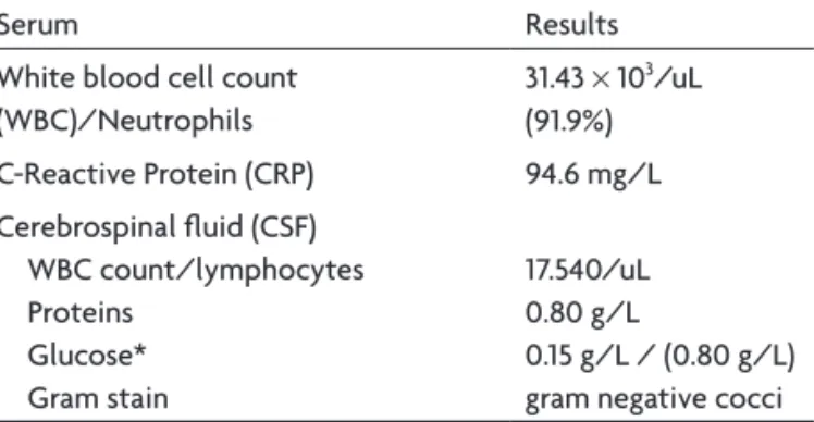
abdomen was soft and nontender, without organomegaly, bow-el sounds were normal. The rbow-elevant basic laboratoy valves are shown in Table 1.
Began empirical antibiotherapy with ceftriaxone 2 gr BID/ IV and corticotherapy (dexamethasone). On the third day of her admission to the hospital the patient developed vesicular skin lesions on the left side of her face suggesting herpes and was treated with acyclovir topical ive times a day.
From the previous CSF study no agent was isolated, except for Neisseria meningitidis. C-Reactive Protein DNA from Herpes simplex virus 1 and 2 (HSV1/2), Cytomegalovirus (CMV), Epstein-Barr (EBV), Varicella zoster virus (VZV), Mycoplasma pneumoniae
(MP) and Mycobacterium tuberculosis were negative. The screen-ing for RNA Enterovirus was also negative. The research for Bor-relia burgdorferi, Wright reaction, Veneral Disease Research Lab-oratory test, Treponema pallidum hemagglutination, as well as for the direct and cultural mycobacteriological exam were neg-ative. Table 2 shows the study results.
Antibodies to hepatitis virus C, HBs atg and the Human Im-munodeiciency Virus (HIV) were negative. And so were ANA and ANCA.
Table 1. Relevant basic laboratory values.
Serum Results White blood cell count
(WBC)/Neutrophils
31.43 × 10³/uL
(91.9%) C-Reactive Protein (CRP) 94.6 mg/L Cerebrospinal luid (CSF)
WBC count/lymphocytes Proteins
Glucose* Gram stain
17.540/uL 0.80 g/L
0.15 g/L / (0.80 g/L) gram negative cocci
Arq Neuropsiquiatr 2009;67(3-A)
713
Bilateral facial palsy Quintas et al.
After 7 days of therapy with ceftriaxone, she still complained of headache, did brain and ear computed tomography scan, that showed no abnormalities and repeated lumbar puncture.
The CSF analysis revealed white blood cell WBC 260/uL, a persisting high protein level of 1.07 g/L, glucose of 0.46 g/L.
A cervical echography and chest X-rays showed no abnormalities. Ceftriaxone was changed to ampicillin for more 5 days. On the eleventh day of admission, the patient complained of difi-culty in articulating some words, but showed no other signs and symptoms. The neurological examination was normal. After clin-ical recovery, she was discharged after 13 days of hospitalization. One week later, she returned to the hospital with dificulty in opening her mouth and articulating words. She was examined by a neurologist and was admitted in the Neurological Department.
Neurological examination
Mental status – Alerted, oriented. Speech: dificulty in artic-ulating words.
Cranial nerves – No alteration in visual acuity. Isocoric, photo-reactive pupils. No ophthalmoparesis.
Normal facial sensibility. Bilaterally diminished nasogenian furrows, mainly on the left side of the face. No face mimics.
All other cranial pairs normal.
The motor and sensitive examination of trunk and extremities was normal. She had no nuchal rigidity or other meningeal signs. Repeated lumbar puncture and the CSF exam revealed: WBC 59/uL, glucose of 0.43 g/L, protein of 0.77 g/L, as shown in Table 3.
All analysis made in the previous admission were repeated showing no abnormalities. PCR for Herpes virus in the CSF was negative, though with positive serologies for HSV1 (Table 4).
Electromyography showed severe signs of axonal lesion of both facial nerves. The brain MRI (magnetic resonance imaging)
showed abnormal bilateral Gadolinium enhancement in the in-tracanalicular part of both facial nerves, and there was no evi-dence of any parenchymal lesions (Fig 1).
The diagnosis of bilateral peripheral facial palsy, probably associated to an infection with Herpes simplex virus was made and treated with acyclovir 800 mg ive times a day Per Os, dur-ing 7 days. Support measures were also taken, namely physio-therapy, artiicial tear eye drops several times a day and occlu-sive eye dressing on both eyes during the night. She improved on this regimen with completely resolution of her facial palsies, some weeks later.
diSCuSSion
The causes of peripheral facial paralysis includes many
conditions such as congenital, traumatic, infectious,
neu-Table 2. Serological study.Serum
VDRL /
TPHA VZV Wright
HSV1 IgM/IgG
HSV2 IgG/IgM
EBV IgG/IgM
CMV IgG/IgM
MP
IgG/IgM Lyme –/– – – 189 /+ –/– +/– +/– –/– –
Table 3. Evolution of the CSF analysis.
Cerebrospinal luid (CSF) Initial study After 7 days of treatment Neurology service (after 21 days) WBC count/lymphocytes 17.540/uL 260/uL 59/uL
Proteins 0.80 g/L 1.07 g/L 0.77g/L Glucose 0.15 g/L 0.46 g/L 0.43g/L
Table 4. Serologic study.
Serum
VDRL /
TPHA VZV Wright
HSV1 IgM/IgG
HSV2 IgG/IgM
EBV IgG/IgM
CMV IgG/IgM
MP
IgG/IgM Lyme 1st study –/– – – 189 /+ –/– +/– +/– –/– –
2nd study –/– – – 188.9/+ –/– +/– +/– –/– –
Arq Neuropsiquiatr 2009;67(3-A)
714
Bilateral facial palsy Quintas et al.
rological/neuromuscular disorders, metabolic, neoplastic,
toxic, vascular, autoimmune, iatrogenic and idiopathic
3,5.
The etiology of Bell’s palsy is not completely
un-derstood
6-9. Unlike unilateral facial paralysis, where the
cause is mostly idiopathic (over 50%), bilateral facial
pal-sy is less often idiopathic (under 20%). Various infectious
agents have been linked to facial palsy, such as the
Her-pes simplex and Varicella zoster viruses, mumps and the
rubella virus, cytomegalovirus and HIV as well as Borrelia
burgdorferi
1,6. Mycoplasma pneumoniae is the other
im-portant pathogen of upper and lower respiratory tract
in-fections, which may cause extrapulmonary complications,
namely in the central nervous system –
meningoencepha-litis and occasionally peripheral facial palsy
10-12.
More recently, the Herpes simplex virus has been
im-plicated in the pathogenesis of Bell’s palsy
13. Neisseria
men-ingitis is a gram-negative diplococcus that can also cause
peripheral facial palsy. It is still a cause of endemic and
ep-idemic disease in developed
14,15and developing nations
16,17.
This infection is a worldwide major public health
prob-lem. Children account for the vast majority of these cases.
A thorough history, a complete physical examination,
and an exhaustive laboratory search are needed to
clari-fy the etiology of bilateral facial paralysis. The entire
pro-cess should include a complete blood cell count, blood
biochemistry, C-reactive Protein, cerebrospinal luid
ex-amination, immunoassay for EBV, CMV, HSV, Lyme disease
and the Syphilis test. A complete microbiologic study and
radiologic examination (computed tomography and/or
magnetic resonance imaging) of temporal bone, brain and
chest should also be done. Effectively, all the
above-men-tioned exams to establish the diagnosis.
In the present case, it was identified the etiologic
agent – Neisseria meningitides, conirmed by the culture
exam in the CSF. Gadolium enhancement was noticed in
the intracanalicular part of both facial nerves. This
ind-ing advocated with neurological examination and
elec-tromyography supports the diagnosis of bilateral
periph-eral facial palsy after infection with bacterial meningitis.
Antibiotherapy and corticotherapy were administered in
the irst four days of hospitalization.
In the second phase, when she was readmitted to the
hospital, herpes simplex virus infection was diagnosed and
she was medicated with acyclovir. She improved over the
next days, and was discharged.
Although it was not identiied by PCR of the HSV-1 in
the CSF, there persisted positive serologies for herpes. If
we consider the time when meningitis and the facial
her-pes appeared we tend to believe that it may be
associat-ed with the patient’s clinical condition.
In summary, this is a report of a young woman with
meningococcal meningitis complicated by bilateral
pe-ripheral facial palsy. Bilateral palsies usually relect an
un-derlying systemic pathology, and the prognosis is
depen-dent on the etiology.
If the etiology can be identiied and successfully dealt
with the prognosis is excellent, but may be worse in
elder-ly people, especialelder-ly in immunocompromised individuals.
Patients with bilateral facial palsies need thorough
as-sessment and follow-up.
reFerenCeS
1. Roberg M, Ecrudh J, Forsberg P. Acute peripheral facial palsy: CSF ind
-ings and etiology. Acta Neurol Scand 1991;83:55-60.
2. Stahl WFT. Recurrent bilateral peripheral facial palsy. J Laryngol Otol 1989;103:117-119.
3. Price T, Fife DG. Bilateral simultaneous facial nerve palsy. J Laryngol Otol 2002;116:46-48.
4. Kilic R, Ozdek A, Felek S, Safak MA, Samin E. A case presentation of bilateral simultaneous Bell’s palsy. Am J Otolaryngol 2003;24:271-273.
5. Gevers GLP. Bilateral simultaneous facial paralysis-differential diagno
-sis and treatment options. A case report and review of literature. Acta Otorhinolaryngol Belg 2003;57:139-146.
6. Morgan M, Nathwani D. Facial palsy and infection: the unfolding sto
-ry. Clin Infect Dis 1992;14:263-271.
7. Abele-Horn M, Franck W, Busch U, Nitschko H, Roos R, Hecsemann J.
Transverse mielitis associated with Mycoplasma pneumoniae. Clin In
-fect Dis 1998;26:909-912.
8. Shaitkin BM, May M, Podrinec M, Ulrich J, Peitersen E, Klein SR. Idio
-pathic (Bell’s) palsy, herpes zoster cephalicus, and other facial nerve dis
-orders of viral origin: In: May M, Shaitkin BM (Eds). The facial nerve,
May’s. 2nd Ed. New York: Thieme Medical Publishers, 2000:319.
9. Mandell GL, Bennett JE, Dolin R. Principles and practice of infectious
diseases. 6th Edition, 2005.
10. Fink CG, Butler L. A cranial nerve palsy associated with Mycoplasma pneumoniae infection. Br J Ophthalmol 1993;77:750-751.
11. Klar A, Gross-Klieselstein E, Hurvitz H, Branski D. Bilateral Bell’s palsy due to Mycoplasma pneumoniae infection. ISR J Med SCI 1985;21:692-694.
12. Wang C-H, Chou M-L, Huang C-H. Benign Isolated abducens nerve pal
-sy in Mycoplasma pneumoniae infection. Pediatr Neurol 1998;18:71-72. 13. Schirm J, Mulkens PSJZ. Bell’s palsy and herpes simplex virus. APMIS
1997;105:815-823.
14. Patel MS, Meriands A, Hanna JN, et al. Epidemic meningococcal men
-ingitis in central Australia. Med J Aust 1993;158:336-340.
15. Durand ML, Calderwood SB, Weber DJ, et al. Acute bacterial meningi
-tis in adults. A review of 493 episodes. N Engl J Med 1993;328:21-28. 16. Girgis NI, Sippel JE, Kilpatrick WR, et al. Meningitis and encephalitis
at the Abbassia Fever Hospital Cairo, Egypt from 1966 to 1989. Am J Trop Med Hyg 1993;48:97-107.
17. Nejmi S, Belhaj A, Guibourdenche M, et al. Study of ninety strains of
serogroup A Neisseria meningitidis isolated from cerebroespinal lu

![Fig 1. T1-weighted coronal sections through the internal auditory ca- ca-nal. [A] Precontrast view and [B] postcontrast view after gadolium administration](https://thumb-eu.123doks.com/thumbv2/123dok_br/15432338.594921/2.955.110.894.400.494/weighted-sections-internal-auditory-precontrast-postcontrast-gadolium-administration.webp)