Arq Bras Cardiol 2004; 82: 217-20.
Silva et al Ebstein´s anomaly. Results of the conic reconstruction of the tricuspid valve
2 1 7
Hospital São Joaquim da Real e Benemérita Sociedade Portuguesa de Beneficência Mailing address: José Pedro da Silva - Rua Maestro Cardim, 769 - S/202 Cep 01323-001 - Paraíso - São Paulo, SP, Brazil - E-mail: [email protected] Received: 12/30/02
Accepted: 04/07/03
English version by Stela Maris C. e Gandour
Arq Bras Cardiol, volume 82 (nº 3), 217-20, 2004
José Pedro da Silva, José Francisco Baumgratz, Luciana da Fonseca, Jorge Yussef Afiune,
Sônia Meiken Franchi, Lílian Maria Lopes, Daniel Marcelo Silva Magalhães, José Henrique Andrade Vila
São Paulo, SP - Brazil
Ebstein’s Anomaly. Results of the Conic Reconstruction of the
Tricuspid Valve
Original Article
Ebstein’s anomaly of the tricuspid valve is a rare con-genital heart disease, treated with various surgical techni-ques since 1958 1,2. Until the 1970s, valvular replacement was the treatment of choice, despite its association with se-vere complications, such as arrhythmias, thrombosis, pros-thetic dysfunction, in addition to a high mortality rate 3.
The Danielson method 4 was the most used tricuspid valvuloplasty procedure for correcting Ebstein’s anomaly in the 1980s. However, as that surgery does not provide good geometry for the right ventricle and is not applicable to a large number of patients, tricuspid valve replacement with a prosthesis is required in 35.6% of the cases.
Carpentier et al 5 classified the various anatomical pre-sentations of the disease and proposed a new technique, which consists of the reconstruction of the right ventricle form and repositioning of the tricuspid valve at the normal level with the routine use of a prosthetic ring. This surgery was carefully described and reported on in 1988 5. Quage-beur et al 6 used a similar surgery, but without the prosthetic ring, in a younger population.
Since 1989, we have been using a surgical technique consisting of total or almost total detachment of the anterior tricuspid megaleaflet from the ventricular wall and valvular ring, transforming it into a cone, whose vertex remained fixed in the right ventricular tip and the base was sutured to the tri-cuspid ring, after its plication, adjusting it to the size of the base of the cone (tricuspid ring), including the septal region. This study aimed at analyzing the late results of this que, whose principle is similar to that of the Carpentier techni-que, but with reconstruction of the septal portion of the tri-cuspid valve, not requiring the use of a prosthetic ring.
Methods
From November 1993 to August 2001, 21 patients with Ebstein’s anomaly of the tricuspid valve were operated on at our institution by using the technique described below. The ages ranged from 20 months to 37 years (mean of 23 years), and 10 (48%) patients were men.
Objective - To assess the results of a technical modifi-cation of tricuspid valvuloplasty in Ebstein’s anomaly.
Methods - From November 1993 to August 2002, 21
patients with Ebstein’s anomaly of the tricuspid valve, with ages ranging from 20 months to 37 years (mean, 23 years), underwent a new technique of tricuspid valvulo-plasty. This technique consisted of total or almost total de-tachment of the anterior tricuspid megaleaflet from the ventricular wall and valvular ring, transforming it into a cone, whose vertex remained fixed in the right ventricular tip, and the base was sutured to the tricuspid ring, after its plication, adjusting it to the size of the base of the cone (tri-cuspid ring), including the septal region.
Results - One (4.7%) patient with cardiomyopathy
caused by chronic hypoxia died in the hospital due to low cardiac output. The mean follow-up lasted 4 years, and the recent echocardiograms showed good morphology of the right ventricle and tricuspid valve with mild or minimal insufficiency in 18 patients and moderate insufficiency in 2 patients. In 2 of the 3 patients with an anomalous bun-dle, it could be located and sectioned during surgery. No atrioventricular block occurred.
Conclusion - The technique used was efficient in
re-pairing tricuspid insufficiency and restoring right ventri-cular morphology, being applicable to all anatomic types of Ebstein’s anomaly, except for Carpentier classification type D.
2 1 8
Silva et al
Ebstein´s anomaly. Results of the conic reconstruction of the tricuspid valve
Arq Bras Cardiol 2004; 82: 217-20.
The clinical presentations were as follows: dyspnea in 17 (80%) patients, cyanosis in 15 (71 %), arrhythmia in 6 (28%), and low cardiac output syndrome in 3 (14%). In re-gard to associated diseases, 8 (21%) patients had patent oval foramen, 9 (42%) had interatrial septal defect, and 3 (14%) had anomalous atrioventricular bundle [Wolf-Parkin-son-White syndrome (WPW)]. Pulmonary stenosis was observed in 1 (4.7%) patient, significant mitral insufficiency in 1, and interventricular septal defect in another patient.
The preoperative echocardiographies showed signifi-cant tricuspid insufficiency in 12 (57.8%) patients and mode-rate insufficiency in 9 (42.7%).
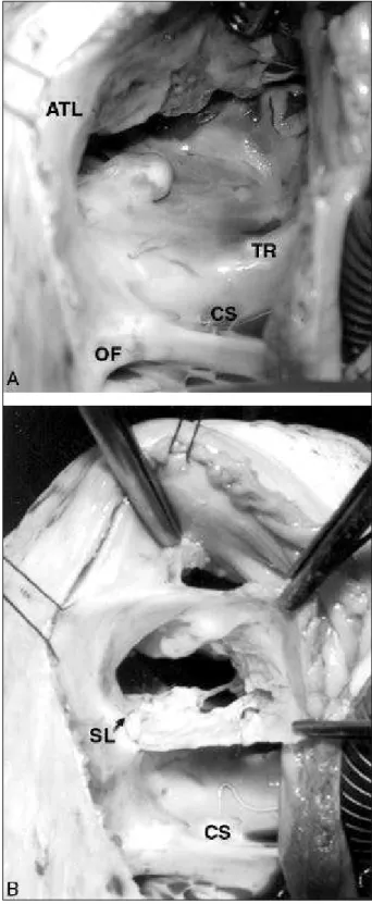
Surgical technique - After installing extracorporeal cir-culation with bicaval cannulation, and infusing blood cardio-plegia for myocardial protection, an oblique right atriotomy was performed (fig. 1). The anterior tricuspid megaleaflet was totally or partially detached from the ventricular wall and val-vular ring, its free margin being rotated clockwise, transfor-ming it into a cone whose vertex remained fixed in the tip of the right ventricle by the chordae tendineae and papillary mus-cles. The base (tricuspid leaflets) was then sutured in the tri-cuspid ring in its true position, in the threshold between the right atrium and the atrialized right ventricle, after plication of the ring, which had its diameter adjusted to the size of the base of the cone (tricuspid ring). This technique also recons-tructs the septal portion of the tricuspid valve, in which a deli-cate and superficial suture is used in the proximity of the atrio-ventricular conduction system. In 5 patients, some fenestra-tions were created in the leaflet to facilitate the entrance of blood into the right ventricle. In 2 cases, misplaced papillary muscles needed to be transferred to the proximities of the right ventricular apex. No prosthetic ring was used for the tri-cuspid valve in this series of patients.
A valved interatrial communication was left, allowing the passage of blood from right to left. This was obtained with the partial closure of the oval foramen and the interatrial septal defect with appropriate techniques and the oblique opening in the superior margin of the fossa ovalis in the ca-ses of intact interatrial septum. The following surgical pro-cedures were associated with tricuspid valvuloplasty: sec-tion of the anomalous bundle in 2 patients; ventriculosepto-plasty in 1 patient; and enlargement of the right ventricular outflow tract with a valved graft (monocuspid) of bovine pericardium and mitral valvuloplasty in another.
Results
One (4.7%) patient with biventricular cardiomyopathy caused by chronic hypoxia died during hospitalization. Al-though the tricuspid valvuloplasty was effective, the biven-tricular insufficiency with low cardiac output led to multi-system organ failure, culminating in death on the fourth postoperative day.
The first patient in this series underwent surgery at the age of 11 years and required reoperation 4 years later, when a new valvuloplasty was performed. Currently, the patient is in excellent clinical condition, with age-appropriate height
and weight development. Another patient developed seve-re biventricular cardiomyopathy after 7 years. The other pa-tients evolved with no intercurrent events and remained asymptomatic.
Fig. 1 - Surgical features of tricuspid valvuloplasty in Ebstein’s anomaly. A) Anato-mic features of the anomalous insertion of the posterior and septal leaflets of the tri-cuspid valve; B) Tritri-cuspid valve partially detached from the ventricular wall, clock-wise rotated, forming a cone.
Arq Bras Cardiol 2004; 82: 217-20.
Silva et al Ebstein´s anomaly. Results of the conic reconstruction of the tricuspid valve
2 1 9
One patient gave birth to a healthy baby after an une-ventful pregnancy 2 years after surgery.
Two patients with WPW in the preoperative period had successful sectioning of the anomalous bundle, while, in a third patient, the bundle could not be located during surgery. No atrioventricular block was observed in the patients.
The recent echocardiograms showed a good surgical result in all patients, with a good morphology of the tricuspid valve and right ventricle, and minimum or mild tricuspid in-sufficiency (fig. 2) after a mean follow-up of 4 years. One (4.7%) patient had moderate tricuspid insufficiency on the postoperative echocardiography, with no clinical symptoms.
Discussion
The surgical indications in Ebstein’s anomaly are well defined for the following patients: 1) symptomatic, in NYHA
functional class III or IV; 2) in functional class I or II, with car-diomegaly and a cardiothoracic index of 0.65 or greater; 3) significant cyanosis and polycythemia; 4) presence of para-doxical embolism; 5) presence of uncontrollable tachycardia and accessory AV bundle.
In the natural history of the disease, a significant increa-se in mortality is obincrea-served in adulthood due to the develop-ment of congestive heart failure or arrhythmias 7,8. The sud-den death caused by arrhythmias not related to WPW oc-curs in 60% (CL 43%-77%) of the patients with Ebstein’s ano-maly who die due to cardiac causes 9,10. Sudden death corre-lates more with cardiomegaly than with functional class (NYHA) 9. Paradoxical embolism or cerebral abscess is an im-portant cause of death, mainly in patients > 50 years 11.
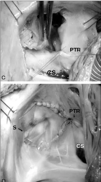
The nonoperated on asymptomatic patients, when Fig. 1 - Surgical features of tricuspid valvuloplasty in Ebstein’s anomaly. C)
Plica-tion of the true tricuspid ring; D) Tricuspid valve sutured in the true ring, correcting the insufficiency.
CS - coronary sinus; PTR- plication of the tricuspid ring.
CS
CS
CS
Fig. 2 - A) Preoperative echocardiogram showing the anomalous insertion of the tri-cuspid valve leaflet. B) Postoperative echocardiogram of the same patient, showing the good position of the tricuspid valve at the same level as that of the mitral valve. RA - right atrium; LA - left atrium; RV - right ventricle; LV - left ventricle; TV - tricus-pid valve; SL - septal leaflet.
2 2 0
Silva et al
Ebstein´s anomaly. Results of the conic reconstruction of the tricuspid valve
Arq Bras Cardiol 2004; 82: 217-20.
compared with healthy people, have a limited tolerance for exercise and a reduction in the capacity to work 12, being, therefore, indicated for serial exercise tests to objectively define their physical limitations and their long-term evolu-tion. The development of simpler techniques of valvulo-plasty, with lower surgical risks, remodeling of the right ven-tricle, and lower index of valvular replacement may broaden the surgical indication for this group of patients.
Anatomical and echocardiographic studies, as well as surgical experience, suggest that the right ventricle (its form, compliance, and contractility) may be a factor as important as tricuspid dysfunction in the pathophysiology of this disease. The main objective of the surgery proposed by Carpentier et al 5 was to restore right ventricular volume and form.
The technique here described uses the innovative principle of Carpentier et al, which consists of detaching the dislocated tricuspid valve from the interior of the right ventricle, bringing it to the normal level of the tricuspid ring, ie, leaving the atrialized portion of the right ventricle in the subvalvular position. However, the technique used in this study is different in regard to the clockwise rotation of the lateral margin of the tricuspid valve megaleaflet, forming a cone, and ignoring the septal leaflet when it is anomalous. The suture of the base of this cone is performed at the nor-mal level of the tricuspid ring, including its septal portion, where the suture line has to be very superficial, avoiding atrioventricular block, a complication not observed in this series of patients. Another difference is the non use of the prosthetic tricuspid ring in any of the cases, while Carpen-tier recommended its use in most cases 5. Quaegebeur et al 6 reported a technique similar to that of Carpentier, which was used in younger patients, with no prosthetic ring, but also without covering the septal portion of the tricuspid ring.
Our technique assures the reconstruction of the tri-cuspid valve in its septal portion, avoiding significant re-gurgitation in the short- and long-term. These results were demonstrated in the postoperative echocardiograms (fig.2), which evidenced good positioning of the tricuspid valve and restoration of the normal ventricular morphology.
No atrioventricular block occurred in any patient, which coincides with the experience of Wu and Huang 13, who, in a more recent series, used fresh autologous pericar-dium in some patients to reconstruct the septal portion of the tricuspid valve, suturing it in the septal part of the ring. Similarly to our case series, no case of atrioventricular block was observed after manipulation of the septal portion. Ho-wever, this suture should be very superficial. Due to the total coverage of the area of the tricuspid orifice and to restora-tion of the tricuspid ring to its normal posirestora-tion and size, the valve became competent, with mild insufficiency in 6 pa-tients, all being in functional class I.
None of the authors showed concern in regard to the interatrial septal defect, performing its routine closure. In our case series, we always left a valved interatrial communi-cation, allowing flow only from right to left when the right atrial pressure increased. This was useful in the postopera-tive period of 3 patients, who had significant right ventricu-lar dysfunction, but maintained good cardiac output due to mild oxygen desaturation.
The results obtained with this technique in this group of patients, from the anatomic and functional points of view, are encouraging. The early death in our series does not rela-te to the surgical rela-technique, but to the preoperative starela-te of the patient, which indicates that surgical intervention should have been taken earlier before deterioration of right and left ventricular function.
In conclusion, the technique reported is efficient for cor-recting tricuspid insufficiency and restoring right ventricular morphology, being applicable to all anatomic types of Ebs-tein’s anomaly, except for Carpentier classification type D.
This technique, which does not require the use of pros-thesis, may extend the surgical indication to asymptomatic or oligosymptomatic patients to prevent the appearance of late complications, such as arrhythmia and ventricular dys-function.
Further studies with larger case series are required to determine the probable advantages of this technique as compared with that of Carpentier.
1. Hunter SW, Lillehei CW. Ebstein malformation of the tricuspid valve. Study of a case together with suggestions of a new form of surgical therapy. Chest Disease 1958; 33: 297-304.
2. Hardy KL, May IA, Webster CA, Kimball KG. Ebstein’s anomaly: a functional con-cept and successful definitive repair. J Thorac Cardiovasc Surg 1964; 48: 927-940. 3. Barbero-Marcial M, Verginnelli G, Awad M, Ferreira.S, Ebaid m, Zerbini EJ: Sur-gical treatment of Ebstein’s anomaly. Early and late results in twenty patients subjected to valve replacement. J Thorac Cardiovasc Surg 1979; 78: 416-422. 4. Danielson GK, Driscoll DJ, Mair DD, Warnes CA, Oliver WC Jr. Operative
treat-ment of Ebstein anomaly. J Thorac Cardiovasc Surg. 1992; 104: 1195-1202. 5. Carpentier A, Chauvaud S, Mace L, Relland J, Mihaileanu S, Marino JP, et al. A
new reconstructive operation for Ebstein anomaly of the tricuspid valve. J Thorac Cardiovasc Surg. 1988; 96: 92-101.
6. Quaegebeur JM, Sreenam N, Fraser AG, Bogers AJ, Stumper OF, Hess J, et al. Sur-gery for Ebstein anomaly: the clinical and echocardiographic evaluation of a new technique. J Am Coll Cardiol. 1991; 17: 722-8.
References
7. Giuliani ER, Fuster V, Brandenburg RO, Mair DD. Ebstein’s anomaly:the clini-cal features and natural history of Ebstein’s anomaly of the tricuspid valve. Mayo Clin Proc 1977; 54: 163-173.
8. Hansen JF, Leth A, Dorp S, Wennevold A. The prognosis in Ebstein’s disease of the heart: long term follow up of 22 patients. Acta Med Scand 1977; 201: 331-335. 9. Gentles TL, Calder AL, Clarkson PM, Neutze JM. Predictors of long term survival with Ebstein’s anomaly of the tricuspid valve. Am J Cardiol 1992; 69: 377-381. 10. Watson H: Natural history of Ebstein’s anomaly of the tricuspid valve in child-hood and adolescence: An international cooperative study of 505 cases. Br Heart J 1974; 36: 417-427.
11. Mathews JL, Pennington WS, Isobe JH, Gaskin TA, Dumas JH, Kahn DR: Para-doxical embolization with Ebstein’s anomaly.Arch Surg 1983; 118: 1101. 12. Barber G, Danielson GK, Heise CT, Driscoll DJ. Cardiorespiratory response to
exercise in Ebstein’s anomaly.Am J Cardiol 1985; 56: 509-514.