2 8 4
Vieira et al
Left atrial myxoma. Three-dimentional echocardiographic assesment
Arq Bras Cardiol 2004; 82: 284-6.
Instituto do Coração do Hospital das Clínicas - FMUSP
Mailing address - Marcelo Luiz Campos Vieira - InCor - Av. Dr. Eneas C. Aguiar, 44 São Paulo, SP, Brasil - Cep 05403-000 - E-mail: [email protected]
Received: 11/11/02 Accepted: 02/24/03
English version by Stela Maris C. e Gandour
Arq Bras Cardiol, volume 82 (nº 3), 284-6, 2004
Marcelo L.C.Vieira, Bárbara M. Ianni, Charles Mady, Javier Encinas, Pablo M. A. Pommerantzeff, Paulo P. Fernandes, Samira B. Leal, Wilson Mathias Jr, José L. Andrade, José A. F. Ramires
São Paulo, SP - Brazil
Left Atrial Myxoma. Three-Dimensional Echocardiographic
Assessment
Case Report
The patient was a 70-year-old female with antece-dents of diabetes mellitus and hypertension, being followed up in the outpatient care clinic due to chronic anemia after corrective surgery for angiodysplasia of the proximal jejunum, in whom an image suggestive of left atrial myxoma was found on routine transthoracic echocardiography. Then multiplanar transesophageal echocardiography and 3-dimensional echocardiography were performed, showing the latter better anatomical details of the tumor. The patient underwent exeresis of the mass with anatomicopathologi-cal confirmation of the tumor. Three-dimensional echocar-diography proved to be a technique that can provide addi-tional contributions to the diagnostic investigation of structural heart diseases.
Left atrial myxoma is the most common primary cardiac tumor 1, occasionally found on routine examinations of
asymptomatic patients. Only a few cases with 3-dimensio-nal echocardiographic images of left atrial myxomas can be found in the literature 2-4. We report the case of a patient
with left atrial myxoma, in whom 3-dimensional echocardio-graphy was useful for detailing the anatomical features of the tumor.
Case report
A 70-year-old white female with a previous history of diabetes mellitus and hypertension, followed up in the out-patient care clinic due to chronic anemia after corrective surgery for angiodysplasia of the proximal jejunum. The pa-tient complained of dyspnea on major exertion, and, on rou-tine transthoracic echocardiography, a left atrial mass was
identified. At the time of hospital admission, the patient was afebrile, in regular general condition, with pale mucosae (++/4). Her blood pressure was 140/90 mmHg, heart rate was 82 bpm, and respiration rate was 16 bpm. On cardiac auscul-tation, a diastolic (++/4) and a systolic (+/4) murmur could be heard in the mitral area. The examinations of the respira-tory and neurological systems and of the abdomen were normal. The laboratory tests were within the normal range. The electrocardiogram showed sinus rhythm with unspeci-fic alterations in ventricular repolarization. Transthoracic echocardiography was repeated 5 days after the first echo-cardiographic investigation and confirmed the presence of a very mobile left atrial mass with homogeneous density and irregular contours, measuring 5.9 cm x 3.2 cm, with a diasto-lic movement through the mitral valve towards the left ven-tricle (fig. 1). A mean transvalvular mitral gradient of 5 mmHg was observed. Left ventricular function was within the normal range. The patient underwent multiplanar transe-sophageal echocardiographic investigation, which confir-med the findings of the transthoracic study and added ana-tomical detailing to the multilobated appearance of the mass and its insertion in the interatrial septum (fig. 2). During the transesophageal echocardiographic examination, images for 3-dimensional reconstruction were obtained (fig. 3). Then, coronary angiography was performed and showed ir-regularities in the coronary arteries. The patient underwent resection of the tumor, which was a left atrial nodular mass of elastic consistency, violaceous to grayish-yellow in co-lor, with a fixating pedicle in the interatrial septum, measu-ring 5 cm x 5 cm x 3 cm, and weighing 32 g (fig. 4). The histo-logical examination showed it to be a myxoma. The patient had an uneventful postoperative evolution, being dischar-ged from the hospital 8 days after surgery.
Arq Bras Cardiol 2004; 82: 284-6.
Vieira et al Left atrial myxoma. Three-dimentional echocardiographic assesment
2 8 5 The multiplanar transesophageal 2-dimensional images
were obtained in the longitudinal and transverse planes, in 4- and 2-chamber views, evidencing the interatrial septum and the mitro-aortic junction, and in the longitudinal gastric plane. Image acquisition was performed with multiplanar 2-dimensional echocardiographic scanning from 0° to 180° every 3°, associated with a capture system determined by the respiratory variation of the patient, captured from the electrocardiographic signal and the variation in the impe-dance of the patient’s chest during the respiratory move-ment. This was integrated in a computer program developed by Philips. The images were recorded and stored on an opti-cal disk, and then transferred to the commercially available reconstruction system and 3-dimensional analysis of volu-metric mapping (TomTec compact 3D model, Omniview pa-ckage, TomTec Imaging Systems Corp., Boulder, CO, USA). The 3-dimensional echocardiographic analysis was perfor-med in the coronal, sagittal, and transverse planes of the structure analyzed, in addition to the parallel and diagonal
planes. The total time for obtaining the 3-dimensional image was 35 minutes, with 3 minutes for acquiring the image, 2 mi-nutes for transferring the information from the optical disk to the work station, and 30 minutes for adjusting and re-constructing the image.
Discussion
Three-dimensional echocardiography was developed in the 1970s, as a method for measuring ventricular volume 5.
Prior to it, a laborious analysis of the images provided by 2-dimensional transthoracic echocardiography was used for obtaining that measure. However, that methodology posed some difficulties, inadequacies, and imprecisions. The evo-lution of the technique led to the use of a mechanical arm for ultrasound mapping, and then to the use of electromagnetic support, parallel mapping, rotational scan mapping, and, more recently, real time volumetric mapping, which is still under development 5-7. This resulted in a progressive
impro-vement in the quality of the images obtained. Three-dimen-sional echocardiography with transesophageal transducers and digital technology enables better cardiac structural de-Fig. 1 - Transthoracic echocardiogram, apical 4-chamber view, showing the presence
of a mass projecting into the left ventricle during diastole (arrows). RA - right atrium; LA - left atrium; RV - right ventricle; LV - left ventricle.
Fig. 4 - Gross appearance of the myxoma found inside the left atrium.
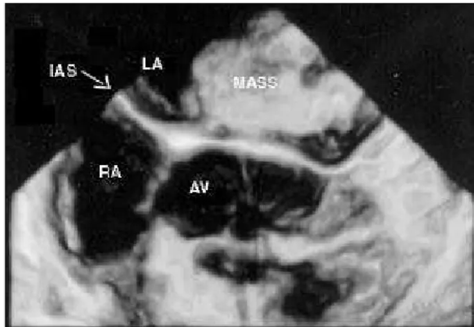
Fig. 3 - Three-dimensional echocardiogram, transverse plane, showing the presence of a mass in the left atrium with a fixating pedicle in the interatrial septum. RA - right atrium; LA - left atrium; AV - aortic valve; IAS - interatrial septum (arrow).
2 8 6
Vieira et al
Left atrial myxoma. Three-dimentional echocardiographic assesment
Arq Bras Cardiol 2004; 82: 284-6.
1. Bulckley BH, Hutchins GM. Atrial myxomas: a thirty-year review. Am Heart J 1979; 97: 639-43.
2. Melzer C, Bartel T, Baumann G. Dynamic 3-D-echo in the preoperative assessment of the left atria nmyxoma in a 51-year-old male. Cardiology, 1997; 59: 167-69. 3. Prêtre R, Vuille C, Diebold-Berger S, Lerch R. Three-dimensional imaging of
atrial myxoma. Circulation 1998; 97: 2186-7.
4. Harada T, Ohtaki T, Sumiyoshi T, Hosoda S. Sucessful three-dimensional recons-truction using transesophageal echocardiography in a patient with left atrial my-xoma. Jpn Heart J 2001; 42: 789-92.
References
5. Roelandt JRT, Yao J, Karsprzak JD. Three-dimensional echocardiography. Curr Opin Cardiol 1998; 13: 386-98.
6. Cheng TO, Xie MX, Wang XF, et al. Evaluation of mitral valve prolapse by four-dimensional echocardiography. Am Heart J 1997; 133: 120-9.
7. Li J, Sanders SP. Three-dimensional echocardiography in congenital heart disea-se. Curr Opin Cardiol 1999; 14: 53-9.
8. De Simone R, Glombitza G, Vahl CF, et al. Three-dimensional Doppler: techniques and clinical applications. Eur Heart J 1999; 20: 619-27. finition. It has been used for assessment prior to surgery
and in the operating room and as diagnostic support in the surgical treatment of cardiac masses; of mitral, aortic, and tricuspid valvular diseases; and in congenital heart disea-ses, such as correction of interatrial septal defects, inter-ventricular septal defects, and cor triatriatum 5-8. In the case
reported, the 3-dimensional reconstruction confirmed the transesophageal echocardiographic data and allowed a better anatomical detailing of the mass in regard to its fixa-ting pedicle in the interatrial septum, approximafixa-ting the fin-dings in the image examinations to the anatomical and sur-gical reality.
We report the case of a patient with left atrial myxoma in which the 3-dimensional echocardiographic reconstruc-tion provided better anatomical detailing, and, consequen-tly, greater safety for the surgical team to perform the
exere-sis of the mass. The 3-dimensional reconstruction confir-med the transesophageal findings and allowed better spa-tial identification of the mass in relation to the interatrial sep-tum. In current clinical practice, the 3-dimensional echocar-diographic technique proved to be potentially useful for the anatomical identification of structural heart diseases. Its routine use will certainly provide the development of more advanced computer programs, greater familiarity of the car-diologist with the method, and a better cost/benefit ratio.