1. M astership in Surgery – Department of Orthopedics and Traumatology – FM C/Unicamp, Laboratory of Biomechanics and Rehabilitation of the Locomotive Device HC-FM C/Unicamp, Campinas State University – Unicamp – Campinas, SP.
2. M aster Professor of the Department of Orthopedics and Traumatology – FM C/Unicamp, Head of the Laboratory of Biomechanics and Reha-bilitation of the Locomotive Device HC-FM C/Unicamp, Campinas State University – Unicamp – Campinas, SP.
3. M astership in Surgery – FCM – Unicamp, Laboratory of Biomechanics and Rehabilitation of the Locomotive Device HC - FM C/Unicamp. 4. Professor Doctor of the Department of Orthopedics and Traumatology
– FCM /Unicamp, Coordinator of the Knee Surgery Group – HC-FM C/ Unicamp, Campinas State University – Unicamp – Campinas, SP. Received in 10/5/05. Final version received in 30/10/05. Approved in 3/11/05.
Correspondence to: Guilherme Lotierso Fehr, Rua João de Bortoli, 354, Jardim Flórida – 14026-330 – Ribeirão Preto, SP. Phone: (16) 3911-1345. E-mail: [email protected]
Effectiveness of the open and closed
kinetic chain exercises in the treatment
of the patellofemoral pain syndrome
Guilherme Lotierso Fehr1, Alberto Cliquet Junior2,
Ênio Walker Azevedo Cacho3 and João Batista de M iranda4
O
RIGINALA
RTICLEKeyw ords: Knees. Electromyography. Exercise. Functional recovery.
ENGLISH VERSION
ABSTRACT
The aim of this study w as to analyze the therapeutic effects of the open kinetic chain (OKC) and closed kinetic chain (CKC) exer-cises to treat the patellofemoral syndrome (PFSD). For this, 24 volunteers, bearers of the PFSD w ere randomly divided in tw o groups: group I (n = 12) performed the OKC exercises; group II (n = 12) performed the CKC exercises. Both groups w ere submitted to eight consecutive w eeks of treatment consisting of three w eekly sessions performed in alternate days. To analyze the activation pattern of the vastus medialis oblique (VM O) and the vastus later-alis (VL) muscles, the electromyographic signals (EM G) w ere col-lected using bipolar surface electrodes quantified by the root mean square (RM S) normalized by the maximal voluntary isometric con-traction of the quadriceps. The pain intensity and the functionality of the volunteers w ere assessed using scales. The analysis of the amounts of the VM O/VL ratio in both groups I and II show ed no significant differences as to the pre- and post-treatment times in the concentric (p > 0.05) and eccentric (p > 0.05) phases of the OKC and CKC exercises. Despite of this, the VM O muscle pre-sented a low er activation rate compared to the VL in the eccentric phase of the CKC exercise. It w as found significant increases in the functionality (p < 0.05), and a reduction in the pain intensity (p < 0.05) betw een the pre- and post-treatment times in both groups, but group II show ed higher amounts compared to group I in both variables. The results found in this study suggest that according to the conditions of the trial, the OKC and CKC exercises provoke no changes in the patterns of the EM G activation in the VM O and VL muscles. How ever, they promoted an improvement in the func-tionality and a reduction in the pain intensity after the eight w eek intervention, and the CKC exercises presented better performances than OKC exercises.
INTRODUCTION
The patellofemoral pain syndrome (PFSD) is a quite often affec-tion seen in the orthopedic practice attacking mainly athletes and young adults. Although its etiology remains unknow n, the force unbalance betw een the vastus medialis oblique (VM O) and the vastus lateralis (VL) muscles, w hich are the main dynamic stabiliz-ers of the patella is considered the main factor that causes the symptoms’ onset. Such unbalance causes changing in the lar kinematics, contributing to increase the strength of the patel-lofemoral’s reaction and compression(1).
The electromyographic (EM G) activation patterns of these mus-cles have been w idely investigated(2,3). Some authors point out the decrease in the EM G activity of the VM O compared to the VL in individuals bearers of the PFSD, and they are searching for ex-ercises to promote its selective activation(1,4).
Aiming the balance and function recovery of the extensor mus-cles of the knees and to re-establish the joint stability, the OKC and CKC exercises have been employed in rehabilitation programs to treat patellofemoral disturbances.
During the accomplishment of the OKC exercises, the femoral quadriceps muscle actuates isolately, thus favoring the increase in the forces of the patellofemoral compression(5). The CKC exer-cises generate muscular co-contraction, propitiating higher stabil-ity of the joint, besides of reproducing commonly performed func-tional movements in the day-to-day activities(6).
Despite of the increasing preference by CKC exercises and the discontinuance of OKC exercises to treat individuals w ith PFSD, there is scarcity of scientific reports show ing w hich method is most effective w hen employed in the form of muscular training.
The aim of this study w as to analyze the therapeutic effect of the OKC and CKC exercises on the pain intensity and the patel-lofemoral pain in individual bearers of the PFSD. For this, the EM G activation patterns of the VM O and VL muscles, the pain intensity, and the functionality of the participants w ere assessed.
M ETHODS
Casuistic
The study w as conducted according to the Resolution 196/96 of the National Health and M edical Sciences Council of the Campi-nas State University (Unicamp). All volunteers signed a free and clarified consent term approved by the committee.
The volunteers w ere randomly distributed in tw o groups: group I (n = 12), and group II (n = 12), as show n on table 1.
Electromyographic assessment
The electrical activity of the VM O and VL muscles w as attained by means of an eight-channel M yosystem electromyography (No-raxon, Scottsdale, Arizona) that presents a 114 dB rejection ratio in the common mode, an entry impedance betw een 20 MΩ and 1 GΩ, and bandw idth betw een 16 and 500 Hz. The electromyographic signals w ere collected at a 1,000 Hz frequency using the M yoRe-search version 2.10 data acquisition softw are (Noraxon, Scotts-dale, Arizona). These signals w ere stored on a PC for later visual-ization and analysis. To collect the electrical activity of the muscles, bipolar surface electrodes constituted by Ag/AgCl (Duo-Trode, M yotronics, Inc.) w ere used w ith tw o centimeters distance be-tw een the detection sites. The electrodes of the VM O and VL muscles w ere fixed over the muscular abdomen, and they w ere oriented at 55 and 15o related to the longitudinal axle of the femur, respectively(12,13). A reference electrode (ground) w as fixed on the tibia’s tuberosity. In order to reproduce the same position of the electrodes in the electromyographic assessment performed after the training program, it w as measured the distance and the an-gles betw een them and the center of the patella. Thus, the amounts initially attained for each volunteer w ere reported and repeated in the final assessment.
To control the angle variation of the knee w hile performing the exercises, it w as used a NorAngle double-axle electrogoniometer (Noraxon, Scottsdale, Arizona) connected to the conditioner mod-ule of the electromyographic signals. To fasten it, it w as used tw o plastic poles w hich w ere positioned on the lateral spots of the thigh (upper pole), and on the lateral portion of the leg (low er pole) of each volunteer(14). The joint positioning and the EM G activity w ere simultaneously recorded w ith the same sampling frequency (1,000 Hz).
The velocity to perform the exercises w as controlled by means of a metronome adjusted to accomplish thirty touches per minute. Before starting the data collection, all volunteers w ere familiar-ized w ith the trial as to the correct performance of the exercises. To the group submitted to the CKC protocol, the collections w ere performed during the semi-squatting exercise in its eccentric phase (dow n) and in the concentric phase (up). From the stand up posi-tion, the volunteers performed five repetitions of the squatting and rising movements, and it w as granted one minute rest be-tw een each repetition. Each repetition lasted four seconds.
To the group submitted to the OKC protocol, the data collec-tions w ere performed on a flexor-extensor bench. The volunteers remained seated having their lumbar and thoracic spine support-ed, hips and knees at a 90o flexion. Next, they performed five rep-etitions of the extension movement (concentric phase), and flex-ion of the knees (eccentric phase), and it w as granted also one minute rest betw een each of them. The load used during the ex-ercise w as of 40% of the M R, and each repetition lasted four sec-onds.
Normalization of the EM G signals
Several studies suggest the normalization of the EM G signal by the maximal voluntary isometric contraction (M VIC)(15). For this, it is demanded an exercise that promotes the M VIC of the assessed muscles, and then, the mean value is attained by the root mean-square (RM S) of that exercise, w hich w ill represent the maximal electrical activity these muscles are able to generate(16). Thus, the means of the amounts in RM S of the other exercises are quanti-fied as a percentage of that amount.
In this study, the M VIC of the knee extensor positioned at a 90o flexion w as used to normalize the data, and having the tibia at a neutral positioning. The M VIC collections w ere performed on a flexor-extensor bench, and each contraction lasted four sec-onds.
TABLE 1
Anthropometric data of groups I and II Group I (N = 12) Group II (N = 12) M ean S.D. M ean S.D. P
Age 022.50 ± 2.94 023.83 ± 2.62 0.23 Weight 067.00 ± 5.51 68.4 ± 4.46 0.77 Height 167.00 ± 3.81 168.20 ± 3.56 0.39 BM I 024.01 ± 1.71 024.23 ± 1.94 0.54
Captions: Group I: treatment w ith OKC exercises; group II: treatment w ith CKC exercises.
Intervention programs
The treatment protocol consisted of three w eekly physiothera-py sessions performed along eight w eeks in alternate days. Table 2 presents the exercises that w ere part of the program for both groups I and II. To perform the isometric OKC and CKC exercises, it w as performed four series of 10 repetitions, and each repetition w as sustained for eight seconds follow ed by one minute resting period. As to the isotonic CKC exercises (45o leg press) and the OKC exercises (flexor-extensor bench), it w as initially analyzed the maximal repetition (M R) of the femoral quadriceps (FQ) for each participant, and next, three series of ten repetitions w ere per-formed: the first at 20% , the second at 40% , and the third at 60% of the M R.
TABLE 2
Intervention protocols for groups I and II Group I (N = 12) Group II (N = 12)
Isometric of the quadriceps Isometric of the quadriceps w ith the knees at a 90° angle* w ith the knees at a 2 0° angle* *
Isometric of the quadriceps Isometric of the quadriceps w ith the knees at a 70° angle* w ith the knees at a 4 0° angle* *
Isometric of the quadriceps Semi-squatting (0o to 50o) w ith the knees at a 50° angle*
Flexor-extensor bench (90o to 50o) Leg press (0o to 50o)
Captions: Group I: treatment w ith OKC exercise; group II: treatment w ith CKC exercises. * Exercises performed on the flexor-extensor bench; * * Exercises performed on the leg press device at 45 cranes.
The OKC exercises w ere performed w ith a knees’ ROM com-prised betw een a 90 and 50o flexion (0o considered as the w hole extension of the knees). To the CKC exercises, the ROM of the knees w as from 0 to 50 grades flexion (0o considered as the w hole extension of the knees). These criteria w ere adopted follow ing the w orks performed by Steimkamp et al.(7) and Escamilla et al.(8), w ho suggested these angles as the most safe to perform the OKC and CKC exercises.
Aiming the isolate assessment of the muscular training effects, all volunteers w ere instructed to perform no activity involving flex-ibility exercises along the training period(9).
Pain and functionality
Statistical analysis
To perform intergroup comparisons as to the amounts attained in the Kujala and VAS scales in the pre- and post-treatment times, as w ell as to verify the homogeneity of the groups, the M ann-Whitney test w as used. As to the intergroup analysis of the VM O/ VL ratio, the Kujala scale and the VAS betw een the pre-and post-treatment, the Wilcoxon test w as used for related sampling. The significance level adopted for the statistical tests w as 5% .
RESULTS
It is verified on table 3 the values of the means and the stan-dard deviations of the normalized EM G activity of the VM O and VL muscles related to the group I during the concentric and eccentric phases of the flexor-extensor exercise of the knees (OKC).
w as detected a higher discrepancy betw een the amounts of the activation percentage of those muscles in the eccentric phase of the CKC exercise.
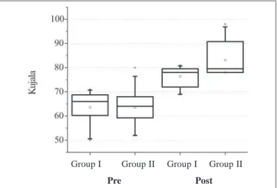
Figures 1 and 2 are box-plots representing graphic depictures of the VAS amounts and the Kujala scale found in both groups I and II in the pre- and post-treatment times.
TABLE 3
Normalized EM G activity of the VM O and VL muscles in concentric contraction (CC) and eccentric contraction (EC) in the pre- and post-treatment times related to the group I
Group I (N = 12)
M ean S.D. P
Pre Post Pre Post
VM O CC 51.57 53.31 ± 8.47 ± 8.83 –
VL CC 55.18 56.67 ± 8.26 ± 8.24 –
VM O/VL CC 00.93 00.94 ± 0.06 ± 0.07 0.79
VM O CE 33.33 35.32 ± 8.25 ± 9.26 –
VL CE 35.65 38.04 ± 8.55 ± 8.71 –
VM O/VL CE 00.93 00.92 ± 0.03 ± 0.07 0.85
M eans and Standard deviations (SD) of the EM G activity in RM S (expressed as a percentage of the M VIC) and the ratio related to the VM O and VL muscles in Concentric Contraction (CC) and Eccentric Contraction (CE) in the flexor-extensor OKC exercise of the knees (n = 12).
There w as no statistically significant differences in the VM O/VL ratio betw een the pre- and post-treatment times in the concentric (p = 0.79) and eccentric (p = 0.85) phases of the exercise.
As to the group II, table 4 show s the means and the standard deviations of the normalized EM G activity of the VM O and VL muscles, as w ell as the VM O/VL ratio in the concentric and eccen-tric phases of the semi-squatting exercise (CKC). These data show that there w ere no significant differences in the amounts of the VM O/VL ratio in the concentric (p = 0.56) and eccentric (p = 0.26) phases of the exercise after the treatment.
TABLE 4
Normalized EM G activity of the VM O and VL muscles in concentric contraction (CC) and eccentric contraction (EC) in the pre- and post-treatment times related to the group II
Group II (N = 12)
M ean S.D. P
Pre Post Pre Post
VM O CC 22.57 23.51 ± 7.99 ± 7.50 –
VL CC 25.31 26.79 ± 8.00 ± 7.87 –
VM O/VL CC 00.89 00.88 ± 0.10 ± 0.10 0.56
VM O CE 15.38 16.37 ± 5.87 ± 5.33 –
VL CE 22.09 22.01 ± 8.04 ± 6.37 –
VM O/VL CE 00.72 00.77 ± 0.17 ± 0.23 0.26
M eans and Standard Deviations (SD) of the EM G activity in RM S (expressed as a percentage of the M VIC) and the ratio related to the VM O and VL muscles in Concentric Contraction (CC) and Eccentric Contraction (EC) in the CKC semi-squatting exercise (n = 12).
Tables 3 and 4 show that the amounts of the VM O/VL ratio w ere low er than 1, indicating a decrease in the VM O activation com-pared to the VL. Such decrease w as observed both in the concen-tric and in the eccenconcen-tric phases in both assessed exercises, and it
Figure 2 – Comparison of the functionality betw een group I (n = 12) and II
(n = 12), measured by the Kujala scale in the pre- and post-treatment
Figure 1 – Comparison of the pain intensity betw een group I (n = 12) and
II (n = 12), measured by the analog-visual 10 cm scale, in the pre- and post-treatment times
In figure 1 it is verified the results related to the pain intensity measured by the VAS before and after the treatment.
The intergroup analysis has show n that before the beginning of the program, there w as no statistically significant difference be-tw een the means of groups I and II as to the pain’s VAS (p = 0.82). How ever, upon the completion of the program, group II attained an accentuated improvement compared to the group I (p = 0.02). The intergroup comparison disclosed that both groups presented a statistically significant improvement after the eight w eek treat-ment (p = 0.0005 for group I; p = 0.0005 for group II).
Figure 2 illustrates the results attained through the functional Kujala scale. As to the intergroup comparison, it w as observed no statistically significant different in the beginning of the treatment betw een them (p = 0.68). But w hen performing the same compar-ison at the end of the treatment, it w as evidenced better results for the group II (p = 0.03). As to the intergroup analysis, both pre-sented significant gains in the functionality after the treatment (p = 0.0005 for group I; p = 0.0005 for group II).
Group I Group II Group I Group II
Pre Post
Group I Group II Group I Group II
DISCUSSION
The results found in this study disclosed that after an eight w eek treatment, the groups attained a significant reduction in the pain intensity and an improvement to perform functional activities. The group II has show n higher results than group I in both assessed variables. These findings are in accordance to the ones found by Witvirouw et al.(17), w ho, after submitting 60 individuals to a five w eek treatment using OKC and CKC exercises, observed an in-crease in the torque peak of the FQ and ischiotibialis muscles, pain reduction and functional gain in both assessed groups.
In another study, Witvirouw et al.(18) reported excellent results related to the pain and functionality after treating individuals re-porting patellofemoral pain, but they did not evidence any effect of such treatment on the reflexive response time of the VM O and VL muscles. Likew ise, Stiene et al.(19) concluded that after an eight w eek treatment, the CKC exercises w ere more effective than the OKC exercises in the functional recovery of individual bearers of patellofemoral disorder.
Bennett & Stauber(20) initially assessed 41 individuals w ith PFSD, and they identified a reduction in the extensor torque of the knees during the eccentric phase of the OKC exercise. Thus, they ap-plied a treatment program using only OKC exercises performed on the isokinetic dynamometer, and they evidenced that in about four w eeks, the individuals had a reduction in the pain, re-estab-lishing the extensor torque of the knees, and they returned to the sportive activities.
The above exposed statement suggests that both the OKC and the CKC exercises can be employed to treat the PFSD. Neverthe-less, before prescribing the activities to strengthen the FQ, it is necessary to understand the biomechanical principles of the pa-tellofemoral joint in order to make an exercise scheduling that com-bines effectiveness and safety. In the present study, the knee’s ROM w as limited during the accomplishment of the exercises that integrated the treatment program, opposed to the previously men-tioned studies. According to Steimkamp et al.(7), w hile performing OKC exercises it must be avoided the last extension grades of the knees, since such angles have a low er joint contact, but the com-pressive strengths are distributed over a small area, thus increas-ing the patellofemoral stress.
As to the CKC exercises, the authors suggest to avoid higher than 45o angles of the knee flexion, since despite the higher joint stability w ith the increment of the flexion, there is also an increase in the compressive strengths and a higher patellofemoral stress. Similar results w ere described by Doucette & Child(21), w ho sug-gest performing OKC exercises w ith higher than 30o angles for the knee flexion, w hile CKC exercises must be performed in an-gles closed to its w hole extension.
The results related to the EM G activity has show n that after the treatment, there w as no significant differences in the VM O/VL ra-tio in the eccentric and concentric phases of the OKC and CKC exercises. How ever, the comparison of these findings w ith prior studies w as difficult, as it w as not found any w ork in the literature assessing the isolate effects of the muscular training in OKC and CKC on the EM G activation patterns of the stabilizer muscles of the patella in PFSD bearers. Nonetheless, even being not influ-enced by the treatment, the EM G activation patterns of the VM O and VL muscles has show n to be distinct in the concentric and eccentric phases in the assessed exercises. The RM S amounts related to the VM O/VL ratio in the pre- and post-treatment times disclosed an accentuated reduction in the VM O activation in the eccentric phase of the CKC exercise. These findings confirm w hat w as found by Shenny et al.(22), w ho reported a decrease in the VM O activation related to the VL in individuals w ith patellofemoral pain w hile descending steps (eccentric contraction). Souza & Gross(23) reported similar findings after assessing the VM O/VL ra-tio during isometric, concentric isotonic, and eccentric isotonic
contraction of the FQ, show ing a reduction in the EM G activity of the VM O compared to the VL in symptomatic individuals.
Ow ings & Grabiner(1) identified a higher EM G activity of the VL compared to the VM O in the eccentric phase of the leg extension OKC exercise, suggesting a deficit in the motor controlling of the patellar stabilizers in PFSD bearers.
On the other hand, Pow ers et al.(24) identified activation pat-terns similar to the VM O and VL muscles in individuals w ith patel-lofemoral pain, and they did not observe any commitment of the VM O activation w hile performing CKC activities. Similar results w ere described by Cerny(25), w ho did not observe significant dif-ferences in the VM O/VL ratio in individuals PFSD bearers per-forming CKC exercises. One possible explanation for the differ-ences in the results of these studies is related to the diversity of methods used to the acquisition and processing the electromyo-graphic signals(26). Furthermore, the inherent variability of the in-dividuals w ith PFSD remains as a challenge to determine the spe-cific activation patters of the stabilizer patella muscles among such population.
Even w hen it does not cause any alteration in the muscular ac-tivation patterns, the interventional schedule adopted in this study has show n to be effective to treat the PFSD. Such fact can be attributed to the strengthening of the FQ as a w hole, once the extensor muscles of the knees absorb part of the strength im-posed to the joint w hile performing activities that cause an over-load to it. Thus, it is believed that the recovery of the quadricipital function is able to re-establish the biomechanical properties of the patellofemoral and femorotibial functions, to increase the exten-sor torque of the knees and to improve the clinical and functional pictures in individual bearers of PFSD(17,20).
Pow ers et al.(27) identified that together w ith a reduction in the torque generated by the FQ, there w as a commitment of the loco-motive function in symptomatic individuals, pointing out the im-portance of the quadricipital strengthening in the PFSD treatment. In recent review s, Wilk & Reinold(28) and Crossley et al.(29) show ed evidences pointing that the FQ strengthening exercises are an in-dispensable part of rehabilitation programs involving patellofemo-ral disorders. But despite these reports, the mechanism by w hich the FQ strengthening promotes an increase in the functionality and reduction of the symptoms in individuals bearers of patellofem-oral pain is not quite clarified.
Before the scarcity of studies assessing the effects of the mus-cular training to treat the PFSD, it is necessary to conduct fur-ther studies involving a higher amount of individuals in the sam-pling, longer intervention periods and using different exercises to help in the collection of information that propitiate to perform and apply a more effective rehabilitation program that favor the improvement of the quality of life in individuals bearers of such affection.
CONCLUSION
In the experimental conditions used in this study, the OKC and CKC exercises promoted a reduction in the pain intensity and im-provement of the functionality in PFSD bearers. The CKC exercis-es have show n to be more effective compared to the CKC exer-cises.
As to the EM G activation patterns, the exercises w ere not able to change the amounts of the VM O/VL ratio. Nevertheless, the VM O muscle presented an accentuated reduction in the ac-tivation related to the VL in the eccentric phase of the CKC exer-cise.
REFERENCES
1. Ow ings TM , Grabiner M . M otor control of the vastus medialis oblique and vastus lateralis is disrupted during eccentric contractions in subjects w ith patellofemoral pain. Am J Sports M ed 2002;30:483-7.
2. Brody LT, Thein JM . Nonoperative treatment for patellofemoral pain. J Orthop Sports Phys Ther 1998;28:336-44.
3. Karst GM , Jew ett PD. Electromyographic analysis of exercises proposed for dif-ferential activation of medial and lateral quadriceps femoris muscle components. Phys Ther 1993;73:286-95.
4. Hanten WP, Schulthies SS. Exercise effect on electromyographic activity of the vastus medialis oblique and vastus lateralis muscles. Phys Ther 1990;70:561-5.
5. Lam PL, Gabriel YF. Activation of the quadriceps muscle during semisquatting w ith different hip and knee positions in patients w ith anterior knee pain. Am J Phys M ed Rahabil 2001;80:804-8.
6. W ilk KE, Escamilla RF, Fleisig GS, Barrentine SW, Andrew s JR, Boyd M L. A com-parison of tibiofemoral joint forces and electromyographic activity during open and closed kinetic chain exercises. Am J Sports M ed 1996;24:518-27.
7. Steinkamp LA, Dillingham M F, M arkel M D, Hill JA, Kaufman KR. Biomechanical considerations in patellofemoral joint rehabilitation. Am J Sports M ed 1993;21: 438-44.
8. Escamilla RF, Fleisig GS, Zheng N, Barrentine SW, Wilk K, Anderw s JR. Biome-chanics of the knee during closed kinetic chain and open kinetic chain exercises. M ed Sci Sports Exerc 1998;30:556-69.
9. Cyrino ES, Oliveira AR, Leite JC, Porto DB, Dias RM R, Segantin AQ, et al. Com-portamento da flexibilidade após 10 semanas de treinamento com pesos. Rev Bras M ed Esporte 2004;10:233-7.
10. Bennell K, Bartram S, Crossley K. Outcome measures in patellofemoral pain syn-drome: test retest reliability and inter-relationships. Phys Ther Sport 2000;1:32-4.
11. Kujala UM , Jaakola LH, Koshinen SK, Taimela S, Hurme M , Nelimarkka O. Scor-ing of patellofemoral disorders. Arthroscopy 1993;9:159-63.
12. De Luca CJ. The use of surface electromyography in biomechanics. J Appl Bio-mech 1997;13:135-63.
13. Lieb FJ, Perry J. Quadriceps function: an anatomical and mechanical study using amputated limbs. J Bone Joint Surg [Am] 1968;50:1535-48.
14. Araujo RC, Amadio AC. Análise biomecânica da ativação das porções superficiais do m. quadríceps femoral durante contrações excêntrica e concêntrica. Rev Bras Fisiot 1996;1:13-20.
15. Turker KS. Electromyography: some methodological problems and issues. Phys Ther 1993;73:698-710.
16. Basmajian JV, De Luca CJ. M uscle alive: their function revealed by electromyo-graphy. 5th ed. Baltimore: Willians & Wilkins, 1985.
17. Witvrouw E, Lysens R, Bellemans J, Peers K Vanderstraeten G. Open versus closed kinetic chain exercises for patellofemoral pain: a prospective, randomized study. Am J Sports M ed 2000;28:687-94.
18. Witvrouw E, Cambier D, Danneels L, Bellemans J, Werner S, Almqvist F, et al. The effect of exercise regimens on reflex response time of the vasti muscles in patients w ith anterior knee pain: a prospective randomized intervention study. Scand J M ed Sci Sports 2003;13:251-8.
19. Stiene HA, Brosky T, Reinking M F, Nyland J, M ason M B. A comparison of closed kinetic chain and isokinetic joint isolation exercise in patients w ith patellofemoral dysfunction. J Orthop Sports Phys Ther 1996;24:136-41.
20. Bennett JG, Stauber WT. Evaluation and treatment of anterior knee pain using eccentric exercise. M ed Sci Sports Exerc 1986;18:526-30.
21. Doucette AS, Child D. The effect of open and closed chain exercise and knee joint position on patellar tracking in lateral patellar compression syndrome. J Or-thop Sports Phys Ther 1996;23:104-10.
22. Sheehy P, Burdett RG, Irrgang JJ, VanSw earingen J. An electromyographic study of vastus medialis oblique and vastus lateralis activity w hile ascending and de-scending steps. J Orthop Sports Phys Ther 1998;27:423-9.
23. Souza DR, Gross M T. Comparison of vastus medialis obliquus: vastus lateralis muscle integrated electromyographic ratios betw een healthy subjects and pa-tients w ith patellofemoral pain. Phys Ther 1991;71:310-6.
24. Pow ers CM , Landel R, Perry J. Timing and intensity of vastus muscle activity during functional activities in subjects w ith and w ithout patellofemoral pain. Phys Ther 1996;76:946-55.
25. Cerny K. Vastus medialis oblique/vastus lateralis muscle activity ratios for select-ed exercises in persons w ith and w ithout patellofemoral pain syndrome. Phys Ther 1995;75:672-83.
26. M erletti R, Rainold A, Farina D. Surface electromyography for noninvasive char-acterization of muscle. Exerc Sport Sci Rev 2001;29:20-5.
27. Pow ers CM , Perry J, Hsu A, Hislop HJ. Are patellofemoral pain and quadriceps femoris muscle torque associated w ith locomotor function? Phys Ther 1997;77: 1063-78.
28. Wilk KE, Reinold M M . Principles of patellofemoral rehabilitation. Sports M ed Ar-throsc 2001;9:325-36.