157
1. Universidade Federal de São Paulo, São Paulo, SP, Brasil. 2. Universidade de São Paulo, São Paulo, SP, Brasil.
C
ASER
EPORTReceived for publication: 24/07/2016 - Accepted for publication: 19/11/2016. The authors declare no conflicts of interest.
Rev Bras Oftalmol. 2017; 76 (3): 157-60
Evaluation of preoperative subconjunctival single
application of mitomycin C in primary pterygium
Avaliação de aplicação única subconjuntival
pré-operatória de mitomicina C em pterígio primário
Thiago Gonçalves dos Santos Martins
1, Ana Luiza Fontes de Azevedo Costa
2, Karina Mie Furuzawa
2,
Milton Ruiz Alves
2, Roger Chammas
2A
BSTRACTPterygia are usually benign lesions that do not require specific treatment. It is a fibrovascular growth onto the nasal side of the cornea. It`s cause has not been fully elucidated yet, but seems to be related to long -term ultraviolet ray exposure. When symptoms are not con-trolled with conservative treatment surgery is considered, but the recurrence rate is still high, and efforts have been made to avoid it. Mitomycin C (MMC) is a fibroblast proliferation inhibitor that can be used as adjuvant to surgery to reduce recurrence. We report here a case that describes pterygium surgery performed in both eyes of the same patient, being one with MMC and the other eye without it. Both pterygium were sent to laboratory analysis. The results and proliferation index were compared between the eyes.
keywords: Mitomycin/therapeutic use; Pterygium/surgery; Pterygium/drug therapy; Recurrence
R
ESUMOPterígios são lesões geralmente benignas que na maioria dos casos não requer tratamento específico. É um crescimento fibrovascular sobre a córnea, geralmente a partir do lado nasal. Sua causa ainda não foi elucidada, mas parece estar relacionada à exposição aos raios ultravioleta. Quando os sintomas não são controlados com tratamento conservador, a cirurgia é indicada, porém o índice de recidiva ainda é alto, e os esforços têm sido no sentido de reduzir esse índice. A mitomicina C (MMC) é uma opção de adjuvante à cirurgia por ser um inibidor da proliferação de fibroblastos, diminuindo o risco de recorrência do pterígio. Relatamos aqui um caso que descreve cirurgia de pterígio realizada em ambos os olhos de uma mesma paciente, sendo um com MMC e outro sem ela. Os resultados e o índice de proliferação celular dos dois olhos foram comparados entre si.
158 Martins TGS, Costa ALFA, Karina Mie Furuzawa KM, Alves MR, Chammas R
Rev Bras Oftalmol. 2017; 76 (3): 157-60
I
NTRODUCTIONP
terygium is classically defined as a degenerative disease of the ocular surface, with a triangular formation of fibro-vascular tissue growing from the conjunctiva towards the surface of the cornea.(1) Although its pathogenesis has not yet beenfully elucidated, it is very likely that the pterygium represents a degenerative response of fibrous connective tissue to different stimuli. Among the risk factors for its development, exposure to ultraviolet radiation seems to play an important role in inducing limbo stem cell damage. As a result, a migration of fibrovascular tissue to the cornea occurs.(2) Other risk factors related to the
development of the pterygium are micro-traumas in the region of limbus and hereditary factors.
The main risk factor is exposure to ultraviolet rays, and a possible explanation for this would be the location of the pterygium, especially in the interpalpebral fissure, which is more exposed to the sun’s rays and dust, which generates inflammation of the ocular surface. It has recently been suggested that there is a mutation in the p53 gene of chromosome 17 related to the development of the pterygium, and also changes in the expression of various growth factors, such as vascular endothelial growth factor A (VEGFA). The pterygium is characterized by the elastomic degeneration of the conjunctival substance itself, with eosinophilic and basophilic deposits in addition to fibroblast proliferation. (1) Pterygium is twice
as common in men.(2) This pathology was first described in 1000 aC
by Susruta, the first ophthalmic surgeon according to the literature.
(3) Over the years, many medical treatments have been used , such
as bile, urine, acids, radiotherapy, thiotepa, 5-fluorouracil and, more recently, mitomycin C (MMC). In the past, the use of horsehair was described to remove pterygium.(4) The surgery is indicated when
the patient feels discomfort despite the use of ocular lubricant, when there is ocular motility restriction, growth restriction in the visual axis, and aesthetic complaints. Currently, the conjunctiva transplantation has been the surgical technique of choice. Some surgical techniques consist in pterygium excision leaving the sclera exposed, but the recurrence rate is up to 88%. (5,6)
The purpose of the use of mitomycin C as adjuvant tre-atment is to avoid the recurrence of pterygium after surgery. (7)
Case Report
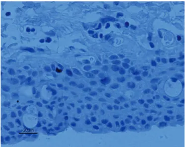
Patient EFM, female, 57 years, searched the ophthalmology service complaining of a progressive growth tissue covering both eyes for 10 years. She denied trauma, ocular surgery or any sys-temic disease. The exam showed the best corrected visual acuity of 20/20 in both eyes. In biomicroscopy, nasal pterygium grade II in RE and grade II in LE. Intraocular pressure of 12 mmHg in both eyes, and fundoscopy with no changes. After a week of appointment in the ophthalmology ambulatory, the patient underwent excision of pterygium in the RE, with conjunctival transplant and tissue sent for laboratory analysis. Two months after the first surgery, 0.1 ml of mitomycin C 0.02% was injected into the pterygium body of the left eye, and two weeks afterwards the pterygium excision was performed and the tissue was sent for analysis. Three months after the first surgery, pterygium of the right eye recurred. The complete follow-up took four years, and there was no recurrence in the left eye. Both surgeries were performed without complications by the same surgeon using the same technique of conjunctival transplant. Immunohistochemical analysis and evaluation of cell proliferation were performed by Ki-67 antibody detection (Clone MIB-1, Dako, M7240, Glostrup, Denmark). The patient developed cell proliferation rate in the right eye of 1.50%, and in the left eye of 4.3%. (Figures 1 and 2). There was no ocular complication due to the use of mitomycin in the four years of follow-up.
D
ISCUSSIONMMC is an alkylating agent that inhibits DNA synthesis. By inhibiting the synthesis of DNA, it leads to cellular death caused by inability to repair the genotoxic damage caused by alkylation. It acts on all cells regardless of the cell cycle, even acting on cells that are not synthesizing DNA. It has activity against all cells regardless of the phase of the cell cycle, and acts even on cells that are not synthesizing DNA. Inhibition of DNA synthesis leads to a reduction in the number of mitoses, especially when mitomycin C comes in contact with cells that
Figure 1. Immunohistochemical analysis and evaluation of cell proli-feration through the detection of Ki-67 antibody from the pterygium sample without the use of preoperative MMC
159 Evaluation of preoperative subconjunctival single application of mitomycin C in primary pterygium
Rev Bras Oftalmol. 2017; 76 (3): 157-60 are in the late G1 and early S phases of the cell cycle. MMC
can be used before, during or after pterygium surgery, locally applied or in the form of eyedrops. The application of injection directly into the pterygium has the advantage of protecting the endothelium and epithelium of the cornea. The subconjuntival injection allows a more precise application of the dose, which normally does not occur with the application of sponges directly on the sclera during the surgery. Its action in the prevention of recurrence occurs by inhibition of proliferation of fibroblasts in the episcleral region. Experimental studies have shown that MMC promotes selective action on the inhibition of fibroblast proliferation restricted to the site of its application and with action remaining for a long time. Increased concentration and duration of mitomycin C application may be associated with to complications such as necrotizing scleritis, scleral calcification, corneal ulceration, iritis, glaucoma, cataracts, hypotonia due to lesion of the ciliary body, besides damage to the corneal epithe-lium and endotheepithe-lium.(8,9)
The administration of MMC in pterygium surgery is con-sidered off-label by the Food and Drug Administration (FDA), but is allowed for the treatment of cancer.
In a study with 25 eyes, the preoperative subconjunctival injection of MMC was shown to be efficient, with two cases of delayed epithelization. Ninety-two percent of eyes with MMC application had no recurrence, and 8% had a two-week delay in corneal epithelization. There were no reports of serious compli-cations.(9) Donnenfeld reported the efficacy and safety of
preo-perative MMC injection of 0.1 ml (0.15 mg/ml) in the pterygium body one month prior to surgery to avoid recurrence. The results showed less vascularity and inflammation within the pterygium one month after injection of MMC, with a recurrence of 6% after 2 years of follow-up.(9)
The risk of preoperative injection is due to the impossibi-lity of washing the MMC that is in the space and subconjuntiva, and it can generate toxicity. Studies showed that the subconjunc-tival injection of MMC 0.2 ml (0.4 mg/mL) injected at 2 mm pos-terior to the limbus causes cellular changes such as flattening and pyknotic nuclei in the ciliary body epithelium, leading to reduced aqueous humor production one month after the injection. (9)
Carrasco et al.(10) reported a case of scleral necrosis in a patient
with severe dry eye who received a subconjuntival injection of MMC 0.15 mg/dL one month before pterygium surgery, being this one of the most feared long-term complications from use of mitomycin C.(11-17)
Among the immunohistochemical methods for evaluation of cell proliferation through the detection of monoclonal antibodies, antibody Ki-67 is the most widely used. It is a monoclonal antibody directed to a nuclear antigen present in proliferating cells. This antigen is expressed throughout the cell cycle, reaching a peak in phase G2 and mitosis. (8)
Knowing the behavior of fibroblasts cultured from ptery-gium, its proliferative potential, its interaction with several medications, its ultrastructure and its behavior compared to fibroblasts from other tissues, brings new perspectives for the understanding of the action mechanisms of medication already used in its treatment, as well as the possibility of using new com-pounds(8,18-20), since this disease whose first report of treatment
dates from the year 1000 aC does not yet have a treatment that can be considered “gold standard”. (4) With the case report, we
can see that mitomycin C presented a good therapeutic option with its preoperative use, reducing the risk of relapse and not showing use complications. The cell proliferation rate of the
material analyzed did not demonstrate a direct correlation with the recurrence, probably due to the action of mitomycin C being also important in other non-analyzed sites, such as in episclera fibroblasts, and being a case of primary pterygium with a low index of cell proliferation. Further studies with larger numbers of patients and long follow-up time can assist in defining the effectiveness of MMC in preventing the recurrence of ptery-gium surgery.
R
EFERENCES1. Bazzazi N, Ramezani A, Rabiee MA. A comparative study of conjunc-tival autograft and minimally invasive pterygium surgery in primary pterygia. Pak J Biol Sci. 2010;13(8):409-12.
2. Akinci A, Zilelioglu O. Comparison of limbal-conjunctival autograft and intraoperative 0.02% mitomycin-C for treatment of primaryp-terygium. Int Ophthalmol. 2007;27(5):281-5.
3. Detorakis ET, Spandidos DA. Pathogenetic mechanisms and tre-atment options for ophthalmic pterygium: trends and perspectives (Review). Int J Mol Med. 2009;23(4):439-47.
4. Kaufman SC, Jacobs DS, Lee WB, Deng SX, Rosenblatt MI, Shtein RM. Options and adjuvants in surgery for pterygium: a report by the American Academy of Ophthalmology. Ophthalmology. 2013;120(1):201-8.
5. Kam KW, Belin MW, Young AL. Monitoring corneal densities following primary pterygium excision with adjuvant topical mitomy-cin-c application-an observational study of corneal scar changes. Cornea. 2015;34(5):530-4.
6. Nassiri N, Farahangiz S, Rahnavardi M, Rahmani L, Nassiri N. Corneal endothelial cell injury induced by mitomycin-C in photorefractive keratectomy: nonrandomized controlled trial. J Cataract Refract Surg. 2008;34(6):902-8.
7. Biswas MC, Shaw C, Mandal R, Islam MN, Chakroborty M. Treatment of pterygium with conjunctival limbal autograft and mitomycin C-a comparative study. J Indian Med Assoc. 2007;105(4):200-4. 8. Zaky KS, Khalifa YM. Efficacy of preoperative injection versus
intra-operative application of mitomycin in recurrent pterygium surgery. Indian J Ophthalmol. 2012;60(4):273-6.
9. Donnenfeld ED, Perry HD, Fromer S, Doshi S, Solomon R, Biser S. Subconjunctival mitomycin C as adjunctive therapy before pterygium excision. Ophthalmology 2003;110(5):1012-26.
10. Carrasco MA, Rapuano CJ, Cohen EJ, Laibson PR. Scleral ulceration after preoperative injection of mitomycin C in the pterygium head. Arch Ophthalmol. 2002;120(11):1585-6.
11. Saldanha MJ, Yang PT, Chan CC .Scleral thinning after I BRITE procedure treated with amniotic membrane graft. Can J Ophthalmol. 2016;51(4):e115-6.
12. Moshirfar M, McCaughey MV, Fenzl CR, Santiago-Caban L, Kramer GD, Mamalis N. Delayed manifestation of bilateral scleral thinning after I-BRITE(®) procedure and review of literature forcosmetic
eye-whitening procedures. Clin Ophthalmol. 2015;9:445-51. Review. Erratum in: Clin Ophthalmol. 2016;10:187.
13. Long T, Li ZBare sclera resection followed by mitomycin C and/or autograft limbus conjunctiva in the surgery forpterygium: a meta-a-nalysis. Int J Ophthalmol. 2015;8(5):1067-73.
14. Rhiu S, Shim J, Kim EK, Chung SK, Lee JS, Lee JB, Seo KY. Complica-tions of cosmetic wide conjunctivectomy combined with postsurgical mitomycin C application. Cornea. 2012;31(3):245-52.
15. Wan Norliza WM, Raihan IS, Azwa JA, Ibrahim M. Scleral melting 16 years after pterygium excision with topical mitomycin C adjuvant therapy. Cont Lens Anterior Eye. 2006;29(4):165-7.
160 Martins TGS, Costa ALFA, Karina Mie Furuzawa KM, Alves MR, Chammas R
Rev Bras Oftalmol. 2017; 76 (3): 157-60
17. Dougherty PJ, Hardten DR, Lindstrom RL. Corneoscleral melt after pterygium surgery using a single intraoperative application of mitomycin-C. Cornea. 1996;15(5):537-40.
18. Almodin J, Almodin F, Almodin E, Minguete-Camara VC, Neves JP, Bezzon AK, et al. Efeitos de algumas drogas sobre a proliferação de fibroblastos de pterígio primário in vitro Rev Bras Oftalmol. 2013;72(2):108-11.
19. Cardilo JA, Alves MR, Jose NK, Camargo JC, Serpa JF, Ambrosio LE, et al. Eficácia da mitomicina C a 0, 04 por cento na prevençäo de recidivas do pterígio primário: aplicaçäo intra-operatória versus uso tópico pós-operatório Rev Bras Oftalmol. 1996;55(6):15-8.
20. Cardillo JA, Alves MR, Jose NK, Tranjan NA, Serpa JF, Ambrosio LF. Uso tópico pós-operatório versus aplicaçäo intra-operatória da mitomicina C a 0, 02 por cento na prevençäo de recidivas pós-ope-ratórias do pterígio primário. Arq Bras Oftalmol. 1995;58(6):413-5.
Corresponding author:
Thiago Gonçalves dos Santos Martins,
Rua Botucatu, 821 Vila Clementino, São Paulo Postal CEP: 04023-062