INTRODUCTION
Address to: Mara S. Benfato. Deptº de Bioísica/UFRGS. Av. Bento Gonçalves 9500/ Prédio 43431, 91501-970 Porto Alegre, RS, Brasil.
Phone: 55 51 3308-7603; Fax: 55 51 3308-7003. e-mail: [email protected]
Received in 08/05/2011 Accepted in 06/02/2012
Glutathione levels in and total anioxidant capacity of Candida sp. cells
exposed to oxidaive stress caused by hydrogen peroxide
Maxwel Adriano Abegg
[1],[2], Paulo Vinícius Gil Alabarse
[1],
Ártur Krumberg Schüller
[3]and
Mara Silveira Benfato
[1],[3][1]. Programa de Pós-Graduação em Biologia Celular e Molecular, Universidade Federal do Rio Grande do Sul, Porto Alegre, RS. [2]. Insituto de Ciências Exatas e Tecnologia, Universidade Federal do Amazonas, Itacoaiara, AM. [3]. Departamento de Bioísica, Universidade Federal do Rio Grande do Sul, Porto Alegre, RS.
Candida albicans, C. glabrata, C. tropicalis, and C. parapsilosis account for approximately 95% of ideniiable Candida infecions. Other species, including C. krusei, C. lusitaniae, and C. guilliermondii, account for less than 5% of cases of invasive candidiasis. The most common causative agent is still C. albicans, but its incidence is declining and the frequencies of other species are increasing. Recently, Furlaneto et al.1 noted that non-albicans Candida was the predominant
species in diferent clinical specimens, with the excepion of urine samples, in a Brazilian teriary-care hospital. Invasive candidiasis has a mortality rate that approaches 40%2,3. Although most people
are colonized by Candida sp., the majority never develop invasive candidiasis. Alteraions in host immunity, physiological features, or normal microlora, rather than the acquisiion of novel or hypervirulent factors by Candida, are suggested to degenerate the commensal-host interacion and lead to an opportunisic infecion4.
During the course of a systemic infection, Candida cells are engulfed by host phagocytes, where they are exposed to reacive oxygen species (ROS)5. ROS contribute to the killing of C. albicans
in both cultured cells and enire organisms6-9. Upon incubaion with
macrophages, C. albicans deoxyribonucleic acid (DNA) repair genes are transcripionally induced, suggesing that DNA damage indeed occurs in the phagosome and that genotoxic hypersensiivity stress would be disadvantageous to the pathogen10. Recently, it was demonstrated that
a large proporion of C. albicans cell surface anigens related to acute candidemia are involved in oxidaive stress4. In C. albicans, hyphal cells
ABSTRACT
Introducion: The capacity to overcome the oxidaive stress imposed by phagocytes seems to be criical for Candida species to cause invasive candidiasis. Methods: To beter characterize the oxidaive stress response (OSR) of 8 clinically relevant Candida sp., glutathione, a vital component of the intracellular redox balance, was measured using the 5,5’-dithiobis-(2-nitrobenzoic acid (DTNB)-glutathione disulide (GSSG) reductase reconversion method; the total anioxidant capacity (TAC) was measured using a modiied method based on the decolorizaion of the 2,2’-azinobis-(3-ethylbenzothiazoline-6-sulfonic) acid radical caion (ABTS*+). Both methods were used with cellular Candida sp. extracts
treated or not with hydrogen peroxide (0.5 mM). Results: Oxidaive stress induced by hydrogen peroxide clearly reduced intracellular glutathione levels. This depleion was stronger in Candida albicans and the levels of glutathione in untreated cells were also higher in this species. The TAC demonstrated intra-speciic variaion. Conclusions: Glutathione levels did not correlate with the measured TAC values, despite this being the most important non-enzymaic intracellular anioxidant molecule. The results indicate that the isolated measurement of TAC does not give a clear picture of the ability of a given Candida sp. to respond to oxidaive stress.
Keywords: Candida sp. Glutathione. Total anioxidant capacity.
are more resistant to oxidaive stress10, and hyphal formaion is higher
in isolates resistant to azole drugs11. Taking into account these data,
overcoming the oxidaive phagocyic challenge seems to be criical for the establishment of candidemia.
Candida species have evolved an anioxidant defensive response in order to withstand ROS atack, which encompasses, among other components, glutathione (GSH, L-γ-glutamyl-L-cysteinyl-glycine) and GSH-related aciviies (i.e., glutathione reductase, glutathione peroxidase, and glucose-6P-dehydrogenase)12. GSH is the most
abundant non-protein thiol in eukaryoic cells and its very low redox potenial (E’o = -240mV) provides the cell with redox bufer properies. In budding yeasts, GSH and its oxidized disulide form (GSSG) are involved in essenial physiological funcions, such as DNA and protein synthesis, transport, and cellular detoxiicaion13. Yeast isolates lacking
glutathione or that have altered glutathione redox states are sensiive to peroxide-induced oxidaive stress, superoxide anions, and lipid peroxidaion products13-16.
Numerous assays have been described to measure antioxidant status, but it seems that no ideal method is available17. Different
anioxidants can be measured separately, but the measurements are ime-consuming, labor-intensive, costly, and oten require complicated techniques18,19. Hence, the concept of a single test that might relect
the total antioxidant capacity (TAC) of biological fluids has elicited interest. The most widely used colorimetric methods to measure TAC are 2,2’-azinobis-(3-ethylbenzothiazoline-6-sulfonic) acid radical caion (ABTS*+)-based methods. Reduced ABTS, a colorless molecule, is oxidized
to ABTS*+, which is characterisically blue-green. When this radical is
mixed with any oxidizable substance, it is reduced to its colorless form18.
Diferent Candida sp. exhibit unequal oxidaive stress resistances in vitro20-22, and diferent in vitro virulence potenials23, and we proposed
that this may contribute to the capacity of each species to cause candidemia22. Taking into account these diferences, total glutathione
RESULTS
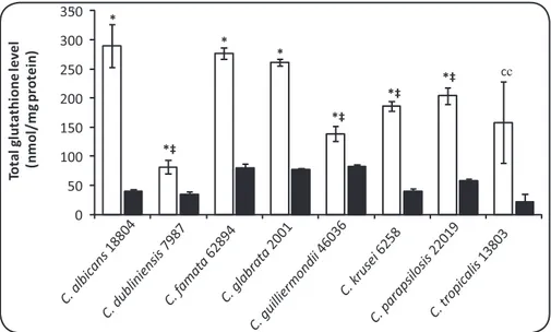
In the present work, the levels of total intracellular GSH following mild oxidaive stress in Candida sp. were determined. GSH levels ranged from 80 to 290nmol/mg of protein in untreated samples and from 21 to 83nmol/mg of protein in treated samples (Figure 1). With excepion of C. tropicalis, all species tested exhibited a signiicant reducion in total GSH levels following exposure to mild oxidaive stress (0.5mM H2O2). C. albicans presented the most dramaic reducion. In untreated samples, C. albicans presented the highest GSH levels and these levels were signiicantly higher than those seen in C. dubliniensis, C. guilliermondii, C. krusei, C. parapsilosis, and C. tropicalis (p < 0.05) (Figure 1). The TAC results were quite varied in each species (Figure 2). One C. albicans isolate (51), 2 C. guilliermondii isolates (6260 and 73), and 1 C. krusei isolate (6258) presented the highest TAC levels. With excepion to C. guilliermondii isolate 73 in comparison with C. tropicalis isolate 55, the isolates cited above exhibited signiicant diferences in TAC levels compared to all other isolates tested (p < 0.05).
Concerning Spearman rank correlaion coeicient, TAC results did not correlate (rho = 0.051) with sensitivity of Candida sp. isolates to oxidaive stress. TAC results also did not correlate with total intracellular GSH levels in untreated (rho = 0.042) and treated (rho = 0.058) samples.
METHODS
Yeast isolates and culivaion
The following yeast isolates were used: C. albicans type strain ATCC 18804, C. dubliniensis type strain from the Centraalbureau voor Schimmelcultures (CBS) 7987, C. famata ATCC 62894, C. glabrata type strain ATCC 2001, C. guilliermondii type strain ATCC 46036, C. guilliermondii ATCC 6260, C. krusei type strain ATCC 6258, C. parapsilosis type strain ATCC 22019, and C. tropicalis ATCC 13803. The clinical isolates used were as follows: C. albicans 1 (isolate from a paient with nosocomial infecion); C. albicans 51 (isolate from the orotracheal tube of an acquired immunodeiciency syndrome (AIDS) paient); C. dubliniensis 23 and C. dubliniensis 25 (both from the oropharynx of AIDS paients); C. famata 1 and C. famata 24 (both clinical isolates from paients with nosocomial infecion); C. glabrata 1, C. glabrata 75, and C. glabrata 118 (all obtained from catheter ips); C. guilliermondii 73 (clinical isolate from a paient with nosocomial infecion); C. krusei 1 and C. krusei 2 (both isolated from skin lesions of diabeic paients); C. parapsilosis 81 and C. parapsilosis 115 (both isolated from paients with onychomycosis); C. tropicalis 1 (isolated from an oral granuloma); and C. tropicalis 55 and C. tropicalis 56 (both clinical isolates from a paient with nosocomial infecion).
The isolates were identified and maintained as previously described22. Viable cells were obtained by culivaion on solid yeast
extract-peptone-dextrose (YPD) medium (1% yeast extract, 2% peptone, 2% glucose, 2% agar), and isolates were then grown in liquid YPD medium in an orbital shaker at 30°C/100 rpm to late exponenial growth (OD600nm = 1.5–1.6). Cells were washed twice with sterile disilled water and diluted to OD600nm = 0.15 in fresh liquid YPD for use. Cells were grown at 30°C rather than at 37°C because C. dubliniensis and C. famata grow beter at 30°C.
Cell-free extracts
Cell suspensions (1.5mL) were centrifuged for 5 min at 8,000g and lysed by adding 0.5mL of lysis bufer (50mM Tris-Cl, 150mM NaCl, 50mM ethylenediamine tetraaceic acid [EDTA], pH 7.2), 50mM phenyl methyl sulfonyl luoride (PMSF; Sigma, St. Louis, MO) and approximately 0.5 g of glass beads (diameter, 425-600µm; Sigma). Lysis was performed by vortexing for 3 mixing cycles of 3 min with 1-min intervals for cooling on ice. Breakage was checked microscopically. The samples were then centrifuged for 10 min at 8,000g to remove cellular debris and beads.
Total glutathione assay
Total intracellular glutathione was determined by the 5,5'-dithiobis-(2-nitrobenzoic acid (DTNB)-glutathione disulide GSSG reductase recycling method24,25.Cell suspensions were left untreated or
were treated with 0.5mM H2O2, incubated for 1h with agitaion at 100rpm/30°C, washed with sterile distilled water, and then resuspended to the same volume in 100mM potassium phosphate bufer (pH 7.0), lysed, and centrifuged. Then, 25µL aliquots of the supernatants were vortexed thoroughly with an equal volume of 2M HClO4 and 4mM EDTA. After 15 min incubation at 0°C, the suspensions were centrifuged for 5 min at 8,000g and 45µL of the supernatant was pH-neutralized by adding 3µL of 2M KOH at 0°C. This
was centrifuged for 1 min at 8,000g and 35µL of the supernatant was added to a mixture containing 174µL of 100mM phosphate bufer (pH 7.0), 17µL of 4mM NADPH, and 7µL of glutathione reductase soluion (6U/mL). This was mixed and incubated for 5 min at 37°C. Then, 18µL of DTNB reagent (0.040g of DTNB [Sigma] dissolved in 10ml of 50mM potassium phosphate bufer, pH 7.0) was added, and the absorbance was read at 412nm ater a 2-min incubaion at 37°C.
Total anioxidant capacity
A modiied method based on ABTS*+ decolorizaion described
by Erel18 was employed. Cell suspensions were treated with 0.5mM
H2O2, washed, lysed, and centrifuged, as previously described22, and
5µL of each supernatant was mixed with 200µL of 0.4 mol/L acetate bufer, pH 5.8. Then, 20µL of ABTS*+ in 30mM acetate bufer, pH
3.6, was added, mixed, and the absorbance measured ater 5 min. The absorbance of a soluion without ABTS*+ was also measured
as the blank. The vitamin E water-soluble analogue 6-hydroxy-2,5,7,8-tetramethylchroman-2-carboxylic acid (Trolox) was used as the standard, and data were expressed in terms of mmol Trolox equivalent per milligram of protein.
Total protein content
The total protein content in lysed cells was determined by the Bradford assay (Bio-Rad, Hercules, CA).
Staisics
0 50 100 150 200 250 300 350
Tot
a
l gl
u
tat
h
io
n
e
le
ve
l
(n
m
o
l/
m
g
p
rot
e
in
)
*
*‡ *
*
*‡
*‡
*‡ cc
c
C. albicans
18804
C. d ublin
iens is 7987
C. famat a 62894
C. glabrat a 2001
C. g uilli
erm ondi
i 46036
C. krusei
6258
C. parapsilosi s 22019
C. tropicalis
13803
FIGURE 1 -Efect of 0.5 mM H2O2 on the total intracellular glutathione concentraion (nmol (GSH + 2GSSG) mg-1 protein) in Candida sp. Cells were treated (black bars) or not (white bars), as described in Methods. Asterisks (*) indicate signiicant diferences (p < 0.01) between untreated and treated samples. The symbol (‡) indicates signiicant diferences between untreated Candida albicans cells and the isolates labeled. The data are the mean ± SD values of 3 independent experiments. C:Candida.
0
10 20 30 40 50 60
T
A
C
(m
m
o
l
Tr
o
lo
x
e
q
u
iv
a
le
n
t/
m
g
p
ro
te
in
)
† †
#
#
FIGURE 2 -Total anioxidant capacity in Candida sp. cells ater 0.5mM H2O2 treatment. Cells were treated as described in Materials and Methods. Each column corresponds to the Candida isolate ideniied below. The symbol (†)indicates signiicant diferences (p < 0.05) between the labeled isolate and all the other isolates tested. The symbol (#) indicates that the isolate Candida guilliermondii was not signiicantly diferent from the isolate Candida tropicalis 55 (p > 0.05), but was signiicantly diferent from all other isolates tested. The data represent the mean ± SD values of 3 independent experiments. C:Candida; TAC: total anioxidant capacity.
DISCUSSION
The virulence of Candida albicans seems to be mulifactorial26, but
the ability of this fungus to mount stress responses is an important aspect, as this promotes survival in the host during systemic infecions27.
In a previous study by our group22, we analyzed the oxidaive efects
(degree of resistance and induction of oxidative damage) and anioxidaive efects (capacity to adapt and inducion of anioxidaive enzymes). Here, we coninued the characterizaion of the oxidaive stress response (OSR) of 8 clinically relevant Candida sp.
Hydrogen peroxide was used to generate oxidaive stress. H2O2 itself is not very reacive, but can be further reduced to extremely damaging hydroxyl radicals. Therefore, all aerobic cells are equipped
with H2O2-removing enzymes. Furthermore, evidence suggests that H2O2 is produced transiently in response to the acivaion of many cell surface receptors and serves as an intracellular messenger. The imely eliminaion of intracellular messengers ater the compleion of their mission is criical for receptor signaling. This would seem especially true for H2O228. According to Ng et al.29 the network of enzymes that
detoxify H2O2 in biological systems has at least 3 nodes: catalase (which is the longest known enzyme for the removal of H2O2 and requires no cofactors), 6 members of the peroxiredoxin family of enzymes, and the glutathione peroxidases that rely on GSH as the electron donor and speciic cofactor.
The GSH levels (90-152) observed by Fekete et al.30 in untreated
Consistent with this, Lemar et al.31 showed that a 30-min C. albicans
exposure to 0.5 mM diallyl disulide (a garlic oxidaive consituent) decreased intracellular GSH and elevated ROS intracellular levels. It was also demonstrated that H2O2 exposure causes a reducion in intracellular GSH levels, paricularly for Saccharomyces cerevisiae, as well as a shit in the GSH redox balance towards the oxidized form, GSSG, as reviewed in Penninckx13. Thomas et al.32 reported a
dramaic decline in the level of intracellular GSH, concomitant with the yeast-to-mycelial conversion, in C. albicans. Consistent with this, Michán and Pueyo33 reported that the GSH levels in C. albicans
hyphae were approximately 50% of those in yeasts. Considering that oxidaive stress diminishes GSH levels33,34, our treatment with H
2O2 was
expected to reduce GSH content. Pacheco et al.25 demonstrated that
cadmium treatment increased ROS producion, depleted intracellular GSH concentraions, and increased external GSH concentraions. Furthermore, González-Párraga et al.12 used the oxidant
1-chloro-2,4-dinitrobenzene to reduce intracellular GSH levels in Candida. Madeo et al.35 also demonstrated that treatment with 3 mM H
2O2 induced
intracellular GSH depleion and apoptosis in S. cerevisiae.
In contrast, Fekete et al.36 found GSH levels of ~160nmol/mg
of protein in untreated isolates of C. albicans and ~260nmol/mg of protein ater treatment with 1mM tert-buil-hydroperoxide, an oxidant. Lee et al.37 found that a 6-h treatment with 0.1mM H
2O2
provoked a 3.14-fold elevaion in GSH levels in Schizosaccharomyces pombe. Manfredini et al.38 reported an increase in GSH levels upon
treatment with 0.5mM H2O2 in wild-type S. cerevisiae cells and a signiicant reducion in those levels with 5mM H2O2. These diferences regarding our results may be related to the duraion of treatment, the use of diferent oxidants or lower doses of hydrogen peroxide, or to diferences in the metabolic aciviies of the species. In the case of S. cerevisiae, it could be related to the higher sensiivity of this species to 0.5mM H2O2 in comparison to that of Candida sp. This concentraion may induce 40% lethality in S. cerevisiae and is normally sublethal (95-100% viability) in the case of Candida sp.22,39.
GSH can occur in yeasts in the reduced form (GSH), the oxidized form (GSSG), and as diferent mixed disulides, for example GS-S-CoA and GS-S-Cys13. The H
2O2 (0.5 mM/1h treatment) used to induce oxidaive
stress was probably detoxiied in part through the acion of the enzyme glutathione-peroxidase (GPx) and the concomitant conversion of GSH into GSSG29. Increasing GSSG levels can potenially inhibit protein
synthesis in animal and plant cells40,41, and because of this, Candida cells
are likely to export GSSG under condiions of oxidaive stress, resuling in a decrease in total intracellular glutathione levels.
In yeasts, peroxide resistance has been associated with intracellular GSH levels42-44. Further, it has been previously proposed
that the rate of removal of H2O2 is a direct funcion of GPx acivity × GSH29. The highest levels of GSH observed and the intense diminuion
of intracellular GSH levels in C. albicans (Figure 1), together with the GPx aciviies previously observed for this species20 (Abegg et
al.unpublished results), may indicate a more eicient detoxiicaion system of H2O2 through GPx/GSH in C. albicans than in other Candida sp. However, the limitaions of the GSH results should be noted, paricularly because of the use of 1 isolate of each species, and further comparisons regarding GSH metabolism should be made.
Total anioxidant capacity assays may be broadly classiied as electron transfer (ET)-based and hydrogen atom transfer (HAT)-based
assays, although these 2 mechanisms may not be difereniated with disinct boundaries in some cases. In fact, most non-enzymaic anioxidant acivity (e.g., scavenging of free radicals and inhibiion of lipid peroxidaion) is mediated by redox reacions. Electron transfer assays include the ABTS, Trolox equivalent antioxidant capacity (TEAC), cupric-reducing anioxidant capacity (CUPRAC), di(phenyl)-(2,4,6-trinitrophenyl)iminoazanium (DPPH), Folin-Ciocalteu, and ferric-reducing anioxidant power (FRAP) methods, each of which use diferent chromogenic redox reagents with diferent standard potenials (reviewed in Apak et al.45).
The ET mechanism of anioxidant acion is based on the following reacions: (1) ROO. + AH/ArOH → ROO- + AH.+/ArOH.+, (2) AH.+/ArOH.+
+ H2O ↔ A./ArO. + H3O+, and (3) ROO- + H
3O+↔ ROOH + H2O; these
reacions are relaively slower than those of HAT-based assays and are solvent- and pH-dependent45. Re et al.46 and Erel18 developed improved
ABTS radical caion decolorizaion assays using persulfate and hydrogen peroxide, respecively, as the oxidant, and thereby compensated for the weaknesses of the original ferryl myoglobulin/ABTS assay. The 3 TEAC tests developed at diferent periods, namely the TEAC assay I (ABTS*+ generated enzymaically with metmyoglobin and hydrogen
peroxide), TEAC II (radical generaion with iltraion over the MnO2 oxidant), and TEAC III (with K2S2O8 oxidant), are enirely diferent from each other, are applicable to diferent solvent media, and their indings for a given anioxidant can vary signiicantly. The ‘pre-addiion technique’ as in TEAC I, involving the addiion of anioxidants before radical generaion, could result in an overesimaion of anioxidant capacity, because many substances interfere with the formaion of the radical; therefore, TEAC I measures the ability to delay radical formaion as well as the scavenging of the radical45.
The advantages of ABTS/TEAC are reported to be operaional simplicity, reproducibility, diversity, and most importantly, lexible usage in multiple media to determine both the hydrophilic and lipophilic anioxidant capaciies of physiological luids, since the reagent is soluble in both aqueous and organic solvent media. Aqueous- and lipid-soluble anioxidants are not separated in the TAC protocol employed; therefore, the combined anioxidant aciviies of all its consituents, including vitamins, proteins, lipids, glutathione, and uric acid, are assessed45.
The intra-speciic TAC variaion found here is in agreement with observed variaions in the sensiiviies of C. albicans isolates to oxidants47.However, the TAC results did not correlate (rho = 0.051) with
the previously reported sensiivity of Candida sp. isolates to oxidaive stress22. One C. albicans isolate (51), 2 C. guilliermondii isolates (6260
and 73), and 1 C. krusei isolate (6258) showed the highest TAC levels. With the excepion of the comparison of C. guilliermondii isolate 73 with C. tropicalis isolate 55, the isolates cited above exhibited signiicant diferences in TAC levels in comparison to all the other isolates tested (p < 0.05).
Lapshina et al.48 compared diferences in the ability to scavenge
ACKNOWLEDGMENTS The TAC based on the scavenging of ABTS free radicals showed a good
correlaion with the radiaion resistance of the yeasts. According to the authors, the results point to the importance of factors other than anioxidaive enzymes and glutathione, in the determinaion of cellular resistance to ionizing radiaion and other types of free-radical stress in S. cerevisiae.
Balcerczyk et al.49 demonstrated that the TAC of cell extracts of
S. cerevisiae showed a stronger dependence on the thiol content as evidenced by the efect of -SH blocking with n-ethylmaleimide (NEM). TAC measured ater 10 s was decreased by 83-90% (in diferent strains) following thiol modiicaion, while TAC measured ater a 1-min reducion of ABTS*+ was decreased by 73-80%. According to the
authors, the results indicate that thiol groups are a major contributor to the TAC of S. cerevisiae and perhaps of other yeast species. These results demonstrate that in cell extracts, in contrast to extracellular luids, thiol groups consitute the dominant determinant of total anioxidant capacity, at least in S. cerevisiae. Depleion of thiols leads to a decrease in TAC. However, cellular adaptaion to oxidaive stress may involve the mobilizaion of mechanisms other than an increase in thiol concentraions. This is especially evident in yeast cells, where strains deicient in anioxidant enzymes show increased values of TAC due mainly to thiol-independent mechanisms. Similarly, the adaptaion of yeast to condiions of staionary culture and reoxygenaion ater growth under anoxia predominantly involves anioxidants other than thiols, as demonstrated by Balcerczyk et al.49.
Considering the mode of acion of the enzymes of the peroxiredoxin (Prx) family, which consists of thiol-dependent peroxidases involved in the removal of various types of hydroperoxides in cells, such as hydrogen peroxide, organic peroxides, and peroxynitrite50,51, and
based on the results described above, these enzymes seem to be criical in determining the TAC of yeast cells. In addiion to detoxifying peroxides, speciic peroxiredoxins have been shown to act as molecular chaperones and to play roles in regulaing hydrogen peroxide-mediated cell signaling events51. In S. cerevisiae, for example, the steady-state
protein level of the peroxiredoxin Tsa1 is 45 imes that of Gpx352,53.
Tsa1 is also the key peroxidase suppressing genome instability and protecing against cell death in S. cerevisiae54,55. Furthermore, in
S. cerevisiae, Demasi et al.56 demonstrated the importance of cytosolic
thioredoxin peroxidase I (cTPxI) and its reductant suliredoxin in the protecion of cells sufering mitochondrial dysfuncion, against H2O2 -induced death.
In S. cerevisiae, there are 5 isoforms of Prx, distributed in diferent cellular compartments57. The 2 most abundant peroxiredoxins in these
species are Ahp1 and Tsa158. The TSA1 gene is present in C. albicans,
C. glabrata, C. tropicalis, and C. dubliniensis and is similar to the TSA1
and TSA2 of S. cerevisiae. AHP 11, AHP 12, and AHP 13 are genes from strain SC5314 of C. albicans and show similarity to the S. cerevisiae
alkyl hydroperoxide reductase AHP1 (YLR109W) 59.
Urban et al.60 reported the ideniicaion of Tsa1p, a protein that is
diferenially localized to the cell wall of C. albicans in hyphal cells but remains in the cytosol and nucleus in yeast-form cells. According to the authors, this is diferent from S. cerevisiae, where the homologous protein solely has been found in the cytoplasm. These authors reported that TSA1 confers resistance towards oxidaive stress in addiion to being involved in the correct composiion of hyphal cell walls. Shin et al.61 also observed that the protein Tsa1p codiied by
this gene was indispensable in the yeast-to-hyphal transiion when C. albicans was cultured under oxidaive stress. In C. albicans, the genes AHP1 and HSP12 are regulated by the response regulator gene SSK1, and those genes are associated with cell wall biosynthesis and adaptaion to oxidaive stress62. Therefore, it seems that the
peroxiredoxin system is criical for the funcioning of the anioxidant system of Candida and is one of the most important contributors to the TAC in Candida cell lysates.
As far as we are aware, this is the irst atempt to use a single test of TAC in Candida. The use of a single marker of anioxidant capacity has drawbacks and these data must be interpreted with cauion. According to Young17, these single markers measure predominantly low molecular
weight and chain-breaking anioxidants, excluding the contribuions of anioxidant enzymes and metal binding proteins. The fact that the TAC results did not correlate with the sensiivity of Candida sp. isolates to oxidaive stress has been reported previously22 (Abegg et
al. unpublished results); the fact that it also did not correlate with total intracellular GSH levels in untreated (rho = 0.042) and treated (rho = 0.058) samples may indicate that a single marker cannot provide a picture of the anioxidant capacity of a Candida sp.
Fekete et al.30 searched for C. albicans isolates that are naturally
resistant to oxidaive stress but did not ind this phenotype. They argued that the selecion of mutants that are tolerant to oxidaive stress in vivo would be beneicial to the pathogen-phagocyte interacion, but would be unlikely because of the concomitant and disadvantageous changes in virulence atributes, like morphological transiions and phospholipase secreion. They also point out that an over-eicient anioxidaive defense system may be disadvantageous for C. albicans by hindering the ROS-triggered acivaion of genomic ageing and cell death programs that promote adaptaion to stresses in the human body. Besides the unlikely selecion of C. albicans mutants that are naturally oxidant-resistant, certain species like C. dubliniensis, C. guilliermondii, and C. famata are probably not evoluionarily prepared to cope with the irst line of immune defense and oxidaive stress, even in moderately immunocompromised individuals. This would be relected in the relaive incidence of this species as a causal agent of invasive candidiasis.
Macrophages and neutrophils use ROS, reacive nitrogen species (RNS), and chlorine species for host protecion6-9,63, but the idea that
ROS exert direct in vivo efects in the fungal killing of Candida and other species is sill controversial. Balish et al.64 studied the deicient producion
of ROS and RNS in mice using gastrointesinal Candida inoculaion. Although these mice died, an exaggerated immune response rather than an overwhelming fungal infecion appeared to cause death. Further, an in vitro study with phagocytes from normal and ROS/RNS-deicient mice revealed equal abiliies of both to kill C. albicans. Wellington et al.65
considered these data to be in agreement with their results of C. albicans suppression of ROS producion in phagocytes. However, it seems to be clear that Candida species have disinct capaciies to deal with oxidaive stress, and the inhibition of specific antioxidant molecules may be therapeuically useful in the future.
The authors declare that there is no conlict of interest.
CONFLICT OF INTEREST
REFERENCES
1. Furlaneto MC, Rota JF, Quesada RMB, Furlaneto-Maia L, Rodrigues R, Oda S, et al. Species distribuion and in vitro luconazole suscepibility of clinical Candida isolates in a Brazilian teriary-care hospital over a 3-year period. Rev Soc Bras Med Trop 2011; 44:595-599.
2. Pfaller MA, Diekema DJ. Epidemiology of invasive candidiasis: a persistent public health problem. Clin Microbiol Rev 2007; 20:133-163.
3. Sobel JD. Changing trends in the epidemiology of Candida blood stream infecions: A mater for concern? Criical Care Medicine 2010; 38:990-992.
4. Mochon AB, Ye J, Kayala MA, Wingard JR, Clancy CJ, Nguyen MH, et al. Serological Proiling of a Candida albicans Protein Microarray Reveals Permanent Host-Pathogen Interplay and Stage-Speciic Responses during Candidemia. Plos Pathog 2010; 6:1-14.
5. Vazquez-Torres A, Balish E. Macrophages in resistance to candidiasis. Microbiol Mol Biol Rev 1997; 61:170-192.
6. Thompson HL, Wilton JMA. Interacion and intracellular killing of Candida albicans blastospores by human polymorphonuclear leukocytes, monocytes and monocyte-derived macrophages in aerobic and anaerobic condiions. Clin Exp Immunol 1992; 87:316-321.
7. Stevenhagen A, Vanfurth R. Interferon-gamma acivates the oxidaive killing of
Candida albicans by human granulocytes. Clin Exp Immunol 1993; 91:170-175.
8. Donini M, Zenaro E, Tamassia N, Dusi S. NADPH oxidase of human dendriic cells: Role in Candida albicans killing and regulaion by interferons, decin-1 and CD206. Eur J Immunol2007; 37:1194-1203.
9. Aratani Y, Kura F, Watanabe H, Akagawa H, Takano Y, Suzuki K, et al. Criical role of myeloperoxidase and nicoinamide adenine dinucleoide phosphate-oxidase in high-burden systemic infecion of mice with Candida albicans. J Infect Dis 2002; 185:1833-1837.
ABSTRACT IN pORTUGUESE
Níveis de glutaiona e capacidade anioxidante total em células de Candida sp. expostas a estresse oxidaivo
causado por peróxido de hidrogênio
Introdução: A capacidade de suportar o estresse oxidaivo imposto por fagócitos parece ser críica para que espécies de Candida causem candidíase invasiva. Métodos: Para melhor caracterizar a resposta ao estresse oxidaivo (REO) de oito Candida sp. clinicamente relevantes, um componente vital do balanço redox intracelular, a glutaiona, foi mensurada pelo método de reconversão DTNB-GSSG redutase e a capacidade anioxidante total (CAT) foi mensurada por um método modiicado baseado na descoloração do ABTS*+. Ambos os métodos
foram utilizados em extratos celulares das espécies de Candida
tratadas ou não com peróxido de hidrogênio (0,5mM). Resultados: O estresse oxidaivo induzido pelo peróxido de hidrogênio claramente reduziu os níveis intracelulares de glutaiona. Esta diminuição foi mais intensa em C. albicans e os níveis de glutaiona em células não tratadas foram também maiores nesta espécie. A capacidade anioxidante total demonstrou variação intraespecíica na capacidade anioxidante. Conclusões: Os níveis de glutaiona não se correlacionaram com a capacidade antioxidante total mensurada, apesar desta ser a defesa anioxidante intracelular não-enzimáica mais importante. Os resultados indicam que a medição isolada da CAT não fornece um quadro claro da habilidade de certa espécie de Candida responder ao estresse oxidaivo.
Palavras-chaves:Candida sp. Glutaiona. Capacidade anioxidante total.
10. Lorenz MC, Bender JA, Fink GR. Transcripional response of Candida albicans upon internalizaion by macrophages. Eukaryot Cell 2004; 3:1076-1087.
11. Costa CR, Souza LKH, Ataídes FS, Ferri PH, Costa MP, Fernandes OFL, et al. Molecular analysis and dimorphism of azole-suscepible and resistant Candida albicans isolates. Rev Soc Bras Med Trop 2011; 44:740-744.
12. Gonzalez-Parraga P, Marin FR, Arguelles JC, Hernandez JA. Correlaion between the intracellular content of glutathione and the formaion of germ-tubes induced by human serum in Candida albicans. Biochim Biophys Acta 2005; 1722:324-330. 13. Penninckx MJ. An overview on glutathione in Saccharomyces versus
non-convenional yeasts. FEMS Yeast Res 2002; 2: 295-305.
14. Carmel-Harel O, Storz G. Roles of the glutathione- and thioredoxin-dependent reducion systems in the Escherichia coli and Saccharomyces cerevisiae responses to oxidaive stress. Annu Rev Microbiol 2000; 54:439-461.
15. Grant CM. Role of the glutathione/glutaredoxin and thioredoxin systems in yeast growth and response to stress condiions. Mol Microbiol 2001; 39:533-541. 16. Belozerskaya TA, Gessler NN. Reactive oxygen species and the strategy
of antioxidant defense in fungi: A review. Appl Biochem Microbiol 2007; 43:506-515.
17. Young IS. Measurement of total anioxidant capacity. J Clin Pathol 2001; 54:339. 18. Erel O. A novel automated direct measurement method for total anioxidant
capacity using a new generation, more stable ABTS radical cation. Clinical Biochemistry 2004; 37:277-285.
19. Erel O. A new automated colorimetric method for measuring total oxidant status. Clin Biochem 2005; 38:1103-1111.
20. Tosello ME, Biasoli MS, Luque AG, Magaro HM, Krapp AR. Oxidaive stress response involving inducion of protecive enzymes in Candida dubliniensis. Med Mycol 2007; 45:535-540.
21. Cuéllar-Cruz M, Briones-Martin-Del-Campo M, Canas-Villamar I, Montalvo-Arredondo J, Riego-Ruiz L, Castaño I, et al. High resistance to oxidaive stress in the fungal pathogen Candida glabrata is mediated by a single catalase, Cta1p, and is controlled by the transcripion factors Yap1p, Skn7p, Msn2p, and Msn4p. Eukaryot Cell 2008; 7:814-825.
22. Abegg MA, Alabarse PVG, Casanova A, Hoscheid J, Salomon TB, Hackenhaar FS, et al. Response to Oxidaive Stress in Eight Pathogenic Yeast Species of the Genus
Candida. Mycopathologia 2010; 170:11-20.
23. Rörig KCO, Colacite J, Abegg MA. Produção de fatores de virulência in vitro por espécies patogênicas do gênero Candida. Rev Soc Bras Med Trop 2009, 42:225-227. 24. Akerboom T, Sies H. Assay of glutathione, glutathione disulide, and glutathione
mixed disulides in biological samples. Methods Enzymol 1981; 77:373-382. 25. Pacheco CC, Passos JF, Castro AR, Moradas-Ferreira P, Marco P. Role of respiraion
and glutathione in cadmium-induced oxidaive stress in Escherichia coli K-12. Arch Microbiol 2008; 189:271-278.
26. Chauhan N, Inglis D, Roman E, Pla J, Li D, Calera JA, et al. Candida albicans response regulator gene SSK1 regulates a subset of genes whose funcions are associated with cell wall biosynthesis and adaptaion to oxidaive stress. Eukaryot Cell 2003; 2:1018-1024.
27. d'Enfert C, Hube B. Candida: comparaive and funcional genomics. Norfolk, U.K: Caister Academic Press; 2007.
28. Rhee SG, Kang SW, Chang TS, Jeong W, Kim K. Peroxiredoxin, a novel family of peroxidases. IUBMB Life 2001; 52:35-41.
29. Ng CF, Schafer FQ, Buetner GR, Rodgers VGJ. The rate of cellular hydrogen peroxide removal shows dependency on GSH: Mathemaical insight into in vivo H2O2 and GPx concentraions. Free Radic Res2007; 41:1201-1211.
30. Fekete A, Pocsi I, Emri T, Gyetvai A, Gazdag Z, Pesti M, et al. Physiological and morphological characterization of tert-butylhydroperoxide tolerant
Candida albicans mutants. J Basic Microbiol 2008; 48:480-487.
31. Lemar KM, Aon MA, Cortassa S, O'Rourke B, Muller CT, Lloyd D. Diallyl disulphide depletes glutathione in Candida albicans: oxidaive stress-mediated cell death studied by two-photon microscopy. Yeast 2007; 24:695-706.
33. Michan C, Pueyo C. Growth phase-dependent variaions in transcript proiles for thioredoxin- and glutathione-dependent redox systems followed by budding and hyphal Candida albicans cultures. FEMS Yeast Res 2009; 9:1078-1090. 34. Schroter C, Hipler UC, Wilmer A, Kunkel W, Wollina U. Generaion of reacive
oxygen species by Candida albicans in relaion to morphogenesis. Arch Dermatol Res2000; 292:260-264.
35. Madeo F, Frohlich E, Ligr M, Grey M, Sigrist SJ, Wolf DH, et al. Oxygen stress: A regulator of apoptosis in yeast. J Cell Biol 1999; 145:757-67.
36. Fekete A, Emri T, Gyetvai A, Gazdag Z, Pesi M, Varga Z, et al. Development of oxidaive stress tolerance resulted in reduced ability to undergo morphologic transiions and decreased pathogenicity in a t-butylhydroperoxide-tolerant mutant
of Candida albicans. FEMS Yeast Res 2007; 7:834-847.
37. Lee YY, Kim SJ, Park EH, Lim CJ. Glutathione content and the aciviies of glutathione-synthesizing enzymes in ission yeast are modulated by oxidaive stress. J Microbiol 2003; 41:248-251.
38. Manfredini V, Roehrs R, Peralba MCR, Henriques JA, Sai J, Ramos AL, et al. Glutathione peroxidase inducion protects Saccharomyces cerevisiae sod1 Delta sod2 Delta double mutants against oxidaive damage. Braz J Med Biol Res2004; 37:159-165.
39. Gonzalez-Parraga P, Sanchez-Fresneda R, Martinez-Esparza M, Arguelles JC. Stress responses in yeasts: what rules apply? Arch Microbiol 2008; 189:293-296.
40. Dhindsa RS. Glutathione status and protein-synthesis during drought and subsequent rehydraion in Tortula ruralis. Plant Physiol 1987; 83:816-819. 41. Schuppekoisinen I, Gerdes R, Moldeus P, Cotgreave IA. Studies on the reversibility
of protein S-thiolaion in human endothelial-cells. Arch Biochem Biophys1994; 315:226-234.
42. Izawa S, Inoue Y, Kimura A. Oxidaive stress-response in yeast - efect of glutathione on adaptaion to hydrogen-peroxide stress in Saccharomyces cerevisiae. FEBS Let 1995; 368:73-76.
43. Kobayashi S, Miyabe S, Izawa S, Inoue Y, Kimura A. Correlaion of the OSR/ZRCI gene product and the intracellular glutathione levels in Saccharomyces cerevisiae. Biotechnol Appl Biochem1996; 23:3-6.
44. Grant CM, Perrone G, Dawes IW. Glutathione and catalase provide overlapping defenses for protecion against hydrogen peroxide in the yeast Saccharomyces
cerevisiae. Biochem Biophys Res Commun 1998; 253:893-898.
45. Apak R, Güçlü K, Demirata B, Özyürek M, Çelik SE, Bektapoolu B, et al. Comparaive evaluation of various total antioxidant capacity assays applied to phenolic compounds with the CUPRAC Assay. Molecules 2007; 12:1496-1547.
46. Re R, Pellegrini N, Proteggente A, Pannala A, Yang M. Anioxidant acivity applying an improved ABTS radical caion decolorizaion assay. Free Radical Biol Med1999; 26:1231-1237.
47. Sampaio P, Nogueira E, Loureiro AS, Delgado-Silva Y, Correia A, Pais C. Increased number of glutamine repeats in the C-terminal of Candida albicans Rlm1p enhances the resistance to stress agents. Antonie Van Leeuwenhoek 2009; 96:395-404. 48. Lapshina EA, Jaruga E, Bilinski T, Bartosz G. What determines the anioxidant
potenial of yeast cells? Biochem Mol Biol Int 1995; 37:903-908.
49. Balcerczyk A, Grzelak A, Janaszewska A, Jakubowski W, Kozio S, Marszalek M, et al. Thiol as major determinants of the total anioxidant capacity. BioFactors 2003; 17:75-82.
50. Dubuisson M, VanderStricht D, Clippe A, Etienne F, Nauser T, Kissner R, et al. Human peroxiredoxin 5 is a peroxynitrite reductase. FEBS Leters 2004; 571:161-165.
51. Ogusucu R, Retori D, Munhoz DC, Neto LES, Augusto O. Reacions of yeast thioredoxin peroxidases I and II with hydrogen peroxide and peroxynitrite: Rate constants by compeiive kineics. Free Radical Biol Med 2007; 42:326-334. 52. Ghaemmaghami S, Huh WK, Bower K, Howson RW, Belle A, Dephoure N,
et al. Global analysis of protein expression in yeast. Nature 2003, 425:737-741. 53. Tachibana T, Okasaki S, Murayama A, Naganuma A, Nomoto A, Kuge S. A major
peroxiredoxin-induced acivaion of Yap1 transcripion factor is mediated by reducion-sensiive disulide bonds and reveals a low level of transcripional acivaion. J Biol Chem 2009, 284:4464-4472.
54. Iraqui I, Kienda G, Soeur J, Faye G, Baldacci G, Kolodner RD, et al. Peroxiredoxin Tsa1 is the key peroxidase suppressing genome instability and protecing against cell death in Saccharomyces cerevisiae. PLoS Geneics 2009, 5:e1000524. 55. Tang HMV, Siu KL, Wong CM, Jin DY. Loss of yeast peroxiredoxin Tsa1p induces
genome instability through acivaion of the DNA damage checkpoint and elevaion of dNTP levels. PLoS Geneics 2009, 5:e1000697.
56. Demasi APD, Pereira GAG, Neto LES. Yeast oxidaive stress response inluences of cytosolic thioredoxin peroxidase I ando f the mitochondrial funcional state. FEBS Journal 2006, 273:805-816.
57. Park SG, Cha MK, Jeong W, Kim IH. Distinct physiological functions of thiol peroxidase isoenzymes in Saccharomyces cerevisiae. J Biol Chem 2000; 275:5723-5732.
58. Faria VG. Ahp1 and Tsa1 of Saccharomyces cerevisiae: genetic regulation, biochemical characterizaion, structure and funcion of the two most abundant yeast peroxiredoxins. [Doctoral Thesis]. [São Paulo]: Insituto de Biociências. Universidade de São Paulo; 2007. 160 p.
59. Jones T, Federspiel NA, Chibana H, Dungan J, Kalman S, Magee BB, et al. The diploid genome sequence of Candida albicans. Proc Natl Acad Sci USA 2004; 101:7329-34. 60. Urban C, Xiong X, Sohn K, Schröppel K, Brunner H, Rupp S. The moonlighing protein Tsa1p is implicated in oxidaive stress response and in cell wall biogenesis
in Candida albicans. Mol Microbiol 2005; 57:1318-1341.
61. Shin DH, Jung S, Park SJ, Kim YJ, Ahn JM, Kim W, et al. Characterizaion of thiol-speciic anioxidant 1 (TSA1) of Candida albicans. Yeast 2005; 22:907-918. 62. Chauhan N, Inglis D, Roman E, Pla J, Li D, Calera JA, et al. Candida albicans response
regulator gene SSK1 regulates a subset of genes whose funcions are associated with cell wall biosynthesis and adaptaion to oxidaive stress. Eukaryot Cell 2003; 2:1018-1024.
63. Missall TA, Lodge JK, McEwen JE. Mechanisms of resistance to oxidative and nitrosaive stress: Implicaions for fungal survival in mammalian hosts. Eukaryot Cell 2004; 3:835-846.
64. Balish E, Warner TF, Nicholas PJ, Paulling EE, Westwater C, Schofield DA. Suscepibility of germfree phagocyte oxidase- and nitric oxide svnthase 2-deicient mice defecive in the producion of reacive metabolites of both oxygen and nitrogen, to mucosal and systemic candidiasis of endogenous origin. Infect Immun 2005; 73:1313-1320.